LABS - WEEKS 1-3
1/56
There's no tags or description
Looks like no tags are added yet.
Name | Mastery | Learn | Test | Matching | Spaced | Call with Kai |
|---|
No analytics yet
Send a link to your students to track their progress
57 Terms
I’m looking at a stained peripheral blood film from a horse using the 40x objective. Half of the field of view looks good, but the other half of the field of view is dark. How can this be fixed?
Ensure the object is completely in place.
40x objective is also called what?
High dry
When evaluating a blood film you use the 100x oil immersion objective. If your microscope has 10x ocular lenses what would the total magnification of the cells on the blood film be?
1000 times
I’m looking at erythrocytes in the monolayer using the 100x oil immersion objective and I keep seeing things that look like bubbles. Sometimes they float by, but sometimes the field of view just becomes blurry and I can’t make it come into focus.
What is causing this?
Air bubbles in the oil
I’m looking at erythrocytes in the monolayer using the 100x oil immersion objective and I keep seeing things that look like bubbles. Sometimes they float by, but sometimes the field of view just becomes blurry and I can’t make it come into focus.
How do I make it better?
Clean and move objective out of place then back in place
The oculars and objectives of a microscope should only be cleaned with what?
Lens paper
What is the part of the microscope we look through called?
Eyepiece
There’s a spot in my viewing field, even when I move to a new field of view the spot stays in the same place. It’s still there, in the same place even when I change objectives.
What part of the microscope has the problem?
eyepiece
There’s a spot in my viewing field, even when I move to a new field of view the spot stays in the same place. It’s still there, in the same place even when I change objectives.
How would you fix the problem?
Clean with lens cleaner and lens paper
When I look into the microscope everything looks like I have double vision. I see two of everything. What needs to be adjusted on the microscope, so that only one image is seen?
Interpupillary distance adjustment ring
True/False: After placing a microscope slide on the microscope stage the coarse focus knob is used to bring the specimen into initial focus.
True
When carrying a microscope is should be held by what?
Base and Arm
Why should you not get oil on any other objective other than the 100x (oil immersion) objective?
It may damage the other objectives, because they aren’t made to be used with immersion oil
I’m looking at a urine sediment using the 10x objective. I can get the sample focused using the coarse focus knob. I can’t seem to get the sample in focus when I move up to 40x objective when I use the coarse focus knob. How would I fix the issue?
Use the fine focus knob and not the coarse focus knob. You already got the sample focused using the 10x objective, so you shouldn’t need to use the course focus knob; you should just fine-focus on the new objective.
I’m looking at a stained peripheral blood film from a dog using the 100x oil immersion objective. The field of view is dark and I’m having trouble seeing anything. What could be done to improve the field of view so it’s not so dark?
Use the light control knob and turn the light up on the microscope
Open the condenser
Raise the condenser
Arm of Microscope
The part of the microscope that supports the tube and connects the tube to the base. When carrying the microscope place one hand on the arm and the other hand under the base
Base of microscope
The bottom support of the microscope
Binocular Microscope
A microscope that has two eyepieces
Coarse focus Knob
This is a rough focus knob, used with the lower power objective to initially bring the object into focus. It moves the objective towards or away from the specimen on the stage
Condenser
Located just below the stage, the function is to focus or condense light onto the specimen. It also allows further control of illumination of the specimen, as well as, enhancement of the resolution
Condenser Knob
Used to raise and lower the height of it
Condenser control lever (iris level, diaphragm aperture lever)
A multi-leaved or rotating disk which can be opened or closed to vary the intensity and size of the cone of light that is projected upward towards the specimen. Usage of this lever depends on the transparency of the specimen, the level of contrast desired and the particular objective being used. For hematological work the condenser control lever is generally left in the fully open position
Eyepieces (oculars)
These are the lenses that you look through. They generally have a magnification of 10x or 12.5x, however, they do come in other magnification powers
Field Diaphragm
A rotating disk, generally located on the base below the condenser that regulates the amount of light entering the condenser
Field diaphragm lever
A lever that rotates from one side to another regulating the amount of light entering the condenser. For hematological work it is generally left fully open
Fine focus knob
Used to fine-tune the focus on a specimen.
Focus adjustment ring
located on the left eyepiece, it allows focus adjustments to be made to the eyepiece to compensate for differences in vision in the two eyes of the user so both eyepieces may be comfortably used.
Interpupillary distance adjustment ring
Genera;;y located in the vicinity between the two eyepieces. It may be present as a focusing ring or in the form of a slide type mechanism that allows adjustment to the distance between the eyepieces. It may or may not be marked with a scale
Monocular microscope
A microscope that has only one eyepiece
Objective lenses
Microscopes generally have 3, 4, or 5 of these with varying powers of magnification. Typically: 4x, 10x, 40x, and 100x
Rheostat light control knob
A knob that allows the user to adjust the intensity of the light entering the field diaphragm
Stage of Microscope
The flat platform where slides are placed.
Slides are held in place by stage clips. There are two knobs, which control the movement of the stage. One knob moves the stage from left to right and the other knob moves the stage forward and backward
Tube of microscope
This connects the eyepieces to the objectives
Turret
“Revolving nosepiece” Holds the objective and it can be rotated to change to a different objective
3 parts of a blood film
Feathered edge
Monolayer
Body
Scanning for in the feathered edge
Big things:
Platelets
Clumps
Microfilaria
Large Cells
Feathered edge
10x- rough then fine focus
May need to go up to 40x
Thinnest area of the blood film; many white spaces
Cells look smeared/smudged
we DO NOT do out 100 WBC differential, RBC morphology, platelet count, or platelet estimate here
Scanning for in the body
Big things
Platelet clumps
Microfilaria
Large cells
Body
10x rough then fine focus
May need to go up to 40x
thickest area of blood film
Highest concentration of erythrocytes
we DO NOT do out 100 WBC differential, RBC morphology, platelet count, or platelet estimate here
Scanning for in the Monolayer
100 WBC differential
Reactive lymphocytes
Toxic neutrophils
Nucleated RBCs
RBC morphology
Platelet estimate
Platelet morphology
Monolayer
“Sweet spot”
100x; this is where you use immersion oil
Blood cells are evenly spaced; no stacking
This is where you do your differential
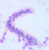
Microfilaria
Heartworms
The viewing field is to dark
Open the iris diaphragm
Increase the light intensity
Be sure that the condenser is up near the slide if the sample is a dry, stained smear.
The view field is partially lit and paritally black
Check the positioning of the objective lens
Be sure that the light is centered on the sample
The view field is cloudy or cannot be perfectly focused using the 40x objective lens
Use lens cleaning solution and lens paper to clean the objective lens
Place a coverslip on the slide to improve the performance of the 40x lens
A coverslip can be placed onto a slide using immersion oil or mounting medium
There is a stationary spot in the field of view that does not move when the slide is moved
Use lens cleaning solution and lens paper to clean the eyepieces
Use lens cleaning solution and lens paper to clean the objective lens
Remove the eyepieces and clean any debris at the bottom of the draw tube with lens paper
Wipe off the condenser with lens paper
Wipe off the light source with lens paper
You just obtained a PCV of 34% on Boo the dog. What is his approximate Hgb value? Include proper units.
11.3 g/dL
If you had a hemoglobin value of 2.5 g/dl on a horse you would expect the packed cell volume to be approximately what? Include proper units
7.5%
Estimate the hemoglobin on a bovine blood sample that has a PCV of 63%. Include proper units.
21 g/dL
Kristy was doing a quick PCV and TP for Dr. Funnell. She noticed that after she had spun the microhematocrit tube the plasma was milky red in color. She reported to Dr. Funnell that the PCV was 36%, the TP was 5.9 g/dl and that the plasma was:
Hemolyzed and lipemic
Peter did a TP on Olive the Samoyed puppy. He noticed that the plasma had a slight yellow tinge to it. What is the term for this yellow tinge?
Icteric
Plasma which is ______ and/or _____ may make it difficult to determine TP. TP may be elevated due to these changes.
Hemolyzed and/or lipemic
I’m looking at a hemacytometer on the microscope and I can’t find anything. The lighting seems a bit bright. How do you recommend resolving this problem? Give at least two solutions. Use the proper name for the microscope part to receive full credit.
Close the condenser some
You can turn down the Rheostat light control knob
Using the following laboratory data calculate the MCV, MCH and MCHC on this dog sample and then describe the blood cell picture.
Total Protein = 7.1 g/dL
WBC Count = 9,200 / mm3
RBC Count = 7.64 x 10⁶ / mm3
Hemoglobin = 16.1 g/dL
Hematocrit = 48 %
MCV = 62.8fL
MCH = 21.1pg
MCHC = 33.5g/dL
Normocytic, Normochromic
Estimate the PCV of a ferret sample that has a hemoglobin of 18 g/dl. Use proper units.
54%
Estimate the hemoglobin of a horse sample that has a PCV of 22%. Use proper units.
7.3 g/dL
Using the following laboratory data calculate the MCV, MCH and MCHC on this dog sample, then describe the blood cell picture
Total Protein = 7.3 g/dl
WBC count = 3,400 / mm3
RBC count = 2.15 x 10^6 / mm3
Hemoglobin = 5.0 g/dl
Hematocrit = 16%
MCV = 74.4fL
MCH = 23.3pg
MCHC = 31.3 g/dL
Macrocytic, hypochromic