Cell Biology - Ch. 9 Cytoskeletal Motor Proteins
1/20
There's no tags or description
Looks like no tags are added yet.
Name | Mastery | Learn | Test | Matching | Spaced |
|---|
No study sessions yet.
21 Terms
How is the energy for molecular motor proteins generated?
convert chemical energy (stored in ATP) into mechanical energy
What are the three categories of molecular motors?
kinesins, dyneins, and myosins
What types of cargo do these motor proteins move?
ribonucleoprotein particles, vesicles, mitochondria, lysosomes, chromosomes, and other cytoskeletal filaments
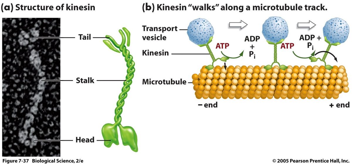
What is the structure of kinesin?

What type of cytoskeletal element does kinesin use for a track?
microtubules
Which direction do kinesins move?
towards the plus end of a microtubule
anterograde transport - from the center of the cell towards the periphery)
How is specificity for cargo achieved?
Many motor proteins don’t directly bind their cargo but rather rely on adaptor or scaffold proteins that link the motor to its cargo
What does highly processive mean?
the motor protein tends to move along an individual microtubule for considerable distances (over 1 µm) without falling off
because at least one of the heads is attached to the microtubule at all times
What are some functions of dynein?
As a force-generating agent in positioning the spindle and moving chromosomes during mitosis
As a minus end–directed microtubular motor with a role in positioning the centrosome and golgi complex and moving organelles, vesicles, and particles through the cytoplasm
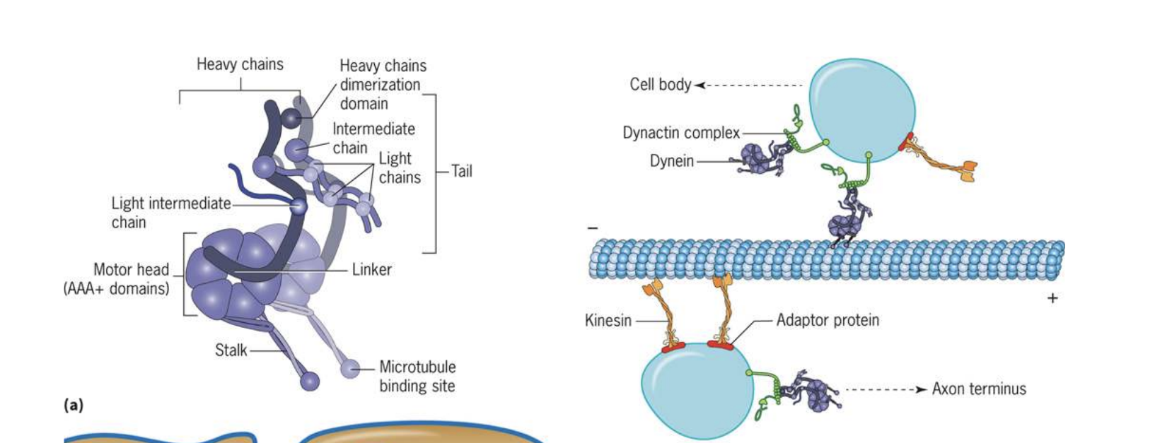
What is the structure of dynein?

Which direction do dyneins move?
towards the minus end of a microtubule
undergoes retrograde transport - moving from peripheral locations to the center of the cell
Why do dyneins need another protein called dynactin?
dyneins don’t interact directly with membrane-bound cargo, so it needs an intervening adaptor to link dynein to cargo via other proteins
helps regulates dynein activity by increasing the processivity of dynein
Describe the mechanism of cilia and flagella locomotion
inside cilia and flagella there are microtubule doublets arranged in a 9+2 structure, the dynein motors attach to one microtubule and "walk" along a neighboring one, pulling it downward
instead of the microtubules sliding past each other, linking proteins hold them together, causing the entire structure to bend
this bending happens in a coordinated, wave-like pattern, creating movement
cilia beat in a back-and-forth (oar-like) motion to move fluids or small particles (like clearing mucus from the lungs)
flagella (like in sperm cells) move in a whip-like or helical motion, propelling the cell forward
What is myosin? What are the two groups?
a type of motor protein that interacts with microfilament tracks to generate force from ATP for movement (plays a crucial role in muscle contraction, intracellular transport, and cell shape changes)
2 groups:
conventional myosins (type II) - first found in muscle tissue, forms thick filaments in sarcomeres, responsible for muscle contraction by sliding actin filaments past each other to shorten muscle fibers
unconventional myosins (everything else) - Involved in intracellular transport, organelle positioning, and membrane dynamics, does not form thick filaments
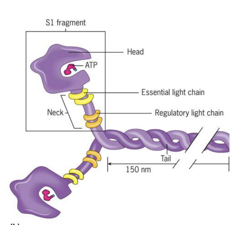
Describe the structure of myosin II

What is a muscle fiber?
a long, cylindrical cell that makes up muscle tissue and is specialized for contraction (basic unit of skeletal muscle)
What is a myofibril?
a long, cylindrical structure inside a muscle fiber composed of hundreds of thinner strands
What is a sarcomere?
a repeating linear array of actin and myosin filaments arranged along a myofibril
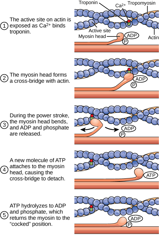
Explain the sliding filament model for muscle contraction. Explain the molecular basis and energetics for this model in detail.
1. Resting State (No Contraction)
Actin-binding sites are blocked by tropomyosin
Troponin holds tropomyosin in place, preventing myosin from binding to actin
ATP is bound to the myosin head, keeping it in a low-energy state
2. Initiation of Contraction (Calcium Activation)
Nerve impulses trigger the release of Ca²⁺ from the sarcoplasmic reticulum into the cytoplasm.
Ca²⁺ binds to troponin, causing a conformational change that shifts tropomyosin, exposing actin's myosin-binding sites
Myosin heads can now attach to actin, initiating contraction
3. Cross-Bridge Formation
The myosin head binds to actin, forming a cross-bridge
Myosin is in a high-energy state after hydrolyzing ATP into ADP + Pi
4. Power Stroke (Force Generation)
ADP and Pi are released, triggering the power stroke
The myosin head pulls actin toward the M-line, shortening the sarcomere
This generates force and brings the Z-discs closer together
5. Cross-Bridge Detachment
A new ATP molecule binds to myosin
This weakens the actin-myosin interaction, causing myosin to detach from actin
6. Myosin Reset (Recovery Stroke)
ATP is hydrolyzed to ADP + Pi, re-positioning the myosin head into its high-energy conformation
Myosin is now ready for another cycle of contraction

How do tropomyosin and troponin function in muscle contraction?
tropomyosin blocks myosin-binding sites on actin, preventing contraction and keeping the muscle relaxed
troponin is a complex of three proteins that binds to calcium ions, moves tropomyosin, allows myosin to bind, which then causes the muscle to contract
What is titin?
a giant elastic protein found in muscle cells that maintain the thick filaments in the center of the sarcomere during contraction