TAMU BIOL 319 LAB Preface and Lab 1
1/128
There's no tags or description
Looks like no tags are added yet.
Name | Mastery | Learn | Test | Matching | Spaced | Call with Kai |
|---|
No analytics yet
Send a link to your students to track their progress
129 Terms
Sagittal plane/section
divide the body or organ into right and left portions
Midsagittal plane/section
divides the body or organ into equal right and left sides
Frontal (coronal) plane/section
divides the body or organ into anterior and posterior portions
Transverse (cross section)
divides the body or organ into superior and inferior sides
Oblique
passes through the body or organ at an angle
Anatomical position
To stand erect with arms at the sides and palms of the hands turned forward
Superior (cranial)
toward the head end or upper part of a structure or the body; above
Inferior (caudal)
away from the head end or toward the lower part of a structure or the body; below
Anterior (ventral)
Nearer to or at the front of the body
Posterior (dorsal)
Nearer to or at the back of the body
Medial
Nearer to the midline of the body
Lateral
Further from the midline of the body
Intermediate
Between two structures
Ipsilateral
On the same side of the body as another structure
Contralateral
On the opposite side of the body as a structure
Proximal
Closer to the trunk or point of attachment
Distal
Farther from the trunk or point of attachment
Superficial
Toward or on surface of the body
Deep
Away from the surface of the body
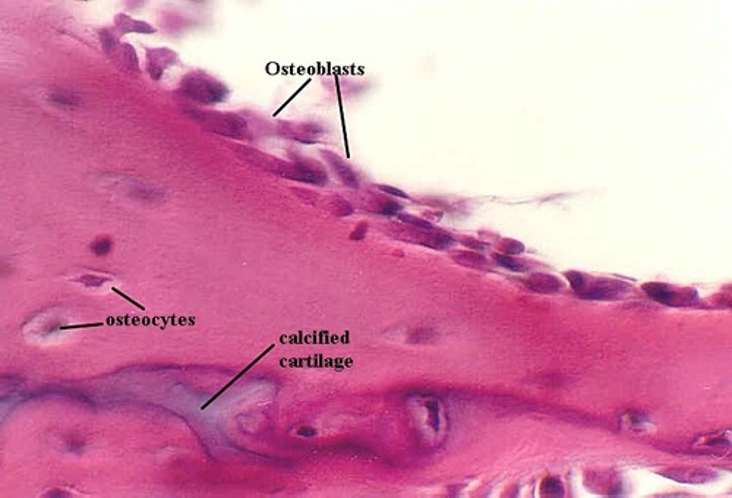
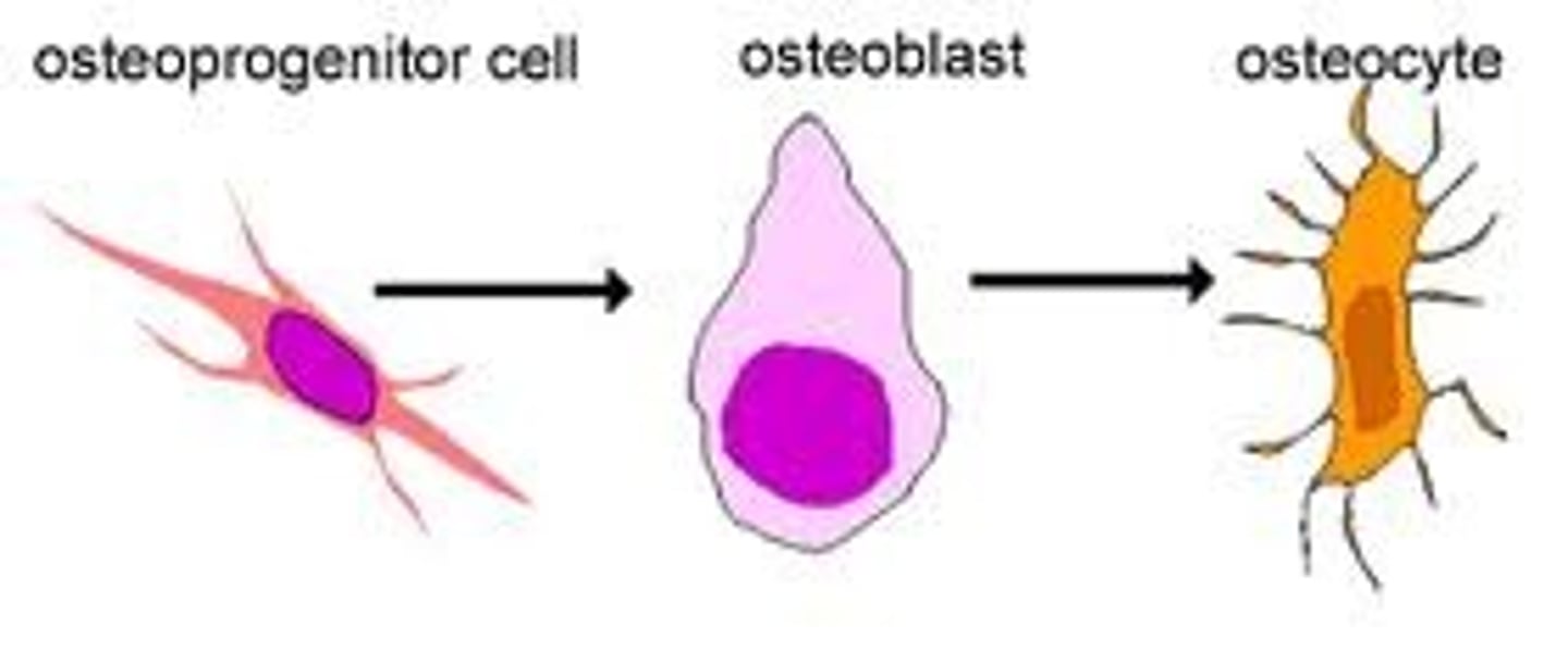
Osteocyte
derived from osteoblasts- involved in bone maintenance
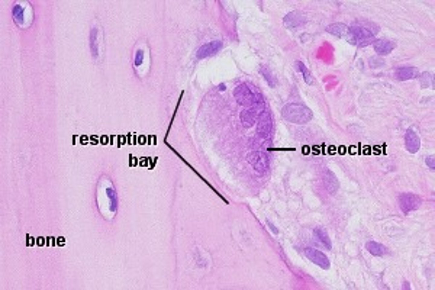
Osteoclast
Multinucleate cell that secretes acids and enzymes to dissolve bone matrix
Osteoblast
Derived from osteoprogenitor cells - secretes organic components of matrix
Osteoprogenitor cells
stem cells that differentiate into osteoblasts
Histology
study of tissues
epithelial tissue
tissue that lines and covers organs as well their internal passageways and forms glands; made up of sheet of cells
Apical (free) surface
Surface of the cells that are exposed to the external environment or to an internal passage way or cavity
Simple epithelium
Single layer of cells
Stratified epithelium
Composed of more than one layer of cells
Regeneration of tissues
How easily the tissues can be regenerated; based on the rate of mitosis and the amount of blood supply
Examples of good to excellent regeneration of tissue
Epithelial tissue, bone, areolar tissue, dense irregular connective tissue, and blood forming tissue
Examples of moderate regeneration of tissue
Smooth muscle and dense regular connective tissue
Examples of weak regeneration of tissue
Skeletal muscle and cartilage
Example of none of almost no regeneration of tissue
Cardiac muscle tissue and (central nervous system) nervous tissue
Reasons why regeneration of tissue can be high
If the rate of mitosis is high and there is adequate or abundant blood supply
Avascular
No blood vessels or supply
Function: simple squamous epithelium
To allow passage of materials by diffusion and filtration in sites where protection is not important; secrets lubricating substances in serosae

Description: simple squamous epithelium
single layer of flattened cells with disc-shaped central nuclei and sparse cytoplasm; the simplest of the epithelia
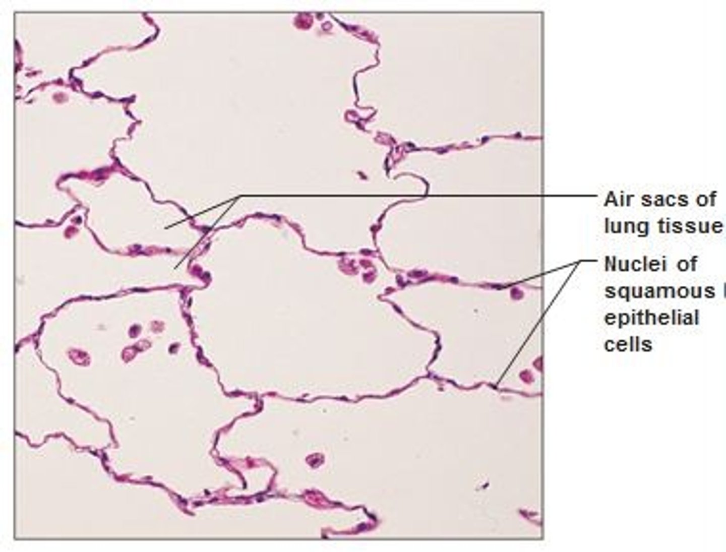
Location: simple squamous epithelium
Kidney glomeruli, air sacs of lungs, lining of heart, blood vessels, lymphatic vessels, lining of ventral body cavity
Avelous
Air sack present in between linings of the simple squamous in the lung

Endothelium
provides a slick, friction-reducing lining in lymphatic vessels and in all hollow organs of the cardiovascular system
Mesothelium
the epithelium found in serous membranes lining the ventral body cavity and covering its organs
Description: simple cuboidal epithelium
single layer of cube-like cells with large, spherical central nuclei

Function: simple cuboidal epithelium
Secretion and absorption
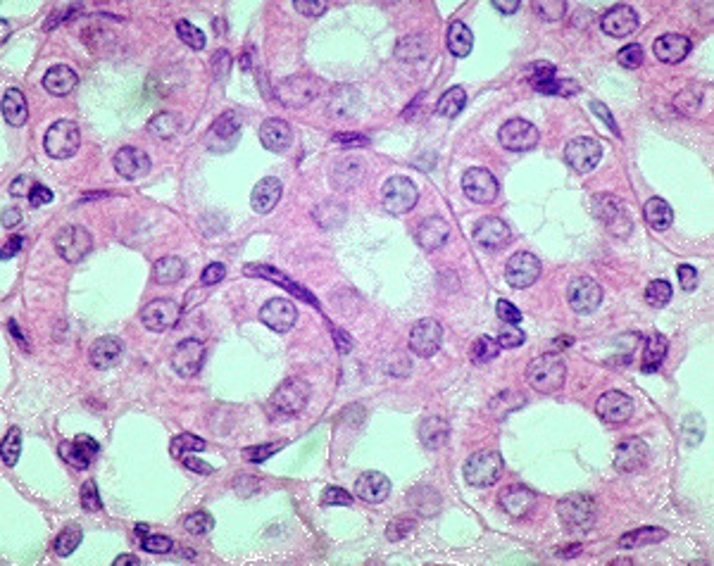
Location: simple cuboidal epithelium
Kidney tubules; ducts and secretory portions of small glands; ovary surface

Tubule lumen
pocket that lies in the center of a circle of simple cuboidal epithelium
Description: simple columnar epithelium
single layer of tall cells with round to oval nuclei; some cells bear cilia; layer may contain mucus-secreting unicellular glands (goblet cells)
Function: simple columnar epithelium
absorption; secretion of mucus, enzymes, and other substances; ciliated type propels mucus (or reproductive cells) by ciliary action
Location: simple columnar epithelium
Nonciliated type lines most of the digestive tract (stomach to rectum), gallbladder, and excretory ducts of some glands; ciliated variety lines small bronchi, uterine tubes, and some regions of the uterus.
Goblet cells
secrete mucus
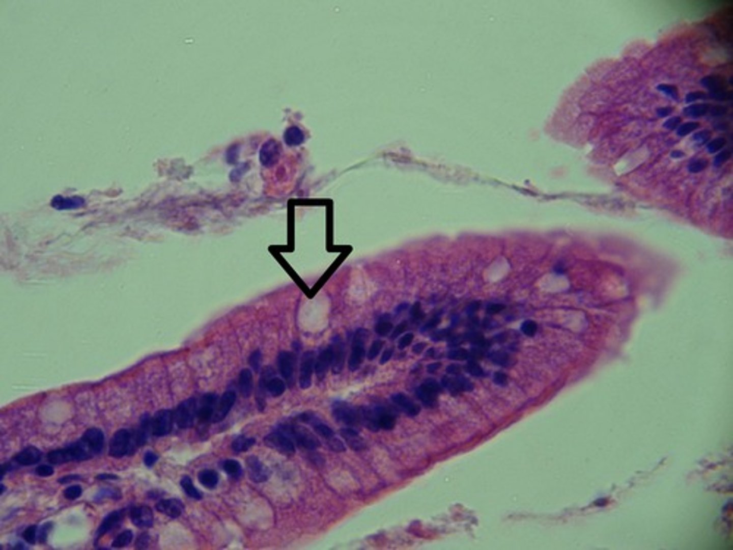
How simple columnar epithelium looks under the microscope
Almost looks like petals with nuclei near the inner rim; the goblet cells look like white pockets
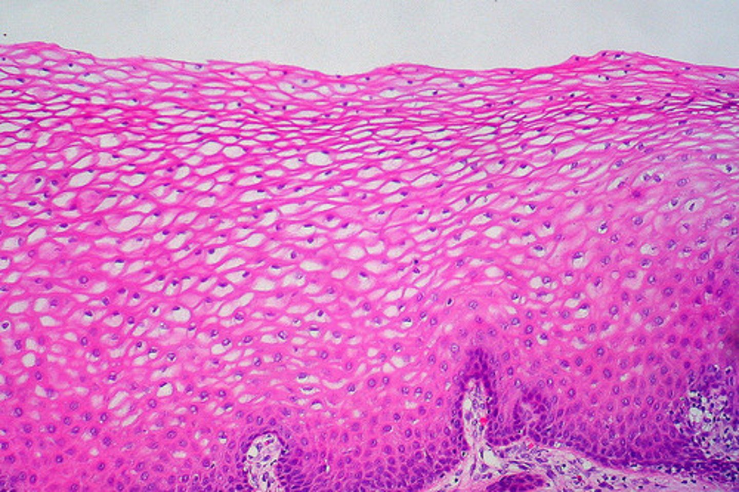
Description: stratified squamous epithelium
thick membrane composed of several cell layers; basal cells are cuboidal or columnar and metabolically active; surface cells are flattened (squamous); in the keratinized type, the surface cells are full of keratin and dead; basal cells are active in mitosis and produce the cells of the more superficial layers
Function: stratified squamous epithelium
protects underlying tissues in areas subjected to abrasion

Location: stratified squamous epithelium
nonkeratinized type forms the moist linings of the esophagus, mouth, and vagina; keratinized variety forms the epidermis of the skin, a dry membrane

Basement membrane
Cells at the base of an epithelial layer are attached to this
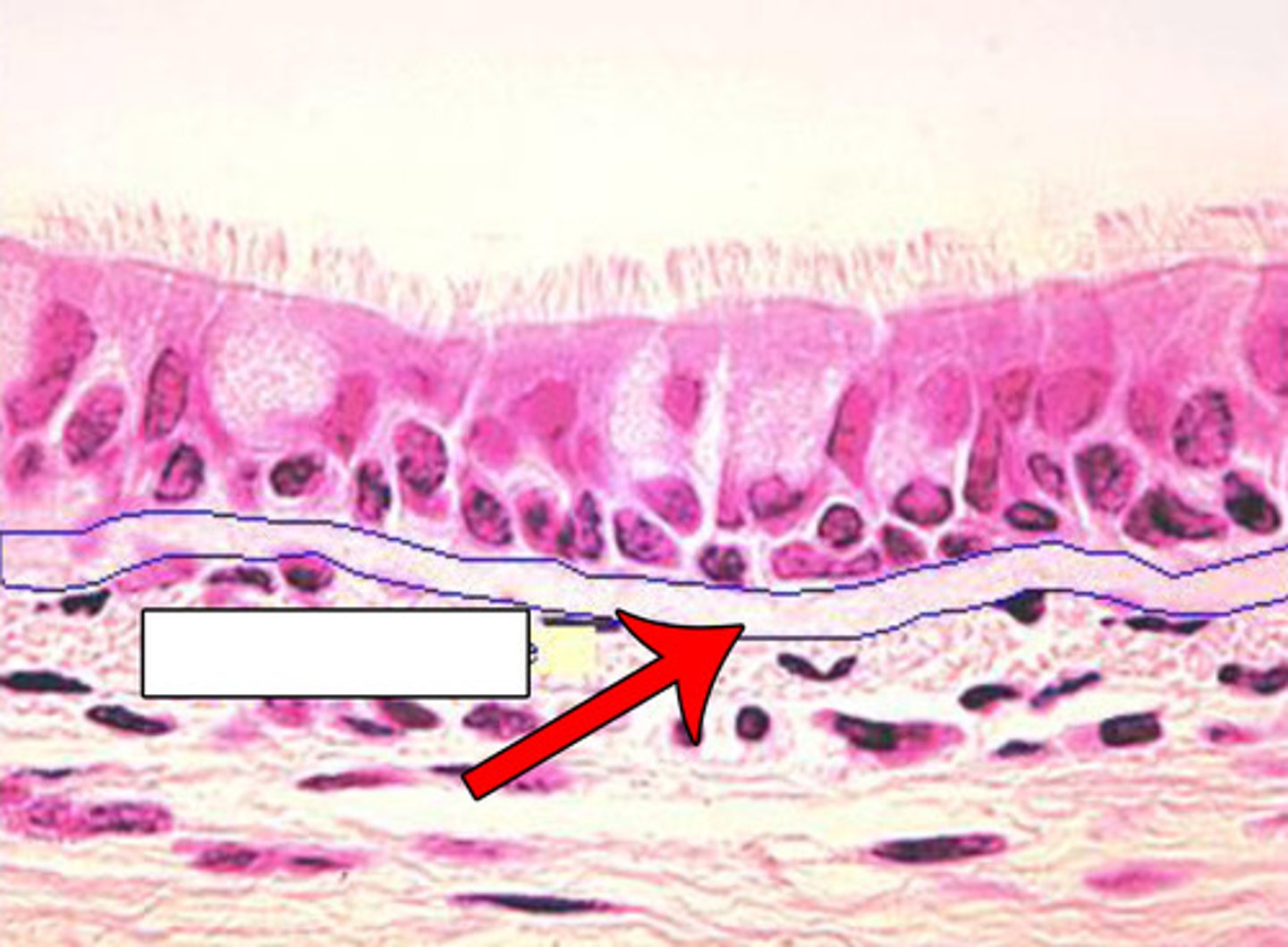
Description: pseudostratified ciliated columnar epithelium
Single layer of cells of differing heights, some not reaching the free surface; nuclei seen at different levels; may contain mucus-secreting cells and bear cilia

Function: pseudostratified ciliated columnar epithelium
Secretion, particularly of mucus; propulsion of mucus by ciliary action
Location: pseudostratified ciliated columnar epithelium
Nonciliated type in male's sperm-carrying ducts and ducts of large glands; ciliated variety lines the trachea, most of the upper respiratory tract

Description: transitional epithelium
resembles both stratified squamous and stratified cuboidal; basal cells cuboidal or columnar; surface cells dome shaped or squamous-like, depending on degree of organ stretch
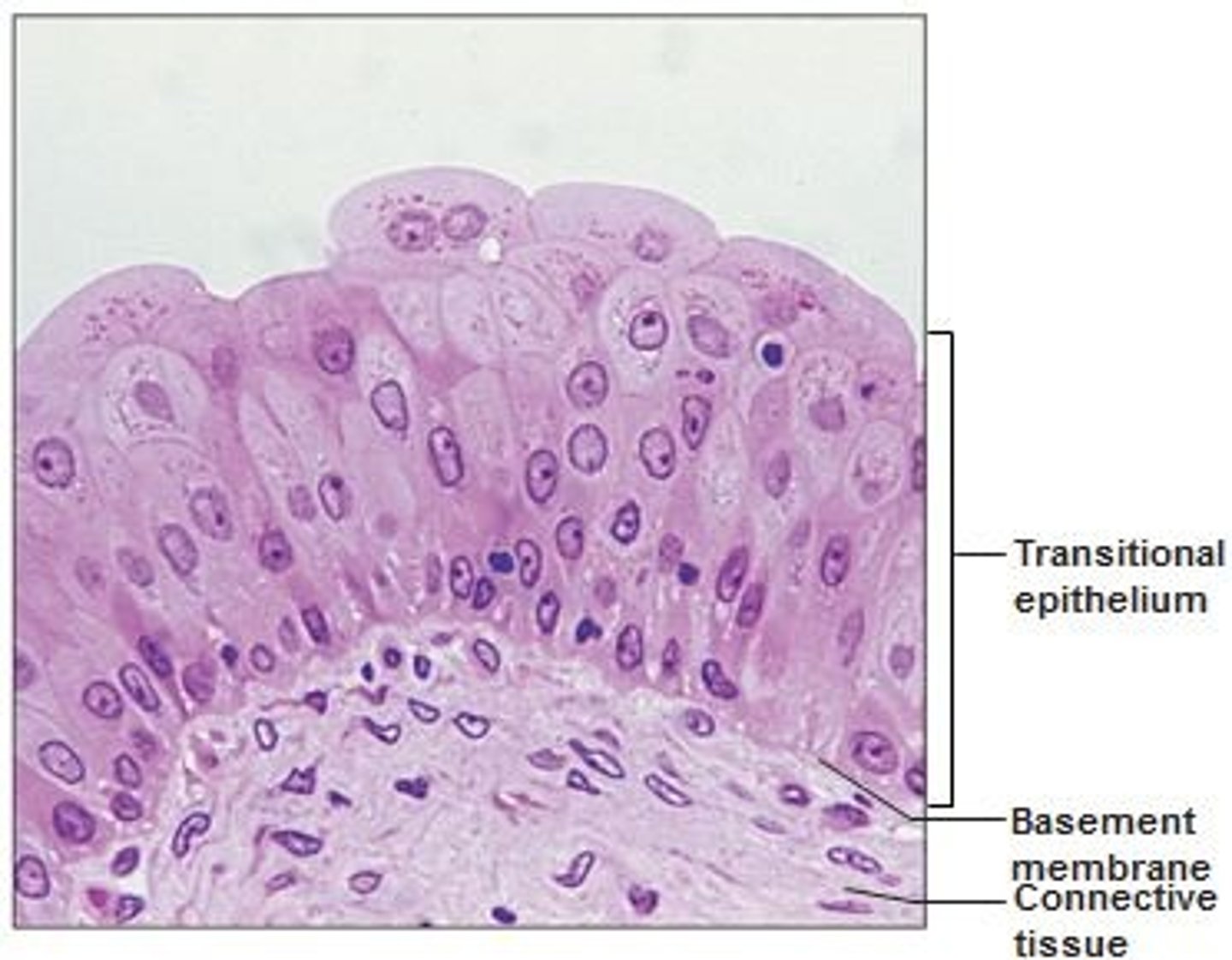
Function: transitional epithelium
stretches readily and permits distension of urinary organ by contained urine

Location: transitional epithelium
lines the ureters, bladder, and part of the urethra

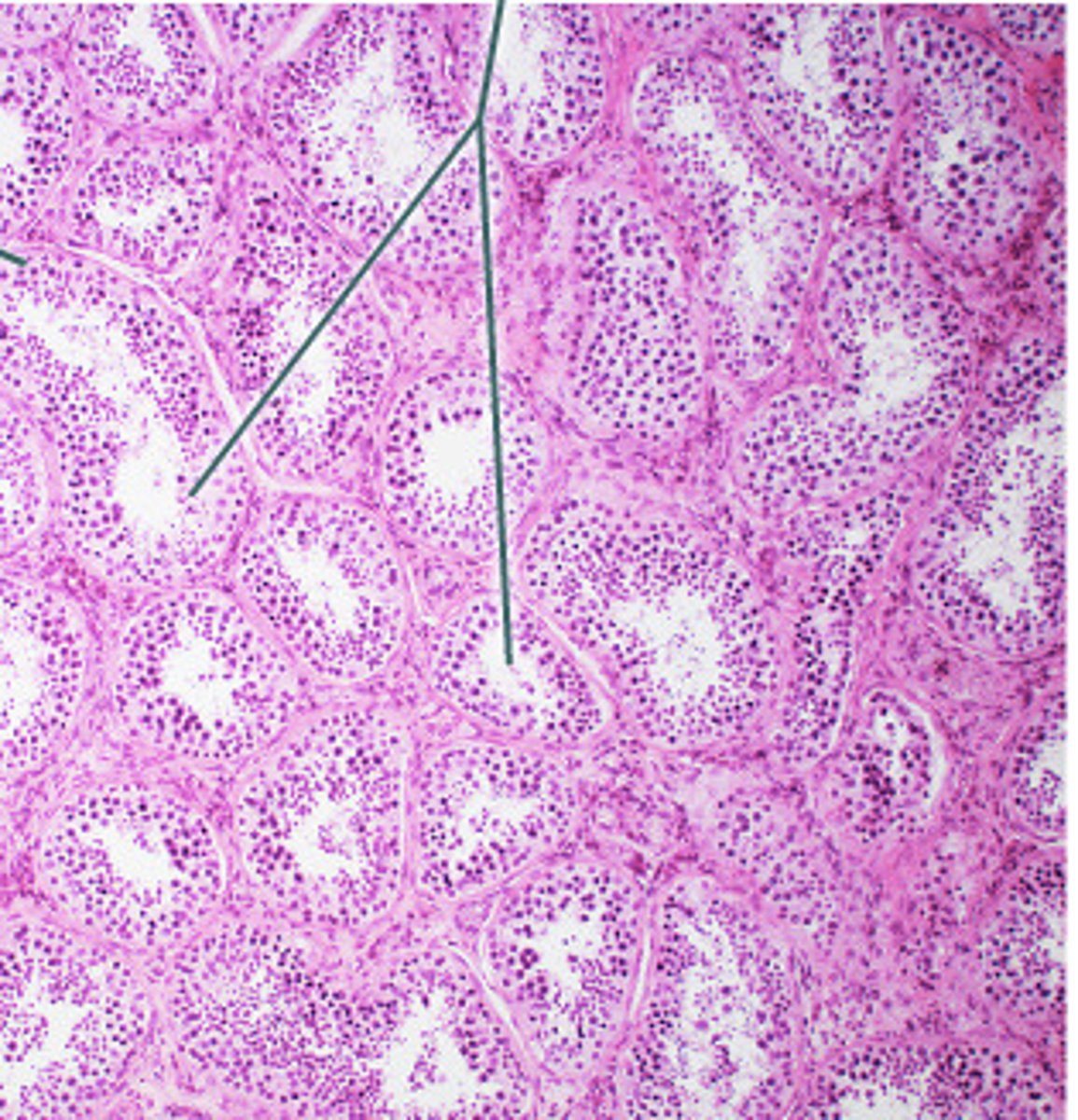
Connective tissue
A body tissue that provides support for the body and connects all of its parts
Fibroblasts
In connective tissue, cells that secrete the proteins of the fibers.
Mast cells
Detect foreign microorganisms and initiate immune responses against them
Adipocytes
fat cells that make up most of the subcutaneous layer
Loose connective tissue
has an open network of protein fibers in a thick, syrupy ground substance and is divided into three groups
Dense connective tissue
Made up of two types of fibers: protein fibers assembled into thick bundles of collagen and elastic fibers with widely scattered cells
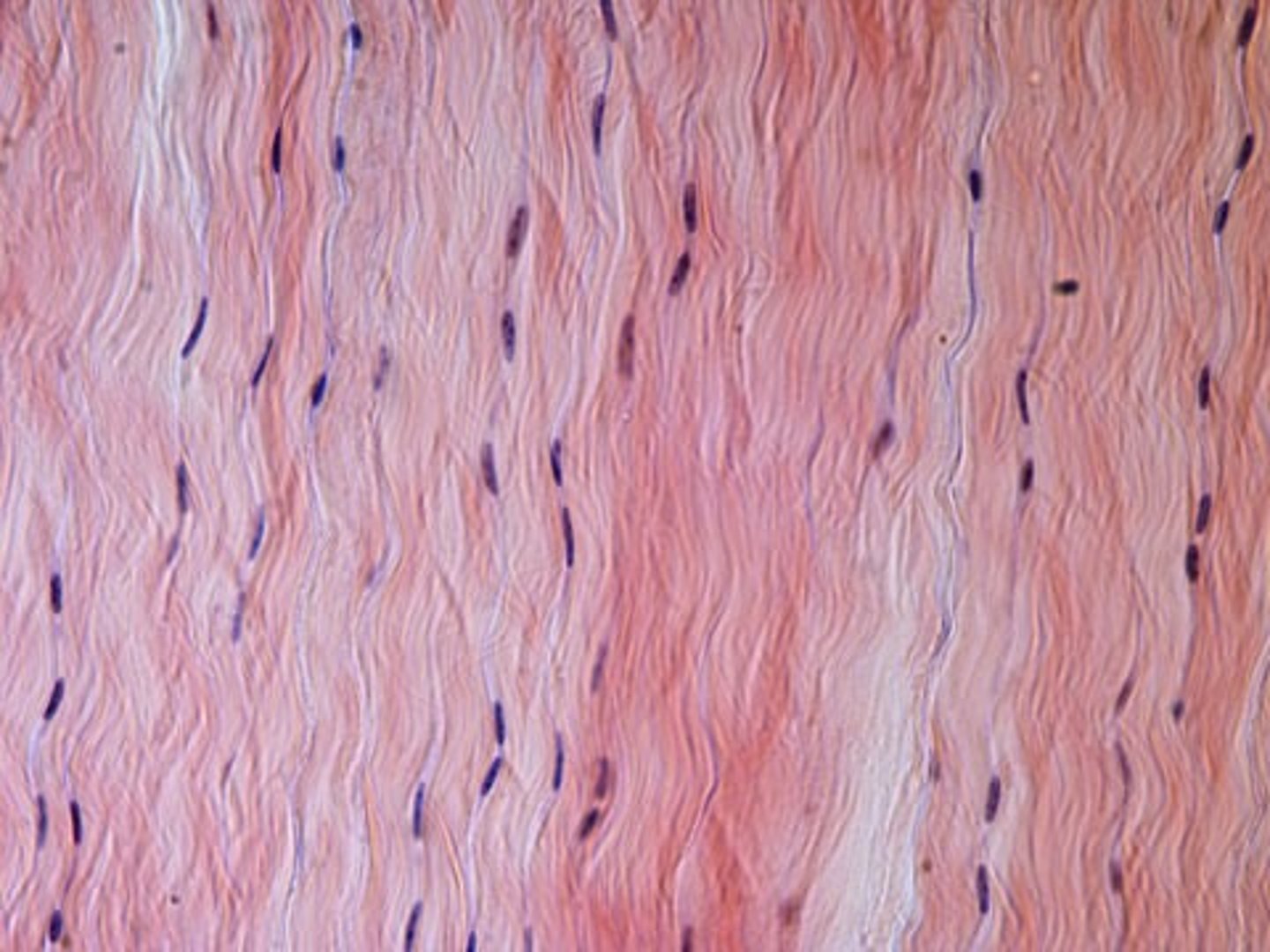
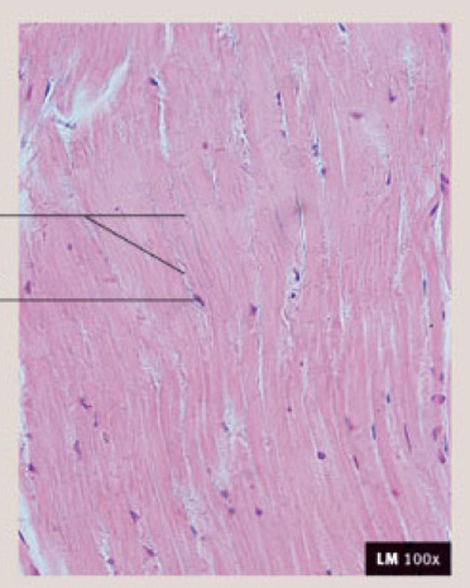
Dense regular
Protein fibers in the matrix are arranged in parallel bands
Dense irregular
fibers are interwoven
What are the two types of fluid connective tissue?
blood and lymph
What are the two types of supporting connective tissue?
bone and cartilage
Perichondrium
surrounds all supporting connective tissue in cartilage and produces chondroplasts
What are the three types of cartilage?
hyaline, elastic, fibrocartilage
Which tissue has all 3 fibers present?
Areolar tissue
Description: Areolar Connective Tissue
gel-like matrix with all three fiber types; cells: fibroblasts, macrophages, mast cells, and some white blood cells; loose arrangement of fibers creating space for water and salt storage

Function: Areolar Connective Tissue
wraps and cushions organs; its macrophages phagocytize bacteria; plays important role in inflammation; holds and conveys tissue fluid
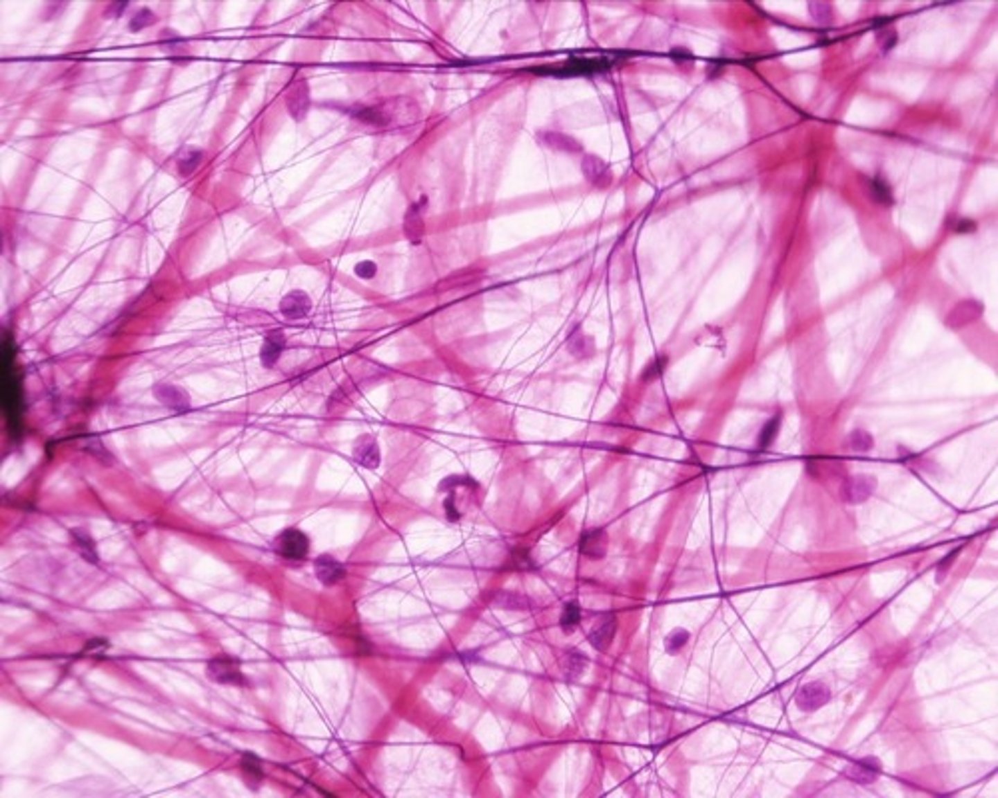
Location: Areolar Connective Tissue
Widely distributed under epithelia of body, e.g., forms lamina propria of mucous membranes; packages organs; surrounds capillaries.

Description: Adipose tissue
matrix as in areolar, but very sparse; closely packed adipocytes, or fat cells, have nucleus pushed to the side by large fat droplet
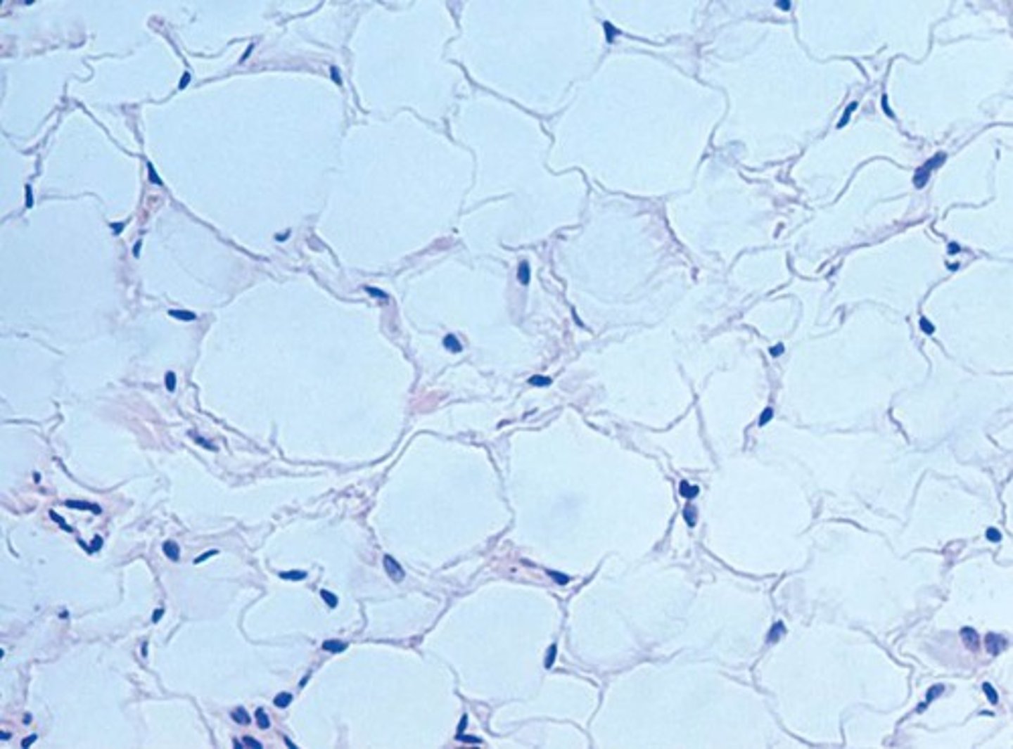
Function: Adipose tissue
provides reserve food fuel; insulates against heat loss; supports and protects organs

Location: Adipose tissue
under skin in subcutaneous tissue; around kidneys and eyeballs; within abdomen; in breasts; around heart and in bone marrow (serves as fuel for high energy demand organs)

Description: Reticular tissue
network of reticular fibers in a typical loose ground substance; reticular cells lie on the network

Function: Reticular tissue
fibers form a soft internal skeleton (stroma) that supports other cell types including white blood cells, mast cells, and macrophages
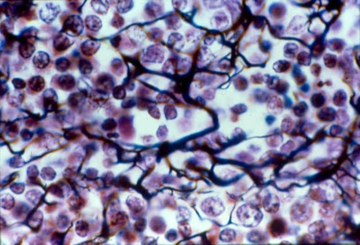
Location: Reticular tissue
lymphoid organs (lymph nodes, bone marrow, and spleen)

Description: Dense regular connective tissue
Primarily parallel collagen fibers; a few elastic fibers; major cell type is the fibroblast
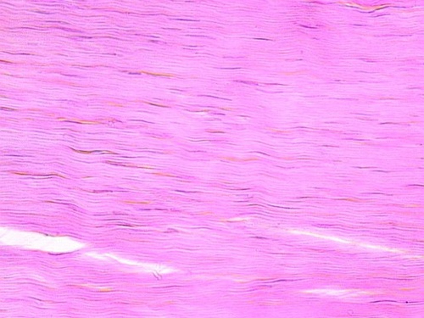
Function: Dense regular connective tissue
attaches muscles to bones or to muscles; attaches bones to bones; withstands great tensile stress when pulling force is applied in one direction
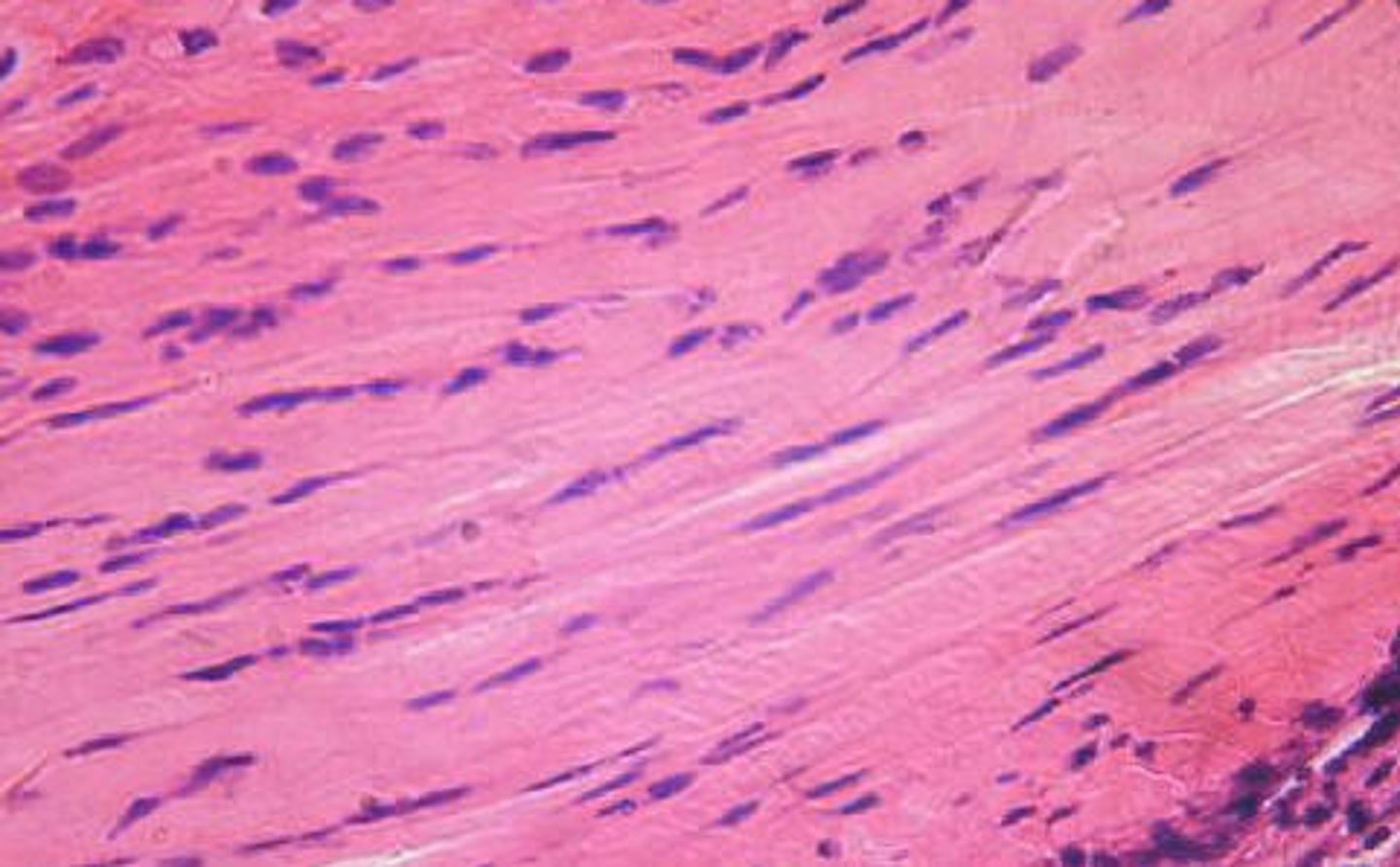
Location: Dense regular connective tissue
Tendons (connect muscle to bone); most ligaments (connect bone to bone); aponeuroses

Description: Dense regular elastic tissue
Dense regular elastic tissue containing a high proportion of elastic fibers
Function: Dense regular elastic tissue
Allows recoil of tissue following stretching; maintains pulsatile flow of blood through arteries; aids passive recoil of lungs following respiration

Location: Dense regular elastic tissue
walls of large arteries; within certain ligaments associated with the vertebral column; within the walls of the bronchial tubes
Description: Blood
Red and white blood cells in fluid matrix

Function: Blood
Transport of respiratory gases, nutrients, wastes and other substances
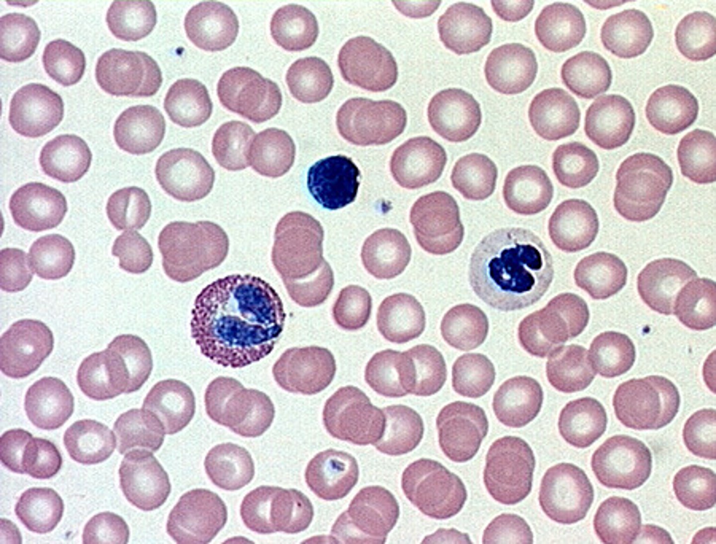
Location: Blood
contained within blood vessels

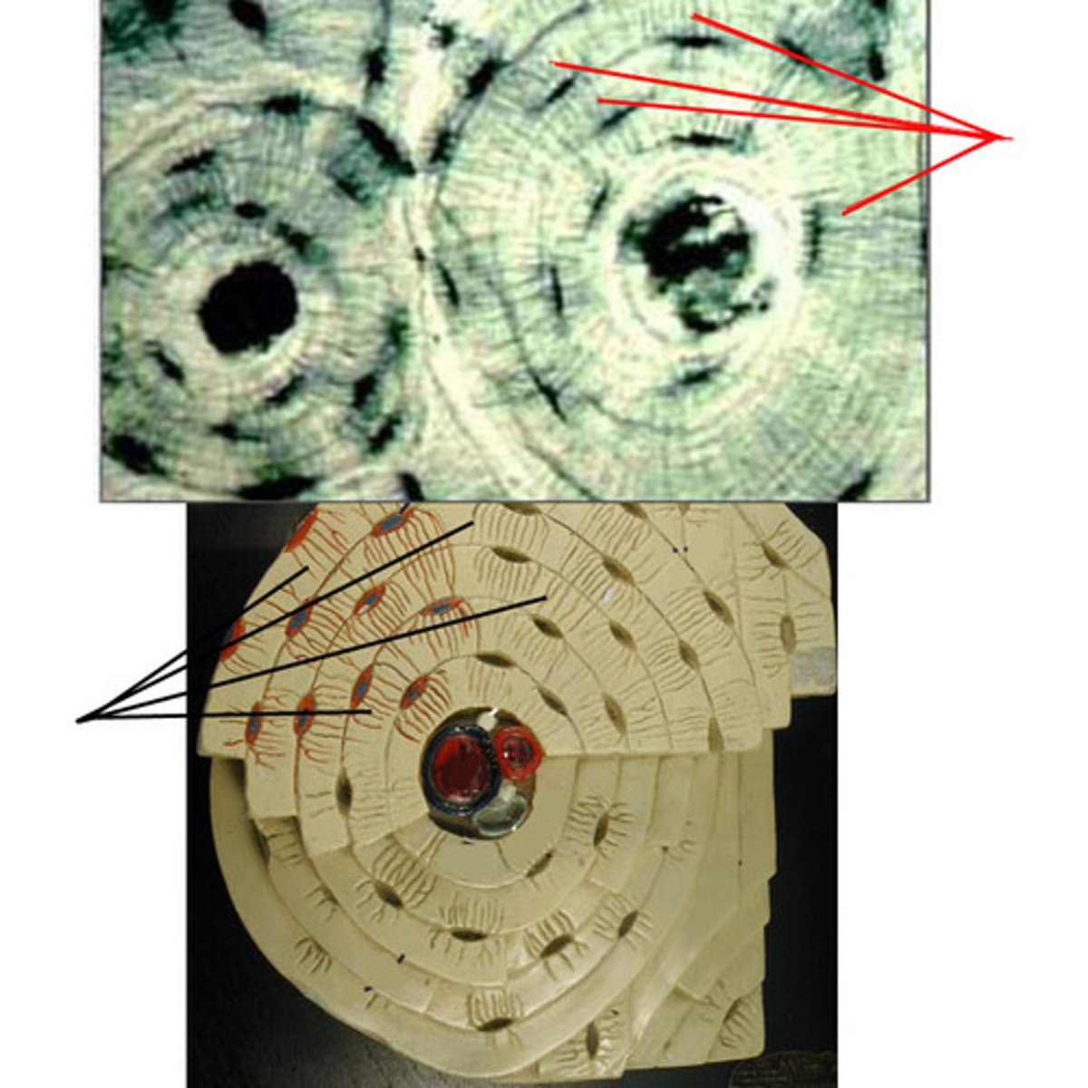
Description: Bone
hard, calcified matrix containing many collagen fibers; osteocytes lie in lacunae

Function: Bone
bone supports and protects (by enclosing); provides levers for the muscles to act on; stores calcium and other minerals and fat; marrow inside bones is the site for blood cell formation (hematopoiesis)

Location: Bone
bones
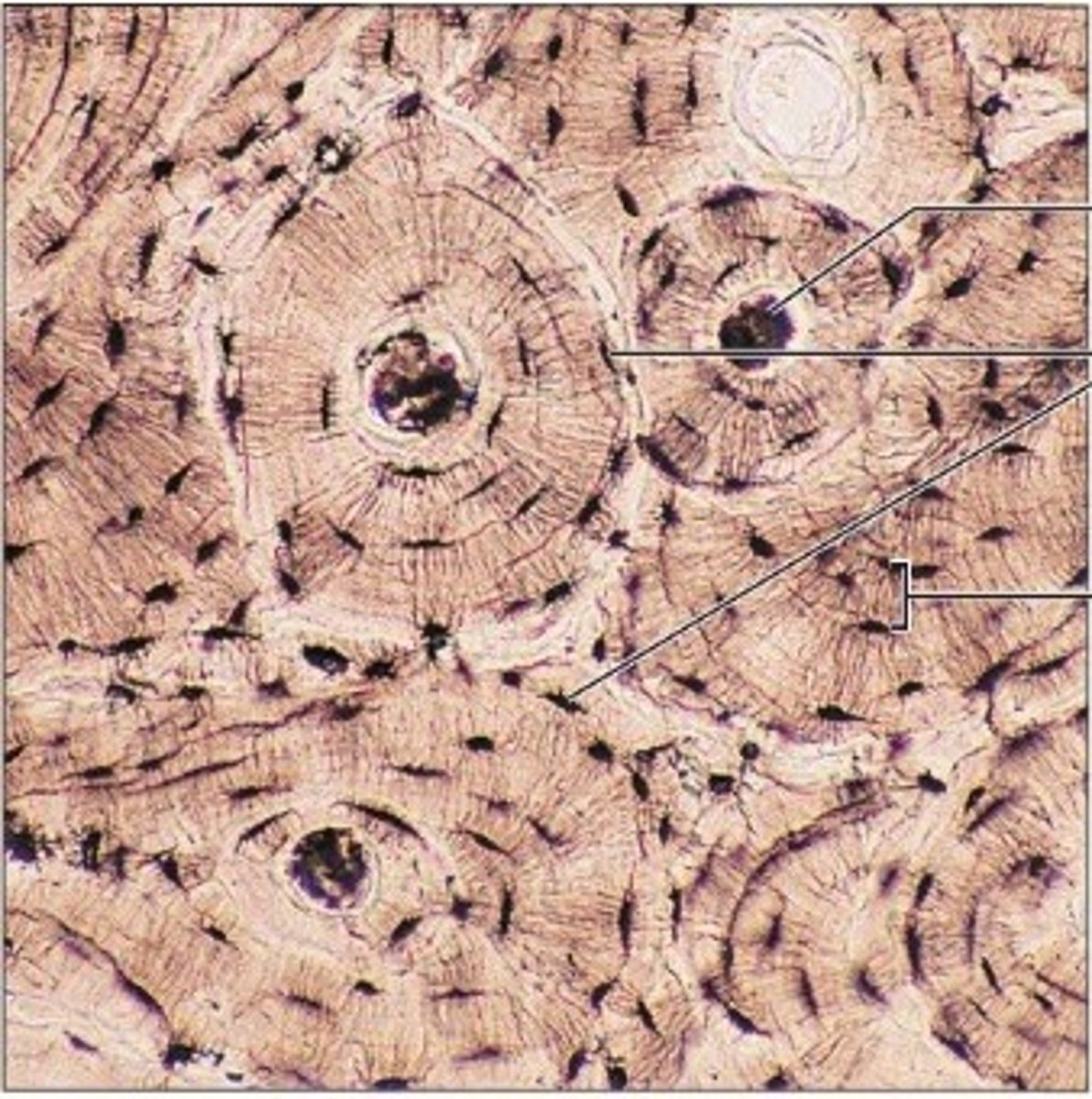
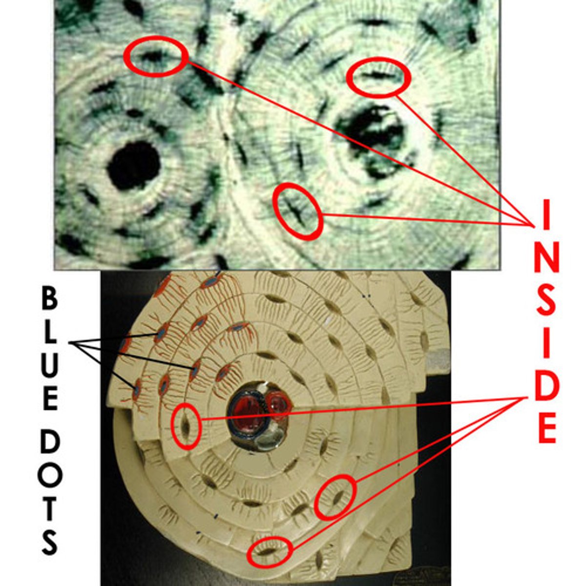
Lamellae
Concentric rings made up of groups of hollow tubes of bone matrix
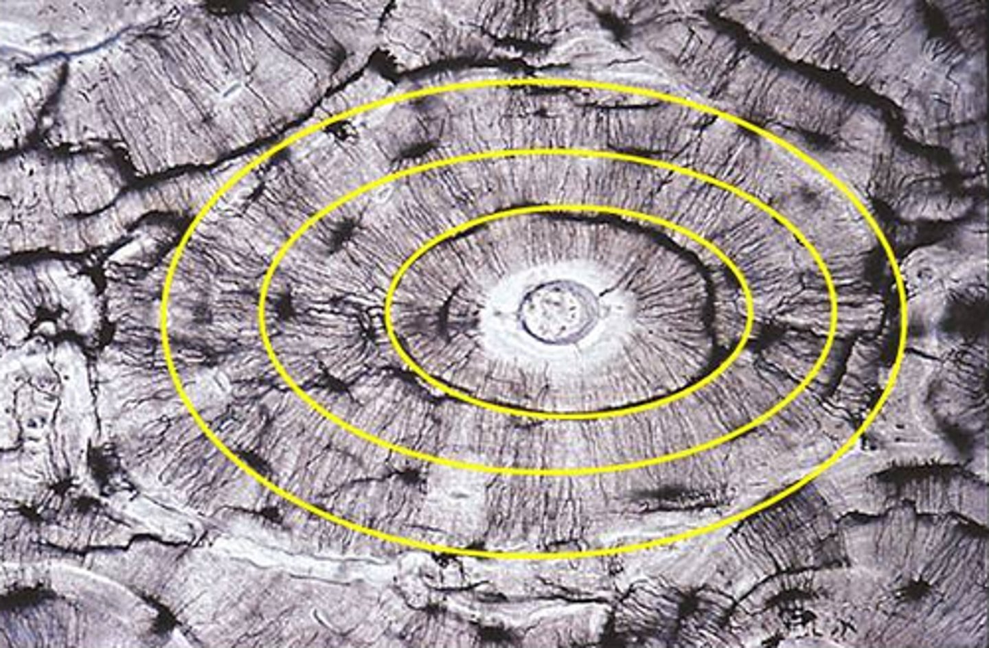
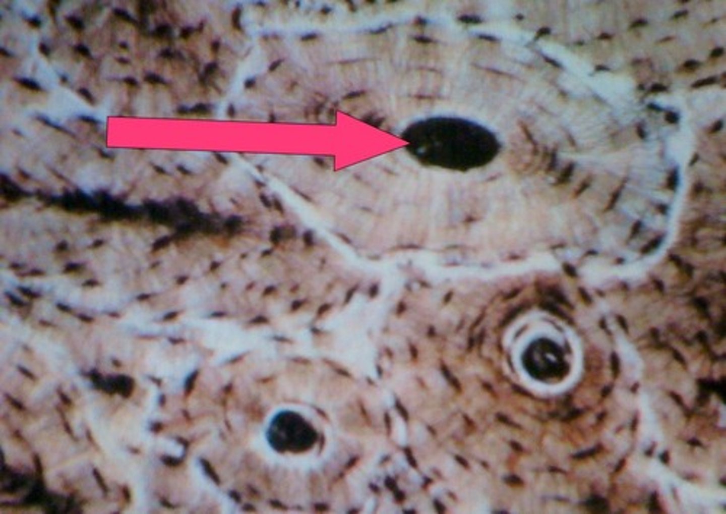
Haversian canal
one of a network of tubes running through compact bone that contains blood vessels and nerves; center dark spot in the osteoblasts
Canaliculi
Hairlike canals that connect lacunae to each other and the central canal
Description: Hyaline cartilage
amorphous but firm matrix; collagen fibers form an imperceptible network; chondroblasts produce the matrix and when mature (chondrocytes) lie in lacunae

Function: Hyaline cartilage
supports and reinforces; serves as resilient cushion; resists compressive stress
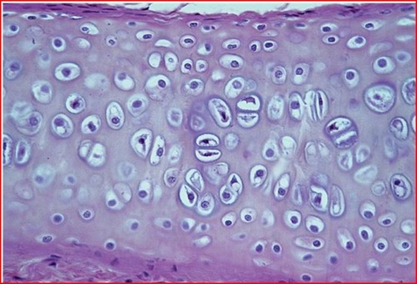
Location: Hyaline cartilage
forms most of the embryonic skeleton; covers the ends of long bones in joint cavities; forms costal cartilages of the ribs; cartilages of the nose, trachea, and larynx
