module 3 exchange and transport
1/22
There's no tags or description
Looks like no tags are added yet.
Name | Mastery | Learn | Test | Matching | Spaced | Call with Kai |
|---|
No analytics yet
Send a link to your students to track their progress
23 Terms
3.1.3 a) the need for transport systems in multicellular plants
multicellular - low SA:vol
diffusion is too slow to meet metabolic needs
substances must be moved over long distances
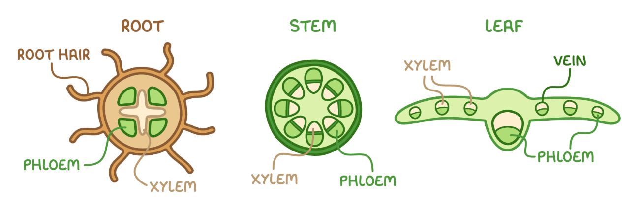
3.1.3 b) i) structure and functions of the vascular system in roots, stems and leaves of herbaceous dicotyledonous plants
roots:
xylem forms central cylinder surrounded by phloem
provides support as root grows through soil
stem:
xylem + phloem in outer region
forms scaffolding to resist bending
leaf:
xylem + phloem form a network of veins
provides support for thin leaves
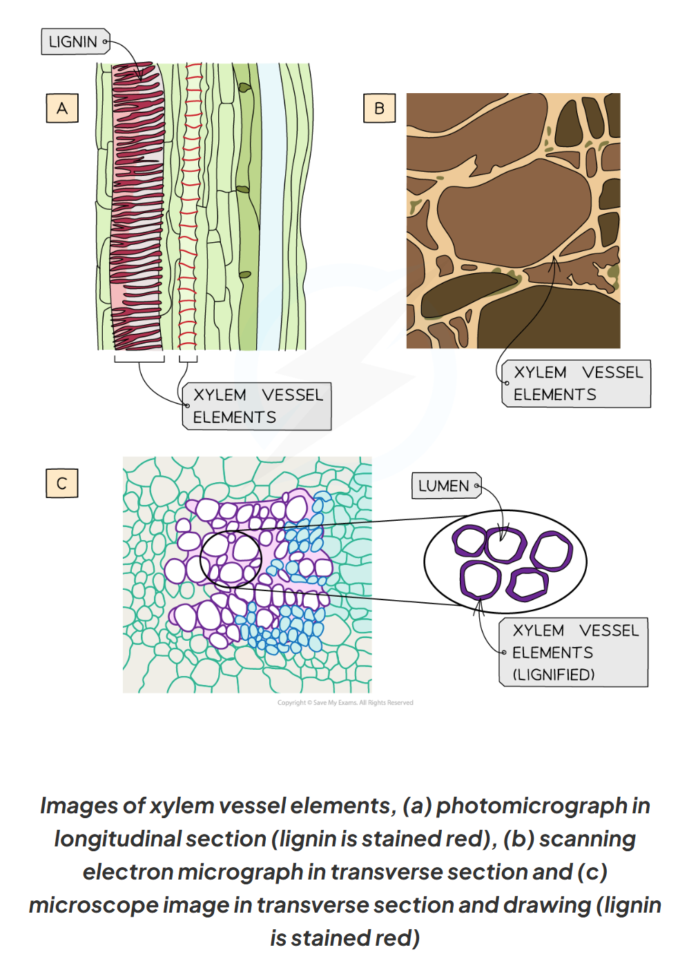
3.1.3 b) ii) the examination and drawing of stained sections of plant tissue to show the distribution of xylem and phloem
3.1.3 b) iii) the dissection of stems, both longitudinally and transversely, and their examination to demonstrate the position and structure of xylem vessels
3.1.3 c) i) the process of transpiration and the environmental factors that affect transpiration rate
light intensity - at high light intensities, stomata open for maximum CO2 absorption for photosynthesis
temperature - at high temperatures, evaporation of water molecules is faster, due to higher KE
humidity - low humidity increases water vapour gradient between the leaf and the atmosphere
wind speed - high wind speeds increase water potential gradient between leaf and atmosphere
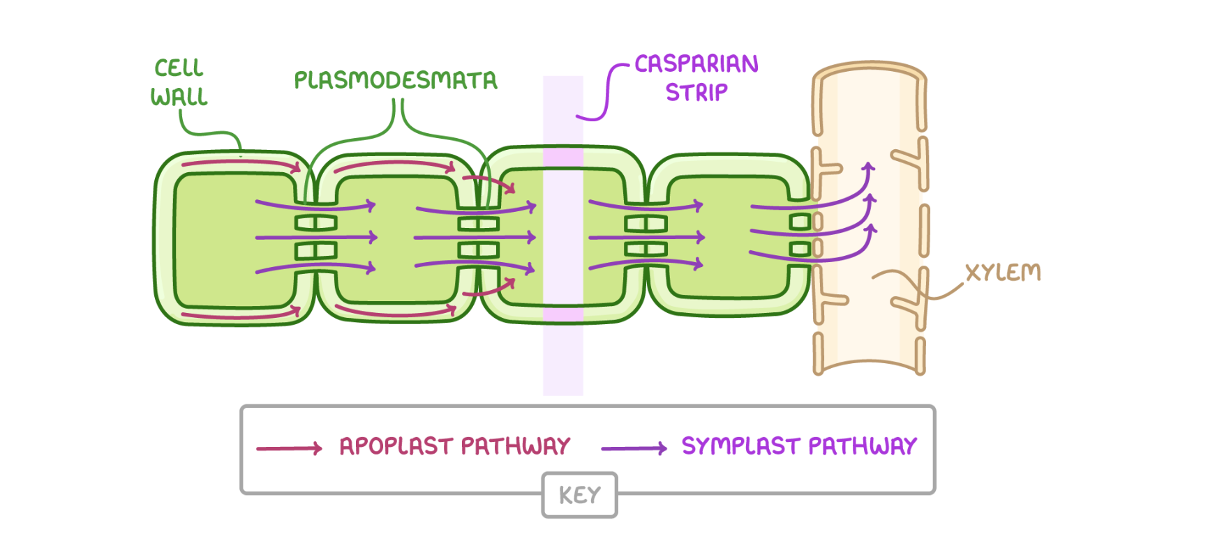
3.1.3 d) the transport of water into the plant, through the plant and to the air surrounding the leaves (part 1)
apoplastic pathway:
water travels within cell walls and in intercellular spaces
water is drawn across the root due to the apoplastic pathway due to cohesive forces between water molecules
cohesion between water molecules means that more water molecules are drawn along the apoplast pathway within the root to replace the water that has moved upwards
symplast pathway:
water travels via cytoplasm and vacuoles by osmosis - or from cell to cell by diffusion via the plasmodesmata
water moves into root hair cells from the soil by osmosis, increasing the water potential of root hair cells
water moves down water potential gradient into neighbouring root cells
3.1.3 d) the transport of water into the plant, through the plant and to the air surrounding the leaves (part 2)
transpiration:
water evaporates from the moist surfaces of mesophyll cells
stomata open so they can absorb CO2 for photosynthesis
provides pathway for water loss through open stomata
water vapour moves down water potential gradient from air spaces in the lead into the atmosphere
cohesion: attraction between molecules of the same substance
allows water molecules to form hydrogen bonds with each other
creates surface tension on the surface of the water
helps maintain the transpiration stream
adhesion: attraction between molecules of different substances
can form hydrogen bonds with other charged surfaces
helps water climb up narrow tubes like xylem vessels
3.1.3 e) adaptations of plants to the availability of water in their environment (xerophytes)
fewer stomata - to avoid dehydration
sunken stomata in pits in the epidermis - traps moist air, decreasing water potential gradient
fine hairs (trichomes) covering the epidermis - traps moist air
curled leaves - stomata sheltered from the wind
leaves and stems covered in a waxy cuticle
3.1.3 e) adaptations of plants to the availability of water in their environment (hydrophytes)
plant - cell wall - no excess water uptake
floating leaves - thin + flat, large air spaces for buoyancy - floating on the water surface allows for more access to light for photosynthesis
stomata on upper surface - allows for gas exchange with air
reduced root systems - can extract nutrients from surrounding water through tissues
reduced veins in the leaves - xylem are reduced as there is no need to transport water through the plant
3.1.3 f) the mechanism of translocation
3.1.2 a) the need for transport systems in multicellular animals
take in: nutrients and oxygen
release: waste products
factors: increased transport systems, SA:vol, increased levels of activity
3.1.2 b) the different types of circulatory systems
single circulatory systems: heaty only has one atrium and one ventricle, blood passes through heart once in a complete circuit of the body
double circulatory systems (mammals): blood passes through the heart twice during a circuit of the body
closed circulatory systems (all vertebrae and many insects): blood pumped around the body is always contains in a network of vessels
open circulatory systems (molluscs and arthropods): blood pumped directly into body cavities
circulatory systems in insects one main blood vessel (dorsal vessel), tubular heart pumps haemolymph (insect blood) into the dorsal vessel
3.1.2 c) the structure and function of arteries, arterioles, capillaries, venules and veins
arteries: transport blood away from the heart (usually at high pressure) to tissues
arterioles: arteries branch into narrower blood vessels called arterioles which transport blood into capillaries
veins: transport blood to the heart (usually at low pressures)
venules: narrower blood vessels which transport blood from capillaries to the veins
capillaries: thin walls, allowing substances to leave the blood to reach the body’s tissues
3.1.2 d) the formation of tissue fluid from plasma
at the arteriole end of a capillary, there is high hydrostatic pressure
the pressure forces water + small solutes out of the capillary
forms tissue fluid, which bathes the cells, delivering nutrients and oxygen
3.1.2 f) the cardiac cycle
series of events that take place in one heartbeat, including muscle contraction and relaxation
contraction: systole - decrease in heart volume, increase in pressure
relaxation: diastole - increase in heart volume, decrease in pressure
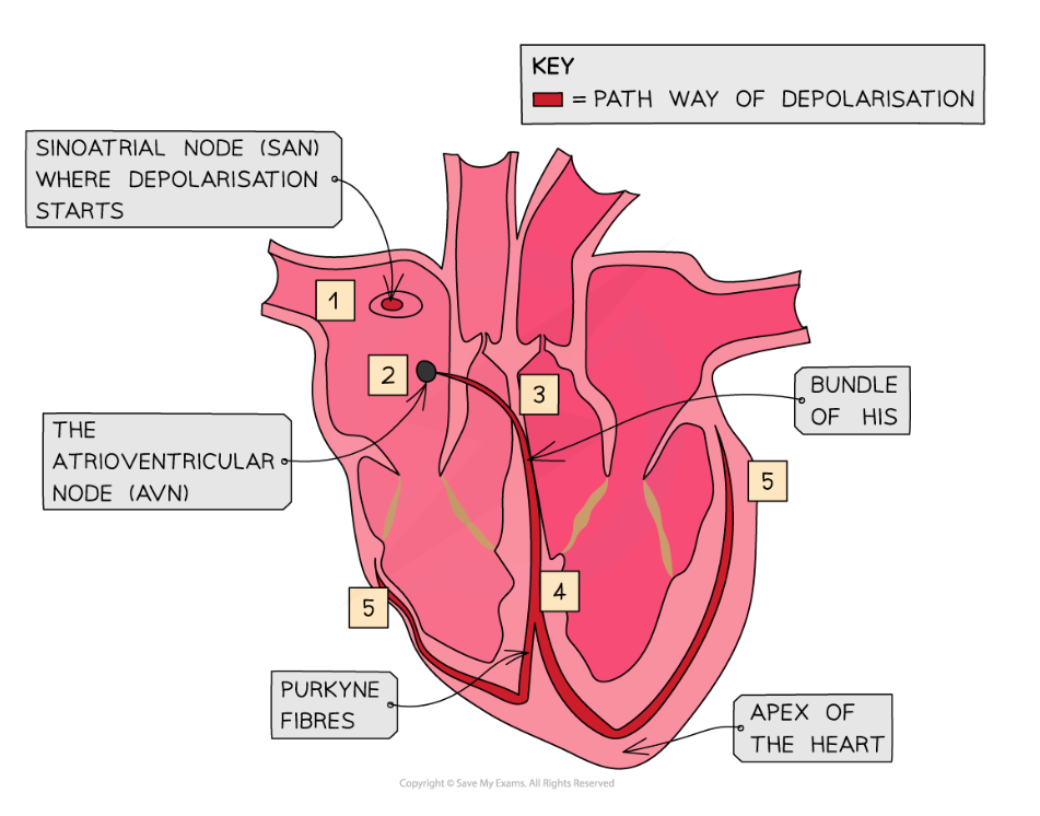
3.1.2 g) how heart action is initiated and coordinated
control of basic heartbeat is myogenic - happens without external stimulus
sinoatrial node sends out wave of excitation
atria contract
atrioventricular node sends out wave of excitation
Purkyne tissue conducts wave of excitation
ventricles contract
3.1.2 h) the use and interpretation of electrocardiogram (ECG) traces (part 1)
electrodes placed on the skin
tachycardia: heartrate is too fast, resting heart rate > 100bpm
bradycardia: heartrate is too slow, resting heart rate < 60bpm (common in athletes)
ectopic: early heartbeat, followed by a pause - relatively common condition, no treatment necessary usually, apart from severe cases
fibrillation: irregular heartbeats, severe cases may be fatal
3.1.2 h) the use and interpretation of electrocardiogram (ECG) traces (part 2)
p wave: caused by the depolarisation of the atria, results in atrial contraction (systole)
QRS complex: caused by depolarisation of ventricles, resulting in ventricular contraction (systole) - largest wave on ECG as ventricles have the largest muscle mass
T wave: caused by repolarisation of ventricles, resulting in ventricular relaxation (diastole)
U wave: cause undetermined, may be the repolarisation of Purkyne fibres
the role of haemoglobin in transporting oxygen and carbon dioxide (part 1)
oxygen + haemoglobin ←→ oxyhaemoglobin
binding of first oxygen molecule results in a confirmational change
easier for each successive oxygen molecule to bind - cooperative binding
the role of haemoglobin in transporting oxygen and carbon dioxide (part 2)
CO2 from respiration diffuses from plasma to RBCs
inside RBC: CO2 + H2 → H2CO3
RBC contains carbonic anhydrase enzyme which catalyses the formation of carbonic acid (H2CO3)
carbonic acid dissociates into H+ and HCI3- ions
H+ can combine with haemoglobin, forming haemoglobinic acid, preventing H+ ions from lowering pH of RBC - haemoglobin is said to act as a buffer
HCO3- diffuses out of RBC into blood plasma where they are transported into solution
the role of haemoglobin in transporting oxygen and carbon dioxide (part 3)
chloride shift: movement of Cl- into RBC that occurs when HCO3- are formed
HCO3- are transported out of erythrocytes via a transport protein in the membrane
to prevent an electrical imbalance, Cl- are transported into the RBC via the same transport protein
if this did not occur, RBC would become positively charged as a result of the build up of H+ formed from the dissociation of carbonic acid
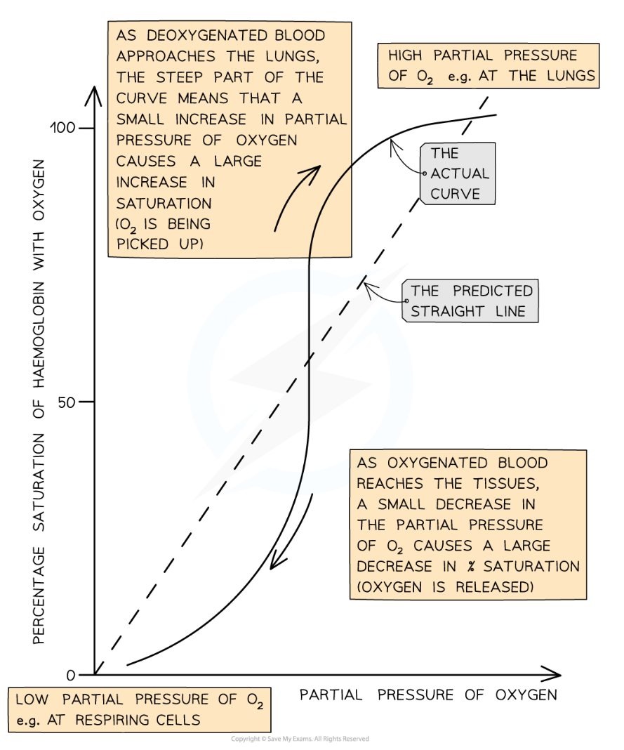
the oxygen dissociation curve for fetal and adult human haemoglobin (part 1)
oxygen dissociation curve: rate at which oxygen associates and dissociates with haemoglobin at different partial pressures of oxygen (pO2)
partial pressure: pressure exerted by oxygen within a mixture of gases
the ease with which haemoglobin binds + dissociates with oxygen molecules is its affinity for oxygen
(diagram) relates to the 4 subunits (haems) of haemoglobin, and cooperative bonding (and vice versa)
the oxygen dissociation curve for fetal and adult human haemoglobin (part 2)
foetal haemoglobin: higher affinity for oxygen than adult haemoglobin
allows foetus to obtain O2 from the mother’s blood in the placenta
foetal haemoglobin can bind to O2 at low pO2
at low pO2 mother’s haemoglobin is dissociative with oxygen
after birth, a baby begins to produce adult haemoglobin which gradually replaces foetal haemoglobin
important for the easy release of oxygen in respiring tissues of more metabolically active individuals