Biopsych+neuropsych
1/64
There's no tags or description
Looks like no tags are added yet.
Name | Mastery | Learn | Test | Matching | Spaced |
|---|
No study sessions yet.
65 Terms
frontal lobe
Largest lobe of the brain
3 areas: Prefrontal cortex, motor cortex, broca’s area
prefrontal cortex areas
Dorsolateral PFC
Medial PFC
Orbitofrontal PFC
Motor cortex
Primary motor cortex
Premotor cortex
Supplementary mc
Frontal eye fields
Broca’s area
DLPFC (dorsolateral PFC)
Functions:
Working memory
A place where past and future meets: looks backward in time to create memories from sensory input and looks forward in time to assemble a motor plan of action.
Lesion: executive function deficit
Orbitofrontal PFC
Emotional processing
Recieves info from amygadala
Emotional perspective taking
Reward contigencies
medial OFC
Anticipation of rewards
Addiction
Lateral OFC
Absences od rewards
Aversion
Medial PFC
The social brain
Long term memory
Emotional processing thru limbic system
Premotor cortex major areas
Dorsal (PMd )
Ventral (PMv)
Inferior frontal gyrus (broca’s area)
Dorsal PMd
Active for choosing movement from its mov lexicon
Ventral (PMv)
Contains mirror neurons that recognise others movment and select similar or different actions.
Fronal eye fields
Stimulus directed eye movmnt
Voluntary control of conjugate (horizontal) eye mov
Saccadic eye movmnt
Lesion to frontal eye fields
Turns eye to opp side
Lose of conjugate eye movmnt
Broca’s area location
Left hemisphere FL
Note: Dominant hemisphere —> speech
non-dominant —> emo component of speech
Motor humunculus
Wilder penfield
Cartoons of body parts to represent the areas of primary motor cortex and pre motor cortex.
Disproportion in the relative sizes of body parts.
Major cortical tracts and motor pathway
Cortico-spinal tract
Corticobulbar tract
Ventro-medial tract
Rubro-spinal tract
Corticospinal tract
2 functionally distinct tract
Lateral tract: distal muscles (forearm, lower limb, hand & finger).
Ventral tract: medial muscles ( trunk, upper limbs…)
Damage to lateral corticospinal tract
Comprimise skilled movments
Damage to ventrak corticospibal tract
Affect posture and ambulation
Cortico-bulbar pathway
Axon descend till pons, where they innervate some ofnthe cranial nerves to control facial, mouth and tongue muscles.
Projection to upper part of the face tend to bi-lateral, where lower face and mouth region tend to be contralateral.
Rubro-spinal pathway
Function: movment of limbs independent of movment of trunk
Supplementary motor area
Buffer store cos its position (in a heirarchy) to cordinate motor plans been ‘approved’ by the big execution via the pyramidal neurons of primary motor strips.
“Bereitshaftpotentials”
Temporal organisation of bhvr
Function of FL
Pfc selection depends on:
Internal cues
External cues
Context cues
Autonoetic awareness
Autonoetic awareness
Tulving
Bhvr is affected by lifetime experiences and goals.
Impairment in AW: Deficits in the self-regulation of bhvr. Seen in DID, PD, SCHIZO, delusional d, ASD
Dorsal visual stream
Object recognition (where pathway)
DLPFC
Dorsal DLPFC - monitoring info in WM
Ventral DLPFC - Regulate the encoding & retrieval of info
Medial PFC
Initiation of activity
Socio-emotional-motivation aspect of bhvr
MPFC
Ventral medial PFC (VMPFC)
Dorsolateral PFC (DMPFC)
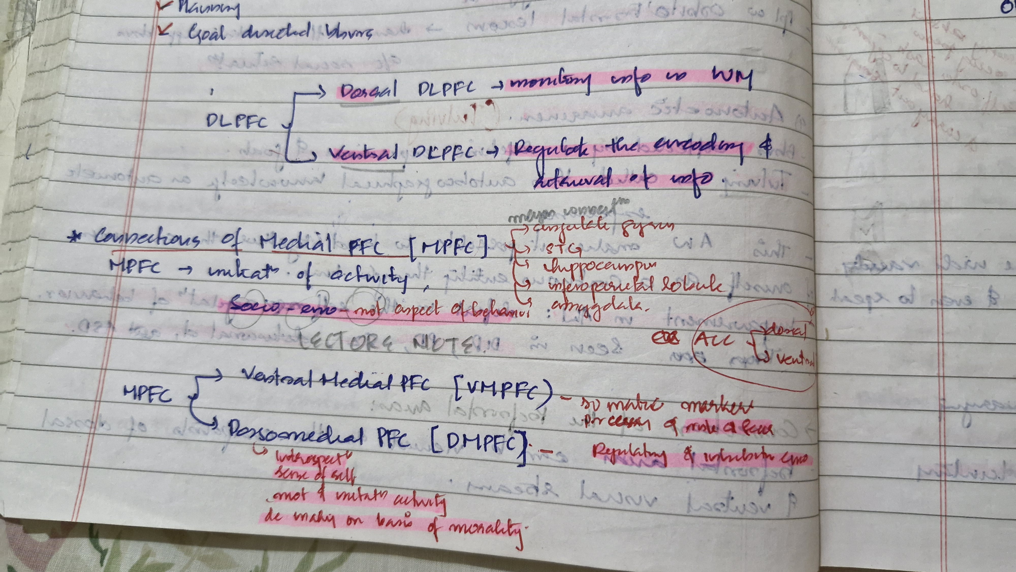
Ventral medial PFC
Somatic markers
Processing of risk and fear
Dorsomedial PFC (DMPFC)
Regulating & inhibiting emotion.
Introspection
Sense of self
Motivation and initiation of activity
Decision making on the basis of morality
Ventral visual stream
Spatial behavior (what pathway)
Symtoms of frontal lobe lesions
Disturbances in motor functions:
Loss of fine movemnts
Movmnt programming
Voluntary gaze
corollary discharge
speech
Loss of divergent thinking
Bhvr spontaneity
Strategy formation
Frequency estimate
Environmental control bhvr
poor response inhibition
impaired associative learning
risk taking & rule breaking
Gambling
self regulating bhvr difficulty
Poor temporal memory
Working mem
Delayed response
Personality changes
Social bhvr
Sexual bhvr
Removal of supplementary motor cortex
Disruption of nearly all voluntary movements
Right & left FL damage (Kolh and Miller study)
Difficulty in copying series of facial movments
Voluntary gaze
Function of frontal eye field
Incase of damage: alteration in voluntary gaze
Corollary discharge ( secondary messages)
A neural signal must produce both the movement and a signal that the movemnt is going to take plade.
When you voluntarily move your eyes, u generate a neural signal that movmnt will happen.
Risk taking & rule breaking
FL damage: failure to comply with instructions.
Role of OFC in risk taking
In gambling: FL lesion pt didnt find ambigous task aversive but controls did.
Tests FL lesions
Response inhibition : WCST, stroop test
Verbal fluency: Thurstone word fluency
Non verbal fluency: design fluency
Working memory : set order
Planning: TOL
Assymmetry of FL function
Left FL : language
Right FL: non verbal mov (eg. Facial expression)
Memory
Encoding: left PFC
Retrievel: Right PFC
Functions of Premotor cortex
Ventral & lateral: movmnt based on external cues
Medial region:
respond to internal signals from brain area.
Relay centr for executing specific physical actions
Contribute to learning, imitation amf support
Social cog skills (eg. Empathy)
Right PMC: spatial activities
Left PMC: tasks that r not spatial in nature.
PMv
PMd
Frontal eye fields
3 main regions of Temporal lobe
Superior TG
Middle TG
Inferior TG
Insular cortex
Primary gustatory cortex
Auditory association cortex
What pathway ( hierarchical sensory pathway)
Stimulus recognition (WHAT IS THATTTT???)
Dorsal pathway ( where pathway)
Spatial location (WHERE IS ITTT)
Polymodal pathway
Stimulus categorisation
Medial temporal projection
long term memory
3 sensory functions and 4 main functions of TL
3 main function:
Processjng auditory input
Visual objecy recognition
Long term storage of sensory inp
4 main functions:
Obj recog
Categorisation
Directed attention
Cross modal matching
Recalling from LTM
Cross modal matching
Cortex of Superior temporal sulcus
Affective response function of
Amygdala
Spatial navigation function of
Hippocampus
Biological motion function of
Superior temporal gyrus.
STS: Involved in social meaning making of gestures.
social perception and social cognition
Two type of memort
Implicit
Explicit
Neural basis of implicit memory
Neocortex —> basal ganglia —> ventral thalamus —> premotor cortex
Neural basis of explicit memory
Entorhinal pathway (integrative forms of memory)
Parahippocampal cortex (visuospatial memory)
Perirhinal cortex (visual object memory)
Types of amnesias
Reterograde
Anterograde
Transient global amnesia
Infantile drug induced
Fugue state
Korsakoff’s syndrome/wernicke-korsakoff sundrome
Alzhiemers
Dissociative amnesia
Neuroanatomical vasus of amnesia
LESIONS IN
Cingulate gyrus (posterior part)
frontal and prefontal lobes
mammilo-thalamic tract (diff in recording of the stimulus, diff in immediate storage function)
Amygdala (memories emotional in nature)
Tests for amnesias
MSE
Galveston orientation & amnesia test (GOAT)
Weschler’s memory scale
Kingshill test or 6 CIT
General tests (learning a word list, learning pairs od words, passage test, corsi’s test “knock cube test”
Galveston Orientation and Amnesia Test (GOAT)
The Galveston Orientation and Amnesia Test (GOAT) is a clinical tool designed to evaluate post-traumatic amnesia (PTA) in patients with head injury. It helps determine when the patient regains orientation and consistent memory function after a traumatic brain injury (TBI).
Key Details:
Developed by: Levin, O’Donnell, and Grossman in 1979.
Purpose: To measure the duration and severity of post-traumatic amnesia (PTA).
Format:
Structured questionnaire with 10 items.
Assesses orientation to time, place, and person as well as memory for events both before and after injury.
Domains Assessed:
1. Orientation to person – e.g., name, age.
2. Orientation to time – date, month, year, time of day.
3. Orientation to place – city, hospital, ward.
4. Memory – events immediately before and after the injury, recall of hospitalization.
Interpretation of Scores:
100: No errors → fully oriented.
76–100: Normal or mild impairment.
66–75: Borderline.
<66: Impaired (still in PTA).
Corsi’s test ( “knock cube test” )
Measure visuospatical LTM
subject must learn sequence of 8 cubes, touched by examiner on corsi’s tablet and remember it after interval of 10 minutes.
Primary auditory cortex of right TL
Make pitch discrimination
“Musical store”
Right auditory association cortex
Amusia (tone defect)
Grant Allan, 1978
Types:
expressive : loss of ability to hum, sing, whistle a tone
perceptive: deficits in ability 6o appreciate various charac of music like pitch, harmony, melody.
Assymmetry of TL funtion
MEMORY
Left TL : verbal
Right TL : Non verbal
Processing of speech sounds: left TL
Processing aspects of musics: Right TL
Social & affective bhvr : TL function little known
Right TL lesion: imp in facial recognition & facial expression.