The Heart
1/32
There's no tags or description
Looks like no tags are added yet.
Name | Mastery | Learn | Test | Matching | Spaced | Call with Kai |
|---|
No analytics yet
Send a link to your students to track their progress
33 Terms
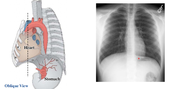
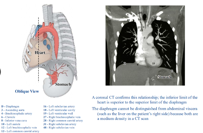
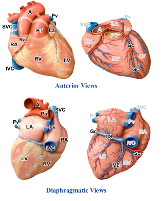
heart in situ
-inferior limit of heart is diaphragm- fused
-heart descends with diaphragm at each inspiration
-diaphragmatic surface of heart is not in a transverse plane, so they look like they overlap in an x-ray
-hemidiaphragms: individual halves of the diaphragm
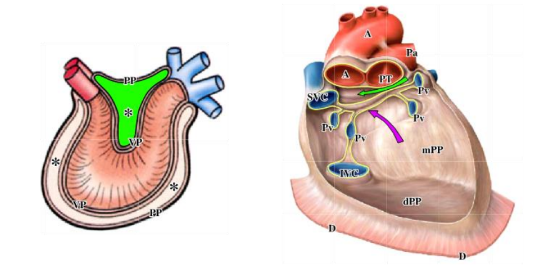
oblique view of heart in situ
____ and _____ parts of the _____ are visible once the heart has been removed
-posterior; inferior; parietal pericardium
-diaphragmatic parietal pericardium fused with the central tendon of the diaphragm
-sites where parietal pericardium reflects on to the surface of the heart are now apparent

pericardial reflections
-site where the arterial and venous ends of the developing heart approached one another remains as the transverse pericardial sinus (green arrow)
-transverse pericardial sinus: facilitates clamping the aorta and pulmonary trunk during cardiac surgery
-oblique pericardial sinus: located posteriorly where the pericardium covering the veins reflects on to the heart (purple arrow)
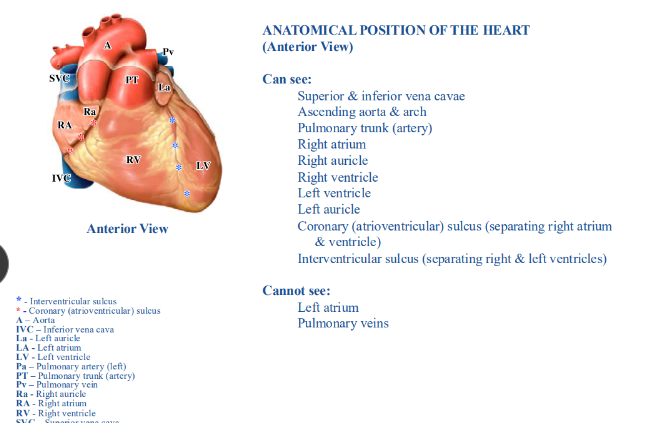
anatomical position of the heart
-superior and inferior vena cavae are in the same vertical axis
-ascending aorta is immediately to the left of the superior vena cava and the pulmonary trunk (artery) is immediately to the left of the ascending aorta- SAP
-right atrium forms the right border
-right ventricle forms most of the anterior surface
-left ventricle forms the left border and apex
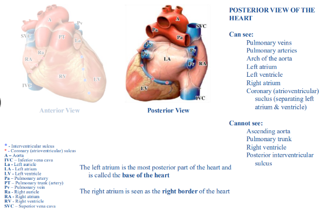
relationships of heart chambers- posterior view of the heart
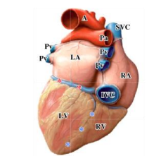
relationships of heart chambers- close to inferior (diaphragmatic) view
-can see: inferior vena cava, right ventricle, left ventricle, interventricular sulcus (separating right and left ventricles)
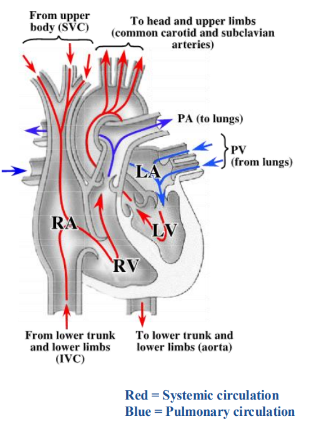
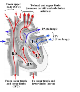
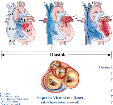
circulation through the heart
-superior and inferior vena cava enter the right atrium
-pulmonary veins enter the left atrium
-blood passes from atria to ventricles by passing through the right (tricuspid) or left (mitral) atrioventricular valves
-blood leaves the heart by passing from the ventricles to the pulmonary trunk (artery) or aorta by passing through the pulmonary or aortic semilunar valves
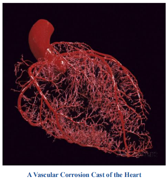
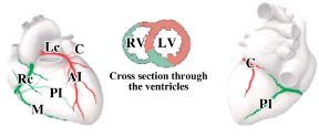
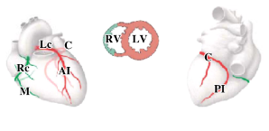
coronary arteries
-coronary (atrioventricular) and interventricular sulci extend around heart
-arteries and veins located in sulci
-all of the arteries are branches of either the right or left coronary arteries
-right and left coronary arteries are branches of the ascending aorta
-coronary arteries supply the heart tissue
right coronary artery
-right coronary artery runs in the coronary (AV) sulcus and gives off the: sinuatrial nodal artery (proximal branch that goes to the SA node near the superior vena cava), marginal artery (runs along the inferior margin of the heart), posterior interventricular artery (runs in posterior interventricular sulcus)
-all vessels have a LOT of branches and tributaries
left coronary artery
-very short and bifurcates into the:
-circumflex artery that runs in the coronary (AV) sulcus
-anterior interventricular artery (aka left anterior descending artery and “widow maker”) that runs in the anterior interventricular sulcus and usually extends to the apex of the heart
coronary arteries
-anastomotic connections between the territories by branches of the right and left coronary arteries typically minimal
-coronary arteries described as being functional end arteries; true end arteries are the only blood supply to a region
-blockages may be asymptomatic, cause angina pectoris with transient pain, or a myocardial infarction where there is prolonged pain and possibly permanent damage to heart muscle
variations in coronary arteries- balanced distribution (~20% of population)
-right coronary artery gives rise to posterior interventricular artery
-both the right and left coronary arteries contribute to the ventricular walls and interventricular septum
variations in coronary arteries- left dominance (~10% of population)
-circumflex branch of left coronary artery gives rise to posterior interventricular artery
-left coronary artery supplies the entire left ventricle, interventricular septum, and part of the right ventricle
variations in coronary arteries- right dominance (~70% of population)
-right coronary artery gives rise to posterior interventricular artery and continues for much of the posterior coronary sulcus
-right coronary artery supplies much of the right ventricle, interventricular septum, and left ventricle
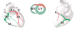
coronary veins
-coronary sinus: large vein in posterior part of coronary sulcus; empties into the right atrium
-great cardiac vein, middle cardiac vein, and small cardiac vein drain into coronary sinus
-great cardiac vein: runs in the anterior interventricular and coronary sulci becoming the coronary sinus on the posterior side of the heart
-middle cardiac vein: runs in the posterior interventricular sulcus draining into coronary sinus just before entering the right atrium
-small cardiac vein: within the posterior atrioventricular sulcus draining into the coronary sinus just before entering the right atrium
blood circulation
-2 circulatory routes: systemic and pulmonary
-both routes originate as arteries, go through a capillary bed, and terminate as veins irrespective of the oxygen content of the vessel
-arteries: defined as vessels leaving the heart
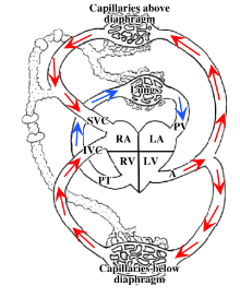
blood route through heart
-superior and inferior vena cava and pulmonary veins enter the right and left atria of the heart (respectively)
-blood passes from the atria to the ventricles by passing through the right (tricuspid) or left (mitral) atrioventricular valves
-blood leaves the heart by passing from the ventricles to the pulmonary trunk or aorta by passing through the pulmonary or aortic semilunar valves
internal features of right atrium
-internal surface of right atrium is smooth posteriorly and raised into ridges anteriorly
-ridges composed of pectinate muscle
-auricle: diverticulum from the right atrium and has pectinate muscle
-superior vena cava, inferior vena cava, and coronary sinus open into the right atrium
-valve: leaflet of tissue surrounding the opening of the inferior vena cava and the coronary sinus
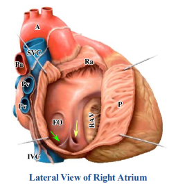
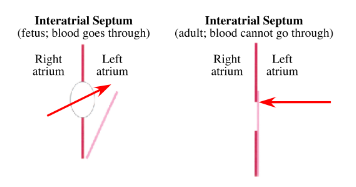
fossa ovalis
-part of interatrial septum
-embryologic remnant of foramen ovale, which shunted oxygenated blood from the inferior vena cava to the left atrium to be distributed to the body of the fetus via the aorta
-foramen still present in ~20% of population- called a patent foramen ovale (functionally not a problem because the pressure is greater on the left side of the heart and the foramen remains closed)
-possible that the valve of the inferior vena cava may preferably shunt blood toward the foramen ovale in the fetus
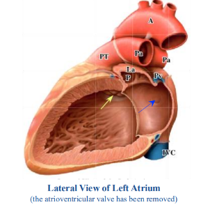
internal features of the left atrium
-internal surface of left atrium entirely smooth except for the left auricle
-four pulmonary veins open into the left atrium
-depression seen in interatrial septum indicating the other side of the fossa ovalis (blue arrow)
-semilunar aortic valve (yellow arrow) has three cusps
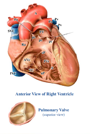
internal features of right ventricle
-trabeculae carneae: internal surface of right ventricle near the atrioventricular (tricuspid) valve raised into ridges
-internal surface of right ventricle approaching the pulmonary valve is smooth
-right AV (tricuspid) valve composed of three cusps that are anchored to papillary muscles by tendinous cords; papillary muscles are extensions of trabeculae carnae; on right side, three groups of papillary muscles (anterior, septal, and posterior)
-septomarginal trabecula (moderator band): band of muscle connecting the interventricular septum and the anterior papillary muscle
-pulmonary valve (type of semilunar valve): composed of three cusps
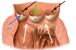
internal features of left ventricle
-internal surface of left ventricle raised into ridges (trabeculae carneae)
-left AV (mitral) valve composed of two cusps that are anchored to anterior and posterior papillary muscles by tendinous cords
-aortic valve not well seen from inside of left ventricle
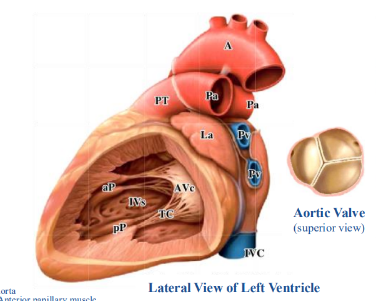
wall of left ventricle usually _______ the thickness than that of the right ventricle because of
-three times; the higher resistance on the left side
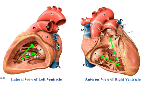
ventricular contraction
-when ventricles contract, blood is forced towards BOTH the atrioventricular and semilunar valves
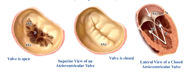
function of atrioventricular valves (tricuspid and mitral)
-opening and closing of an atrioventricular valve is a passive process
-blood going from the atria to the ventricles results in the opening of the valves, while blood going away from the ventricles results in the valves closing
-contraction of the papillary muscles pulls on the cusps via the tendinous cords, preventing the cusps from prolapsing and allowing blood to enter the atria
-incompetent valves will essentially blow inside out like an umbrella
function of the semilunar valves
-opening and closing of semilunar valves is a passive process; similar to valves within veins
-blood forced out of the ventricles results in opening of valves
-valves close due to elastic recoil of the pulmonary trunk or aorta that propels blood away from the heart as well as back to the heart
-thickening at the edge of each cusp called a lunule (blue arrow) adds stability to the valve
-aortic valve differs from the pulmonary valve because the coronary arteries take origin from a depression (sinus) in the wall of the aorta at the level of the cusps (black arrows)
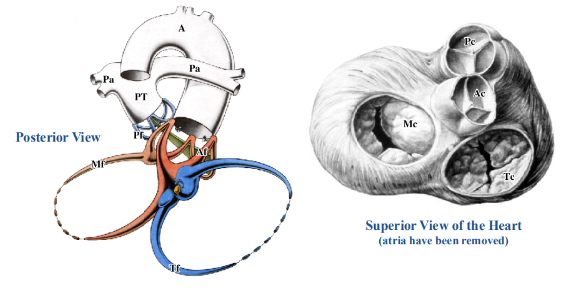
skeleton of the heart
-consists of a series of fibrous rings surrounding the atrioventricular and semilunar valves
-these rings provide stability for the valves ensuring their patency and circular shape; cusps also attach to these rings
-skeleton also serves as an attachment site for cardiac muscle fibers and electrically isolates the atria from the ventricles
heart in situ- anterior view
-right atrium, right ventricle, and part of left ventricle can be seen
-heart is NOT symmetrical; extends further laterally on the left side
-heart is posterior to the inferior part of the body of the sternum and costal cartilages of ribs 3/4/5/6
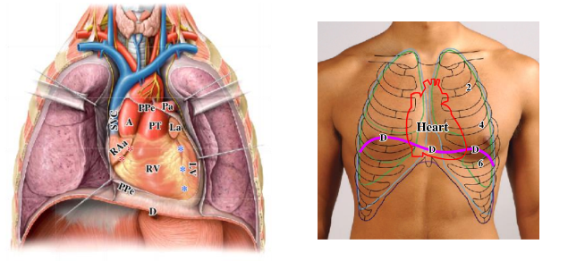
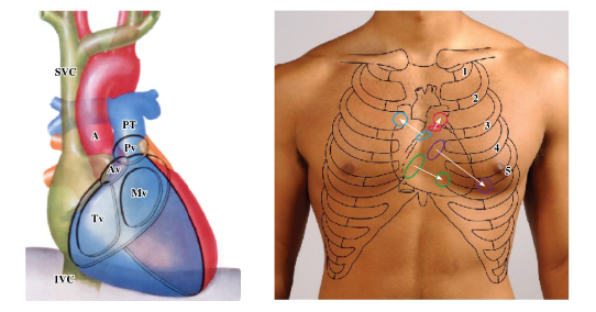
heart sounds
-valves of heart NOT best heard where they are located
-best heard with minimal interference:
-aortic valve: second intercostal space to the right of the sternum
-pulmonary valve: second intercostal space to the left of the sternum
-mitral valve: fifth intercostal space in midclavicular line
-tricuspid valve: fifth intercostal space to the left of the sternum
cardiac cycle
-blood pressure = systolic pressure/diastolic pressure (mm Hg)
-diastole: ventricles relax and fill (blood coming from superior and inferior vena cava, and the pulmonary veins) and the aorta and pulmonary trunk undergo elastic recoil
-systole: ventricles contract and empty (blood leaving via the aorta and pulmonary trunk)
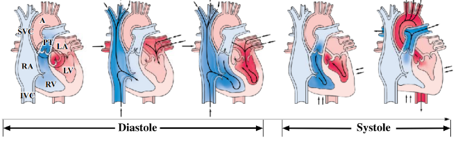
during diastole
-ventricles relax and fill
-there is elastic recoil of the aorta and pulmonary trunk
-semilunar valves close
-atrioventricular valves open
-coronary arteries fill
during systole
-ventricles contract and empty
-blood is forced superiorly
-atrioventricular valves close
-semilunar valves open
