Transport in humans
1/10
Name | Mastery | Learn | Test | Matching | Spaced |
|---|
No study sessions yet.
11 Terms
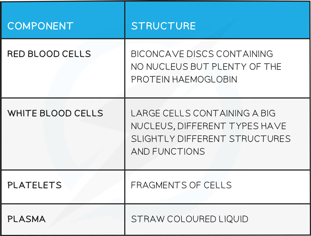
What is the composition of blood?
Blood consists of red blood cells, white blood cells, platelets and plasma
Over half of the volume of the blood is made up of plasma
The majority of the other half is made up of red blood cells
The remaining fraction consists of white blood cells and platelets
What is the role of plasma in transport?
Plasma is a straw coloured liquid which the other components of the blood are suspended within
Plasma is important for the transport of many substances including:
Carbon dioxide - the waste product of respiration, dissolved in the plasma as hydrogencarbonate ions and transported from respiring cells to the lungs
Digested food and mineral ions - dissolved particles absorbed from the small intestine and delivered to requiring cells around the body
Urea - the waste substance produced in the breakdown of proteins by the liver. Urea is dissolved in the plasma and transported to the kidneys
Hormones - chemical messengers released into the blood from the endocrine organs (glands) and delivered to target tissues/organs of the body
Heat energy - created in respiration (an exothermic reaction), heat energy is transferred to cooler parts of the body or to the skin where heat can be lost
What are the adaptations of red blood cells?
They are full of haemoglobin, a protein that binds to oxygen to form oxyhaemoglobin
They have no nucleus which allows more space for haemoglobin to be packed in
The shape of a red blood cell is described as being a 'biconcave disc' this shape gives them a large surface area to volume ratio to maximise diffusion of oxygen in and out
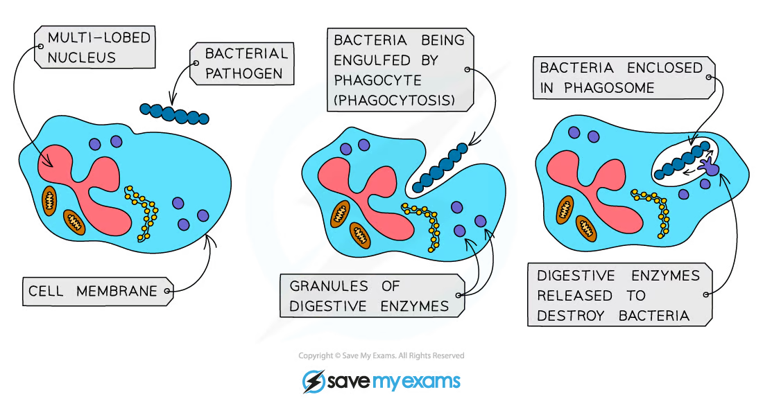
How do Phagocytes respond to disease?
Phagocytes
Phagocytes carry out phagocytosis by engulfing and digesting pathogens
Phagocytes have a sensitive cell surface membrane that can detect chemicals produced by pathogenic cells
Once they encounter the pathogenic cell, they will engulf it and release digestive enzymes to digest it
This is a non-specific immune response
How do Lymphocytes respond to disease?
Lymphocytes produce antibodies
Antibodies are Y-shaped proteins with a shape that is specific (complementary) to the antigens on the surface of the pathogen
This is a specific type of immune response as the antibodies produced will only fit one type of antigen on a pathogen

The lymphocytes produce antibodies that are specific to the antigen on the pathogen
Antibodies attach to the antigens and cause agglutination (clumping together)
This means the pathogenic cells cannot move very easily
At the same time, chemicals are released that signal to phagocytes that there are cells present that need to be destroyed

Agglutinated pathogens cannot move easily
Lymphocytes also produce antitoxins to neutralise toxins released by pathogens
How do vaccinations result in the manufacture of memory cells, which enable future antibody production to the pathogen to occur sooner, faster and in greater quantity?
Vaccines are used to induce immunity to infectious diseases
A vaccine contains harmless versions of a pathogen
There are several different methods by which scientists ensure that vaccines contain harmless pathogens such as:
Killing the pathogen
Making the pathogen unable to grow or divide (attenuated vaccine)
Using fragments of pathogens, rather than whole cells
A vaccine may be administered orally, nasally or via an injection
How vaccines work
Once in the bloodstream, the antigens contained within the vaccine can trigger an immune response in the following way:
Lymphocytes recognise the antigens in the bloodstream
The activated lymphocytes produce antibodies specific to the antigen encountered
Memory cells and antibodies subsequently remain circulating in the blood stream
 The process of long-term immunity by vaccination
Future infection by the same pathogen will trigger a response that is much faster and much larger compared to the initial response
Due to the rapid nature of the response, the pathogen is unable to cause disease and the individual is said to be immune

Graph showing the number of measles antibodies in the blood following vaccination. The secondary response is much faster and a greater number of antibodies are produced.
The importance of vaccination
Vaccines not only protect the vaccinated individuals, they also reduce the likelihood that an infected individual will spread the pathogen to others
If a large proportion of the population is vaccinated, it is unlikely that an unvaccinated individual will become infected with the pathogen (this concept is referred to as herd immunity)
This can prevent the spread of the disease
Vaccines have reduced the cases of certain diseases drastically or even eradicated many diseases worldwide
This includes smallpox, measles, mumps and tetanus amongst many others
There are hopes for the future eradication of several other diseases through vaccination programs
This includes polio, HIV, malaria and of course COVID-19
Disadvantages of vaccinations
There are some disadvantages to vaccinations that reduce how effective vaccination programs can be
Mutations in the pathogen's DNA/RNA can result in significant changes to the antigen of the pathogen meaning that lymphocytes no longer recognise the pathogen
Side-effects of vaccinations can reduce the uptake in the population
Advantages & Disadvantages of Vaccination Table
How are platelets involved in blood clotting?
Platelets are involved in helping the blood clot
Platelets are fragments of cells that are involved in blood clotting and forming scabs where the skin has been cut or punctured
When the skin is broken (i.e. there is a wound) platelets arrive to stop the bleeding
A series of reactions occur within the blood plasma
Platelets release chemicals that cause soluble fibrinogen proteins to convert into insoluble fibrin and form an insoluble mesh across the wound, trapping red blood cells and therefore forming a clot
The clot eventually dries and develops into a scab to protect the wound from bacteria entering
 How the blood clots
The importance of blood clotting
Blood clotting prevents continued / significant blood loss from wounds
Scab formation seals the wound with an insoluble patch that prevents entry of microorganisms that could cause infection
It remains in place until new skin has grown underneath it, sealing the skin again
How does the heart function?
The heart organ is a double pump
Oxygenated blood from the lungs enters the left side of the heart and is pumped to the rest of the body (the systemic circuit)
The left ventricle has a thicker muscle wall than the right ventricle as it has to pump blood at high pressure around the entire body,
Deoxygenated blood from the body enters the right side of the heart and is pumped to the lungs (the pulmonary circuit)
The right ventricle is pumping blood at lower pressure to the lungs
A muscle wall called the septum separates the two sides of the heart
Blood is pumped towards the heart in veins and away from the heart in arteries
The coronary arteries supply the cardiac muscle tissue of the heart with oxygenated blood
As the heart is a muscle it needs a constant supply of oxygen (and glucose) for aerobic respiration to release energy to allow continued muscle contraction
Valves are present to prevent blood flowing backwards

Structure of the Heart
The pathway of blood through the heart
Deoxygenated blood coming from the body flows through the vena cava and into the right atrium
The atrium contracts and the blood is forced through the tricuspid (atrioventricular) valve into the right ventricle
The ventricle contracts and the blood is pushed through the semilunar valve into the pulmonary artery
The blood travels to the lungs and moves through the capillaries past the alveoli where gas exchange takes place
Low pressure blood flow on this side of the heart prevents damage to the capillaries in the lungs
Oxygenated blood returns via the pulmonary vein to the left atrium
The atrium contracts and forces the blood through the bicuspid (atrioventricular) valve into the left ventricle
The ventricle contracts and the blood is forced through the semilunar valve and out through the aorta
Thicker muscle walls of the left ventricle produce a high enough pressure for the blood to travel around the whole body
How does heart rate change during exercise and the influence of adrenaline?
A heart rate is measured by counting the number of times a heart beats in a minute (bpm)
The natural resting heart rate is controlled by a group of cells located in the right atrium called the pacemaker
The role of the pacemaker is to coordinate the contraction of the heart muscle and regulate the heart rate
Pacemaker cells send out electrical impulses which initiate a contraction in the cardiac muscle
Other factors can also influence the heart rate, such as the hormone adrenaline
Exercise and heart rate
The heart pumps blood around the body in order to supply oxygen and glucose to respiring cells
The blood also removes waste products from the respiring cells
During exercise, the cells of the muscles respire more rapidly in order to provide energy for muscle contraction
Respiration may be aerobic if exercise is moderate, or anaerobic is exercise is more intense
An increase in respiration means an increase in requirement for oxygen and glucose as well as an increase in production of waste products that need to be removed
The nervous system responds to this requirement by stimulating the following changes
Heart rate increases to deliver oxygen and glucose and remove waste more frequently
The volume of blood pumped out of the heart also increases to deliver bigger quantities of oxygen and glucose
Production of the hormone adrenaline increases heart rate as part of a 'fight or flight' response
At the end of a period of exercise, the heart rate may remain high for a period of time as oxygen is required in the muscles to break down the lactic acid from anaerobic respiration
This is how the oxygen debt is paid off
The time taken for the heart rate to return to the resting rate is called the recovery time
A physically fit person will have a lower resting heart rate and a shorter recovery time compared to an unfit person
How do the structure of arteries, veins and capillaries relate to their function?
Smaller vessels that branch off from arteries are called arterioles (small arteries) and those that branch into veins are called venules (small veins)
Each vessel has a particular function and is specifically adapted to carry out that function efficiently
Arteries
Key features:
Carry blood at high pressure away from the heart
Carry oxygenated blood (except the pulmonary artery)
Have thick muscular walls containing elastic fibres
Have a narrow lumen
Blood flows through at a fast speed
The structure of an artery is adapted to its function in the following ways
Thick muscular walls containing elastic fibres withstand the high pressure of blood and maintain the blood pressure as it recoils after the blood has passed through
A narrow lumen also helps to maintain high pressure
Veins
Key features:
Carry blood at low pressure towards the heart
Carry deoxygenated blood (other than the pulmonary vein)
Have thin walls
Have a large lumen
Contain valves
Blood flows through at a slow speed
The structure of a vein is adapted to its function in the following ways:
A large lumen reduces resistance to blood flow under low pressure
Valves prevent the backflow of blood as it is under low pressure
 Comparing the structure of arteries and veins
Capillaries
Key features:
Carry blood at low pressure within tissues
Carry both oxygenated and deoxygenated blood
Have walls that are one cell thick
Have ‘leaky’ walls
Speed of blood flow is slow
The structure of a capillary is adapted to its function in the following ways:
Capillaries have walls that are one cell thick (short diffusion distance) so substances can easily diffuse in and out of them
The ‘leaky’ walls allow blood plasma to leak out and form tissue fluid surrounding cells
 Structure of a capillary
Arterioles and venules
As arteries get further away from the heart, they divide more and get narrower
The narrow vessels that connect arteries to capillaries are called arterioles
Veins also get narrower the further away they are from the heart
The narrow vessels that connect capillaries to veins are called venules
 The blood vessel network
What factors may increase the risk of developing coronary heart disease?
Causes of coronary heart disease
Like all cells in the body, cardiac muscle cells need a supply of blood to deliver oxygen and glucose and to remove waste products such as carbon dioxide
The blood is supplied to the heart by the coronary arteries which branch off directly from the aorta
The heart needs to constantly respire, so it is vital that it receives oxygen

The coronary arteries supply the heart with oxygenated blood
In coronary heart disease (CHD), layers of fatty material (plaque) build up inside the coronary arteries
These fatty deposits are mainly formed from cholesterol
There are two sources of cholesterol in the body:
Dietary cholesterol (from animal products eaten)
Cholesterol synthesised by the liver

Buildup of plaque in the coronary arteries narrows the lumen
If a coronary artery becomes partially or completely blocked by these fatty deposits, it loses its elasticity and cannot stretch to accommodate the blood which is being forced through every time the heart contracts
The flow of blood through the arteries is reduced, resulting in a lack of oxygen for the heart muscle
Partial blockage of the coronary arteries creates a restricted blood flow to the cardiac muscle cells and results in severe chest pains called angina
Complete blockage means cells in that area of the heart will not be able to respire aerobically, leading to a heart attack
Treatment of CHD involves either increasing the width of the lumen of the coronary arteries using a stent, or prescribing statins to lower blood cholesterol

The effect of a narrowed lumen in a coronary artery is reduced blood flow to the heart
Risk factors of coronary heart disease
There are several risk factors which will increase the chances of coronary heart disease:
Obesity
Carrying extra weight puts a strain on the heart
Increased weight can lead to Type 2 diabetes which further damages your blood vessels
High blood pressure
This increases the force of the blood against the artery walls and consequently leads to damage of the vessels
High cholesterol
Speeds up the build up of fatty plaques in the arteries leading to blockages
Smoking
Chemicals in smoke cause an increase in plaque build up and an increase in blood pressure
Carbon monoxide also reduces the oxygen carrying capacity of the red blood cells