Parasitology All Labs (BMS171)
1/143
Earn XP
Description and Tags
Parasitology labs. goodluck
Name | Mastery | Learn | Test | Matching | Spaced | Call with Kai | Chat |
|---|
No analytics yet
Send a link to your students to track their progress
144 Terms
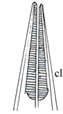
Flattened unsegmented leaf-like = Trematodes
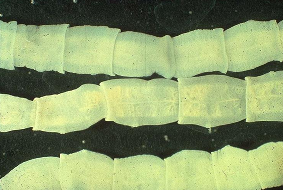
Flattened segmented tape-like = Cestodes
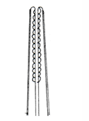
Cylindrical rounded unsegmented = Nematodes
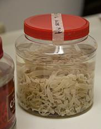
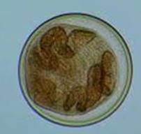
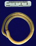
Adult Taenia worm in Jar
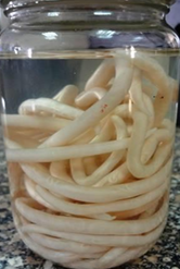
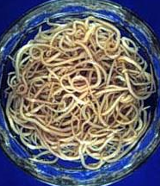
Adult Ascaris in jar
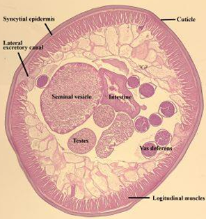
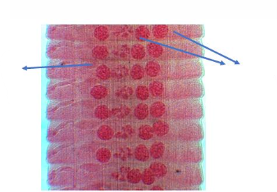
Cross section in Ascaris male
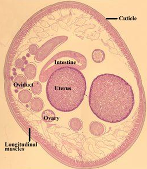
Cross section in Ascaris female
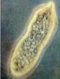
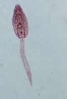
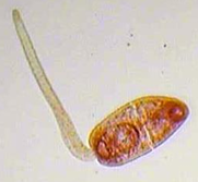
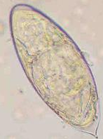
Miracidium
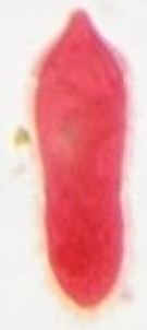
Miracidium of schistosomes
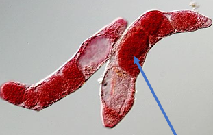
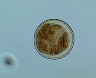
Sporocysts
Arrow: Daughter sporocysts
Sporocyst
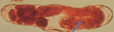
Arrow: Redia
Redia
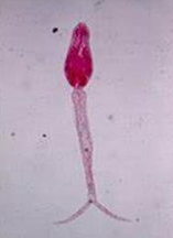
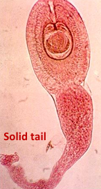
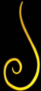
Leptocercus cercaria: with Simple tail
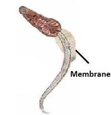
Lophocercus cercaria: tail is surrounded by a membrane
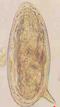
Furcocercus cercaria: with forked tail
Microcercus cercaria): tail is a a knob like structure
Encysted metacercaria
Encysted metacercaria
Encysted metacercaria
Cysticercoid
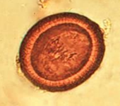
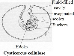
Cysticercus larva
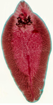
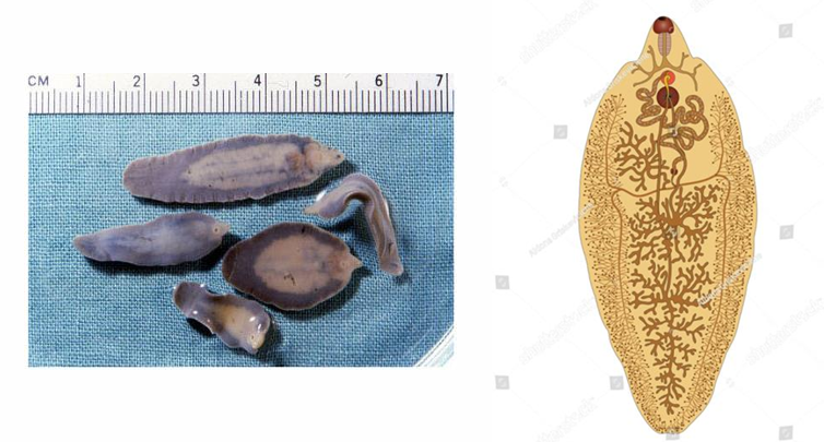
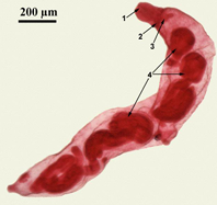
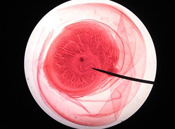
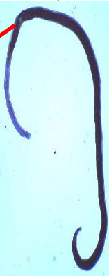
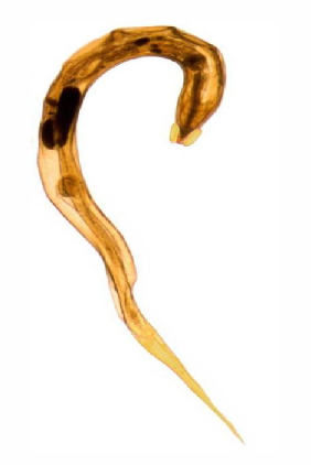
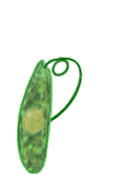
Fasciola Adult
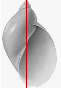
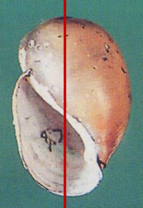
Snail Intermediate host of Fasciola spp, what is it called?
Lymnaea cailliaudi
Dextral, Thin, fragile Prominent apex Short spire, few whorls Aperture 2/3 of length Fresh water IH of Fasciola gigantica Surface feeder Susceptible to molluscicides
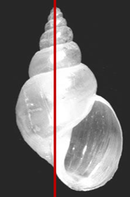
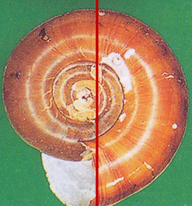
Snail Intermediate host of Fasciola spp, what is it called?
Lymnaea truncatula
Dextral, Thin, fragile Prominent apex Few whorls Aperture 1/2 of length IH of Fasciola hepatica Fresh water Surface feeder Susceptible to molluscicides
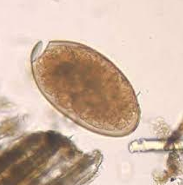
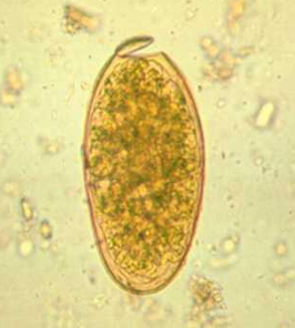
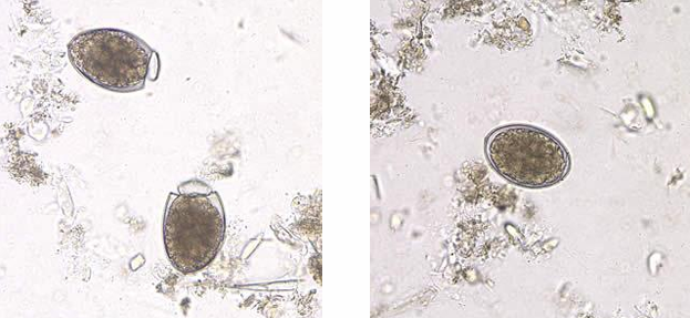
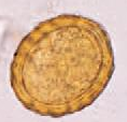
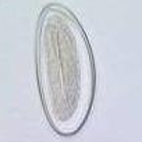
Fasciola Egg (diagnostic stage)
Oval Thin 150 x 70 µ Yellow immature Operculated
Fasciola Egg (diagnostic stage)
Oval Thin 150 x 70 µ Yellow immature Operculated
Leptocercus cercaria: with Simple tail
Fasciola encysted metacercaria (infective stage)
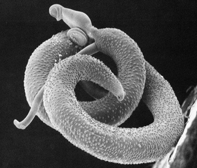
Schistosoma
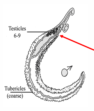
Schistosoma haematobium male
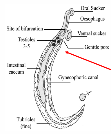
Schistosoma haematobium male
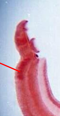
Schistosoma mansoni male
Schistosoma mansoni male
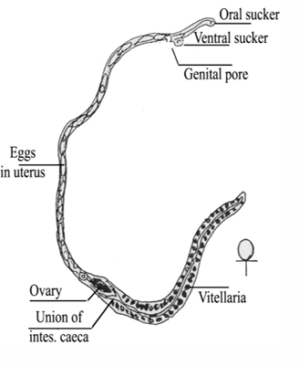
Schistosoma haematobium adult female
Schistosoma haematobium adult female
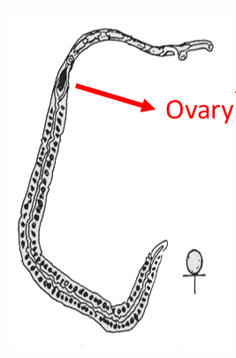
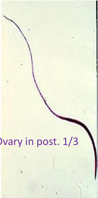
Schistosoma mansoni adult female
Schistosoma mansoni adult female
Snail Intermediate hosts of Schistosoma spp
Bulinus truncatus
Sinistral Blunt apex with shoulder With umbilicus IH of Schistosoma haematobium Fresh water Various level of water Susceptible to molluscicides
Snail Intermediate hosts of Schistosoma spp
Biomphalaria alexandrina
Sinistral Discoid IH of Schistosoma mansoni Fresh water Surface feeder Susceptible to molluscicides
Schistosoma Egg, Terminal Spine
Shape: oval
Shell: Thin
Color: Colorless
Content: Mature with fully formed miracidium
Pass in urine, rarely in stool
Schistosoma Egg, Lateral Spine
Shape: oval
Shell: Thin
Color: Yellow
Content: Mature with fully formed miracidium
Pass in STOOL, rarely in urine
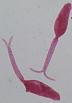
Furcocercus cercaria (infective stage) of schistosoma
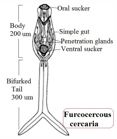
Furcocercus cercaria (infective stage) of schistosoma
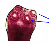
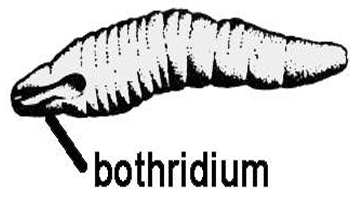
Scolex of cestoda - Pseudophyllidea
arrow pointing at Bothria
Scolex of cestoda - Cyclophyllidea
arrow pointing at suckers
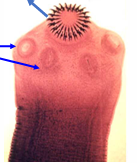
Scolex of cestoda - Cyclophyllidea
arrow pointing at suckers
4-cup shaped suckers-with rostellum and hooks
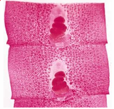
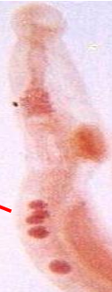
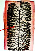
Reproductive system of Pseudophyllidea (Mature Segment)
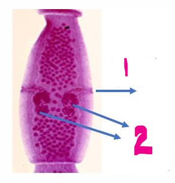
Reproductive system of Cyclophyllidea (Mature Segment)
1- Genital pore
2- Double set of genitalia
Reproductive system of Cyclophyllidea (Mature Segment)
arrows = testis
Reproductive system of Cyclophyllidea
Lateral Branches
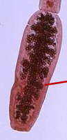
Reproductive system of Cyclophyllidea
Lateral Pouches
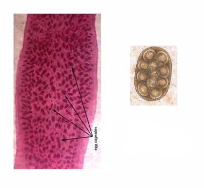
Reproductive system of Cyclophyllidea
Egg Capsules
Developmental stages of Pseudophyllidea
Egg
Developmental stages of Pseudophyllidea
Coracidium
Developmental stages of Pseudophyllidea
Procercoid
Developmental stages of Pseudophyllidea
Plerocercoid
Developmental stages of Cyclophyllidea
Egg
Developmental stages of Cyclophyllidea
Cycticercus
Developmental stages of Cyclophyllidea
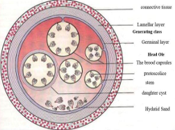
Larva; Hydatid Cyst
Developmental stages of Cyclophyllidea
Cysticercoid
Eggs of Pseudophyllide
Eggs are immature, they are not infective to man
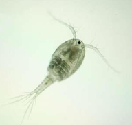
IH of Pseudophyllide
Cyclops
Infective stage of Pseudophyllide
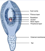
Plerocercoid in fish muscle
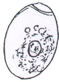
Echinococcus granulosus adult
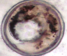
Hydatid sand (Diagnostic Stage of Cyclophyllidea)
Hydatid sand is many miniature tapeworm scolices (heads), these will form adult tapeworms in the DH (dog)
What’s the infective stage of Cyclophyllidea?
Egg
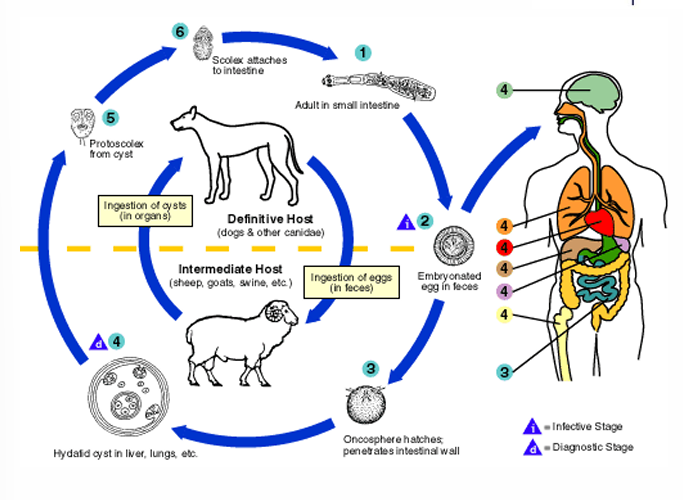
What’s the mode of infection of Cyclophyllidea?
Eating food contaminated by excreta of dogs

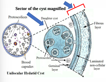
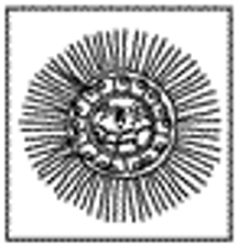
Wall of Unilocular Hydatid Cyst
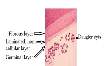
part of wall of Unilocular Hydatid Cyst

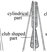
Nematode Cylindrical Myosyringata (Muscular oesophagus)
Nematode Club-Shaped Myosyringata (Muscular oesophagus)
Nematode Rhabditiform Myosyringata (Muscular oesophagus)
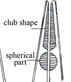
Nematode Double-bulbed Myosyringata (Muscular oesophagus)
Nematode Cellular Oesophagus Trichosyringata
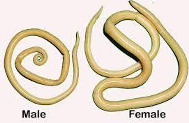
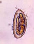
Ascaris lumbricoides
(Giant Intestinal Roundworm)
Ascaris lumbricoides
(Giant Intestinal Roundworm)
Ascaris lumbricoides
(Giant Intestinal Roundworm)
Ascaris lumbricoides
egg
Diagnostic Stage
Ascaris lumbricoides
egg
Diagnostic Stage
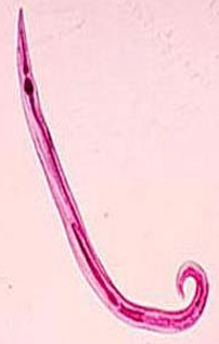
Enterobius vermicularis (Oxyuris, Pinworm)
Female
pointed tail hence the name pinworm
Enterobius vermicularis (Oxyuris, Pinworm)
Male
curved ventrally carries a single spicule.
Infective stage of Enterobius vermicularis?
Mature egg (egg containing larva)
Mode of infection of Enterobius vermicularis?
Autoinfection: Ingestion of eggs lodged on contaminated fingers of the infected persons
Ingestion
Inhalation
Retro Infection: Re-entrance of larvae that sometimes hatch from eggs on the peri anal skin to the large intestine
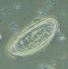
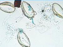
Eggs of Enterobius vermicularis
Plano-convex, flattened on one side and curved in the other (D shaped).
Thick colorless shell
Doubly refractive wall
50 x 25 um
Mature, immediately infective
Eggs of Enterobius vermicularis
Plano-convex, flattened on one side and curved in the other (D shaped).
Thick colorless shell
Doubly refractive wall
50 x 25 um
Mature, immediately infective
Eggs of Enterobius vermicularis
Plano-convex, flattened on one side and curved in the other (D shaped).
Thick colorless shell
Doubly refractive wall
50 x 25 um
Mature, immediately infective
Eggs of Enterobius vermicularis
Plano-convex, flattened on one side and curved in the other (D shaped).
Thick colorless shell
Doubly refractive wall
50 x 25 um
Mature, immediately infective
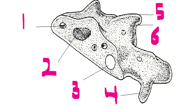
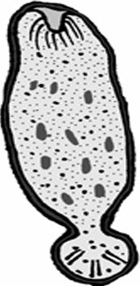
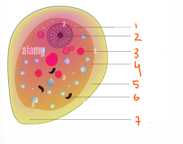
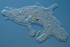
General Characters of Protozoa
1- Food vacuole
2- Nucleus
3- Contractile vacuole
4- Pseudopodium
5- Ectoplasm
6- Endoplasm
1- Nucleus
2- Endosome
3- Ingested RBC
4- Endoplasm
5- Ectoplasm
6- Ingested bacteria
7- Pseudopodium

Reproduction of Protozoa
Asexual: Binary fission
It occurs either longitudinally or transversally. Mitotic division of nucleus is followed by division of the cytoplasm.
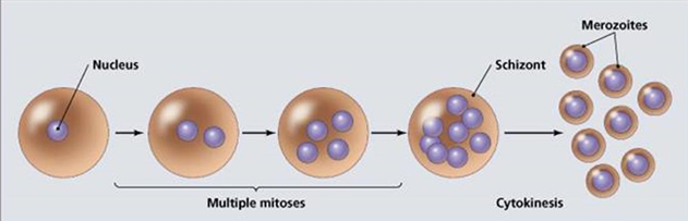
Reproduction of Protozoa
Asexual: Multiple fission or schizogony
The nucleus undergoes several successive divisions within the schizont to produce large number of merozoites
Reproduction of Protozoa
Asexual: Endodyogeny
Two daughter cells form within the parent cell —> Ruptures —> the smaller progeny grow to full size before repeating the process
Reproduction of Protozoa
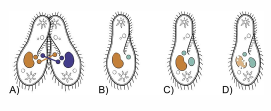
Sexual: Conjugation
where two organisms fuse together and exchange nuclear matter as in Balantidium coli.
Locomotion of protozoa
Flagella
long thread-like cytoplasmic extensions. • Arises in the endoplasm from basal body • Kinetoplast is at the base of the basal body of the flagellum • It consists of a curved, electron-dense structure containing DNA
Locomotion of protozoa
Cilia
Numerous short hair-like threads covering the whole organism. Each arises from a basal granule just below the surface of the cell
Locomotion of protozoa
Pseudopodia
Extension of the ectoplasm followed by the endoplasm resulting in amoeboid movement
Functions: 1- locomotion 2-Engulfing of food
Pseudopodia
Extension of the ectoplasm followed by the endoplasm resulting in amoeboid movement
Functions: 1- locomotion 2-Engulfing of food
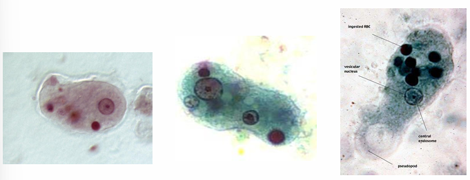
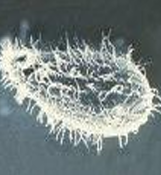
Entamoeba histolytica tissue form (Magna form)
It DOSEN’T form precyst or cyst
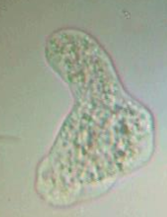
Entamoeba histolytica lumen form (Minuta form)
It DOES form precyst or cyst
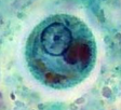
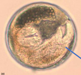
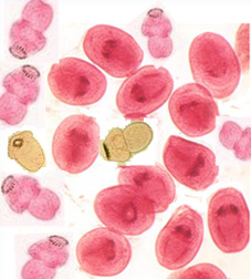
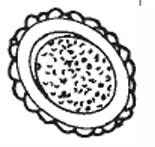
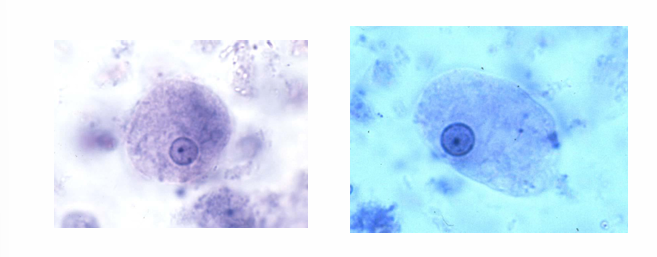
Cyst of Entamoeba histolytica
Rounded with well developed cyst wall, 12-15 µ, it contains 1-4 nuclei, glycogen and chromatoid bodies