BIOL 221 - Chapter 13 - Cardiovascular System
1/36
There's no tags or description
Looks like no tags are added yet.
Name | Mastery | Learn | Test | Matching | Spaced | Call with Kai |
|---|
No analytics yet
Send a link to your students to track their progress
37 Terms
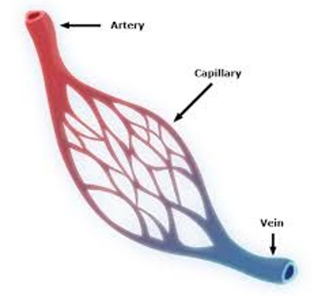
Arteries
Transports blood AWAY from the heart
Veins
Transport blood TOWARDS the heart
Capillaries
Vessels that run between the arteries and veins
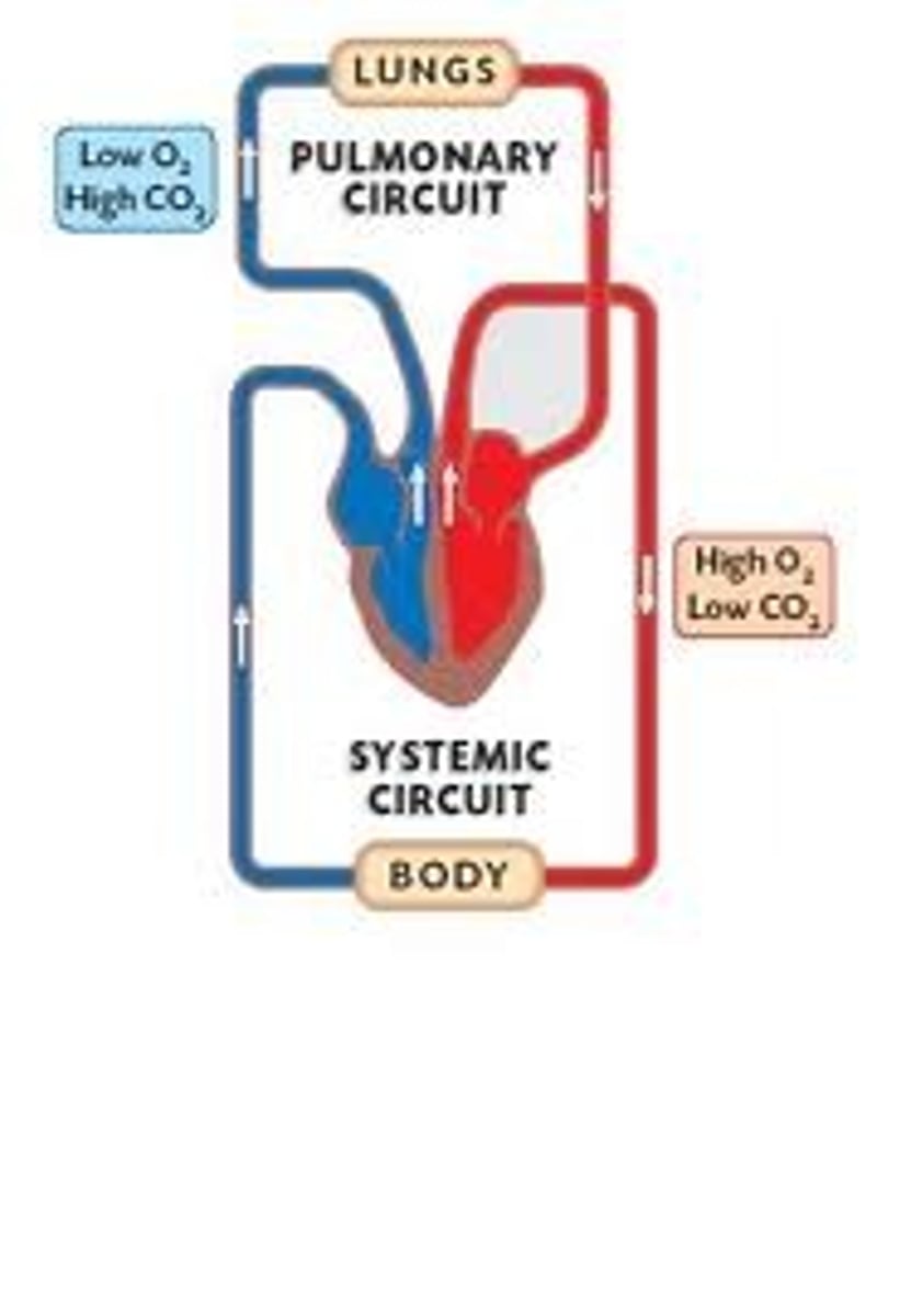
Pulmonary Circuit
Blood flow between the heart and LUNGS
Systematic circuit
Blood flow between the heart and BODY TISSUES
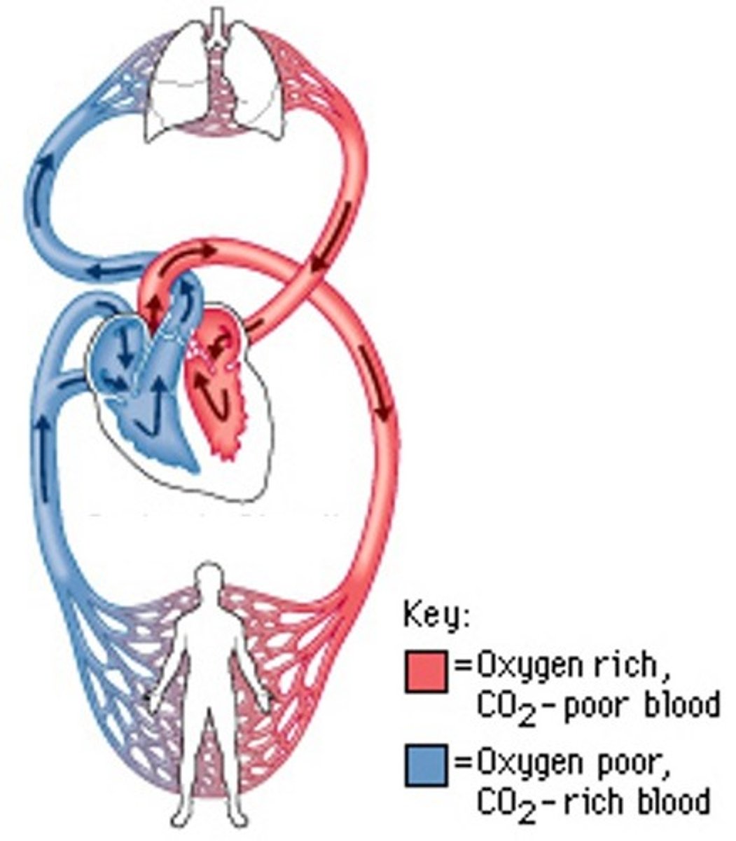
Pulmonary Veins
Deliver oxygen rich blood from the lungs to the left atrium
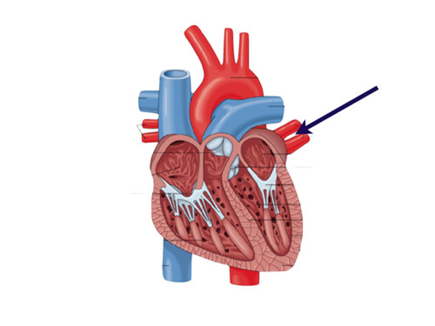
Pulmonary arteries
Carry deoxygenated blood out of the right ventricle and into the lungs
Blood path of travel
Right side of the heart sends blood to the lungs to get oxygenated, it then comes back to the left side to be circulated around the body
Albeolus
Where gas exchange takes place
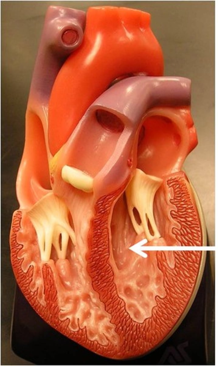
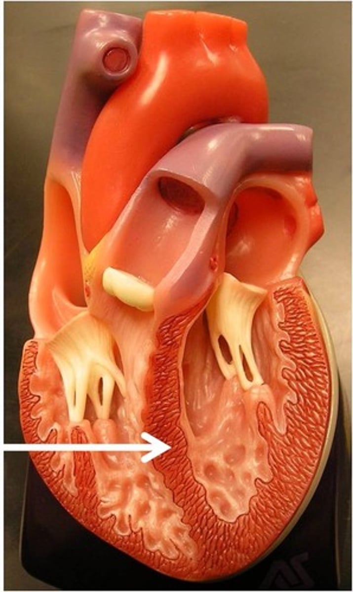
Atria
The top of the heart (L and R); fills with blood returning to the heart; thin
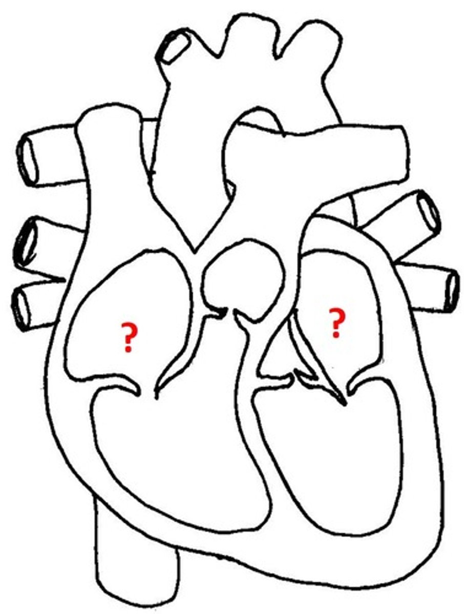
Ventricles
The bottom of the heart (L and R); Pump blood out of the heart; thick
Interventricular Septum
Separates the atrium and the L and R ventricles
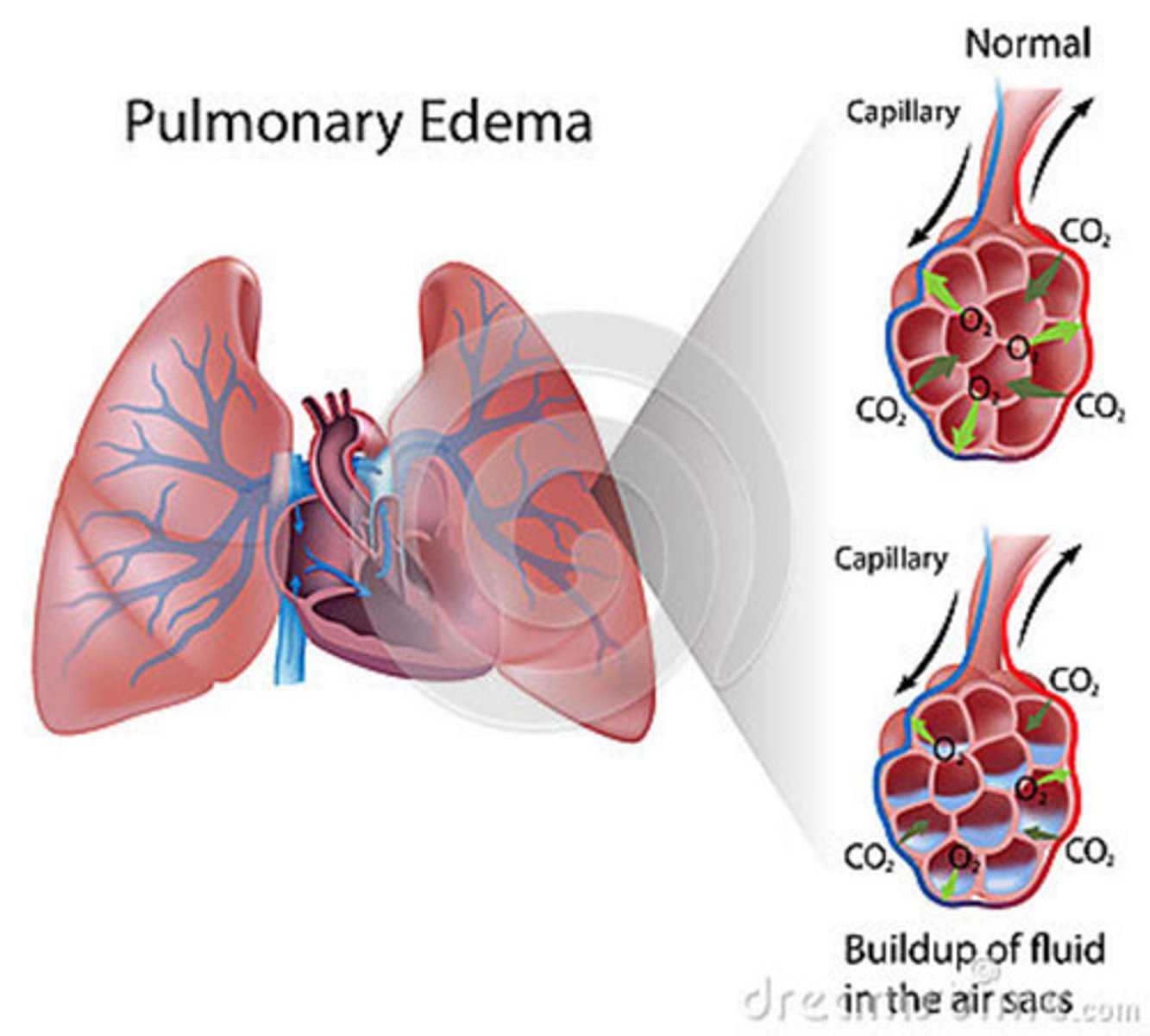
Pulmonary Edema
Fluid in the lungs
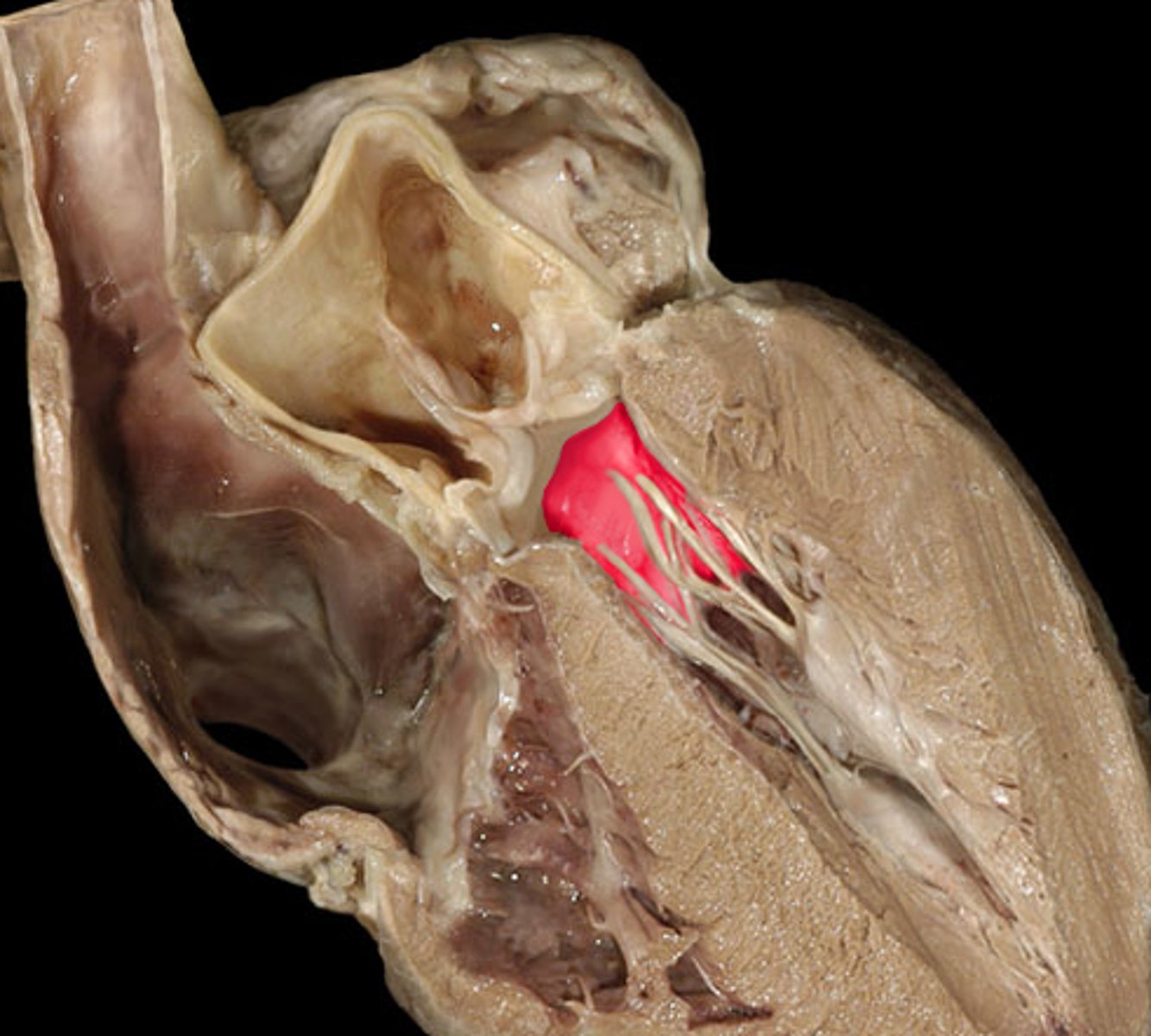
Right AV Valve (Tricuspid Valve)
Located between the R atrium and R ventricle
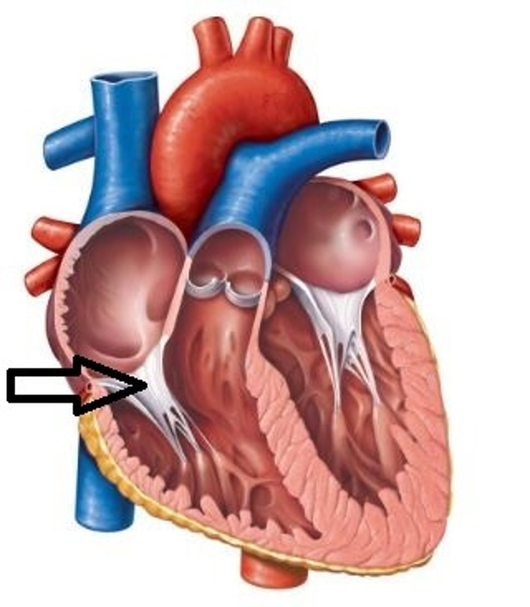
Left AV Valve (Bicuspid or Mitral Valve)
Located between the L atrium and L ventricle
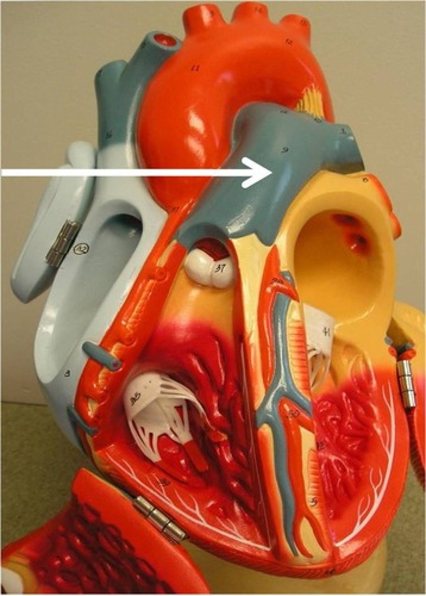
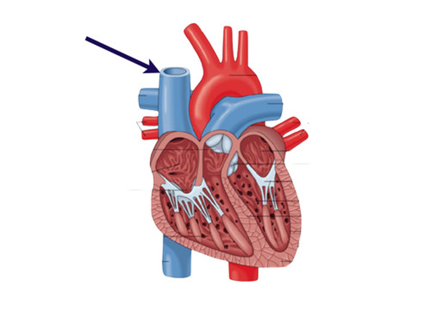
Superior and Inferior Vena Cava
Veins that carry deoxygenated blood to the right atrium from the systemic circuit
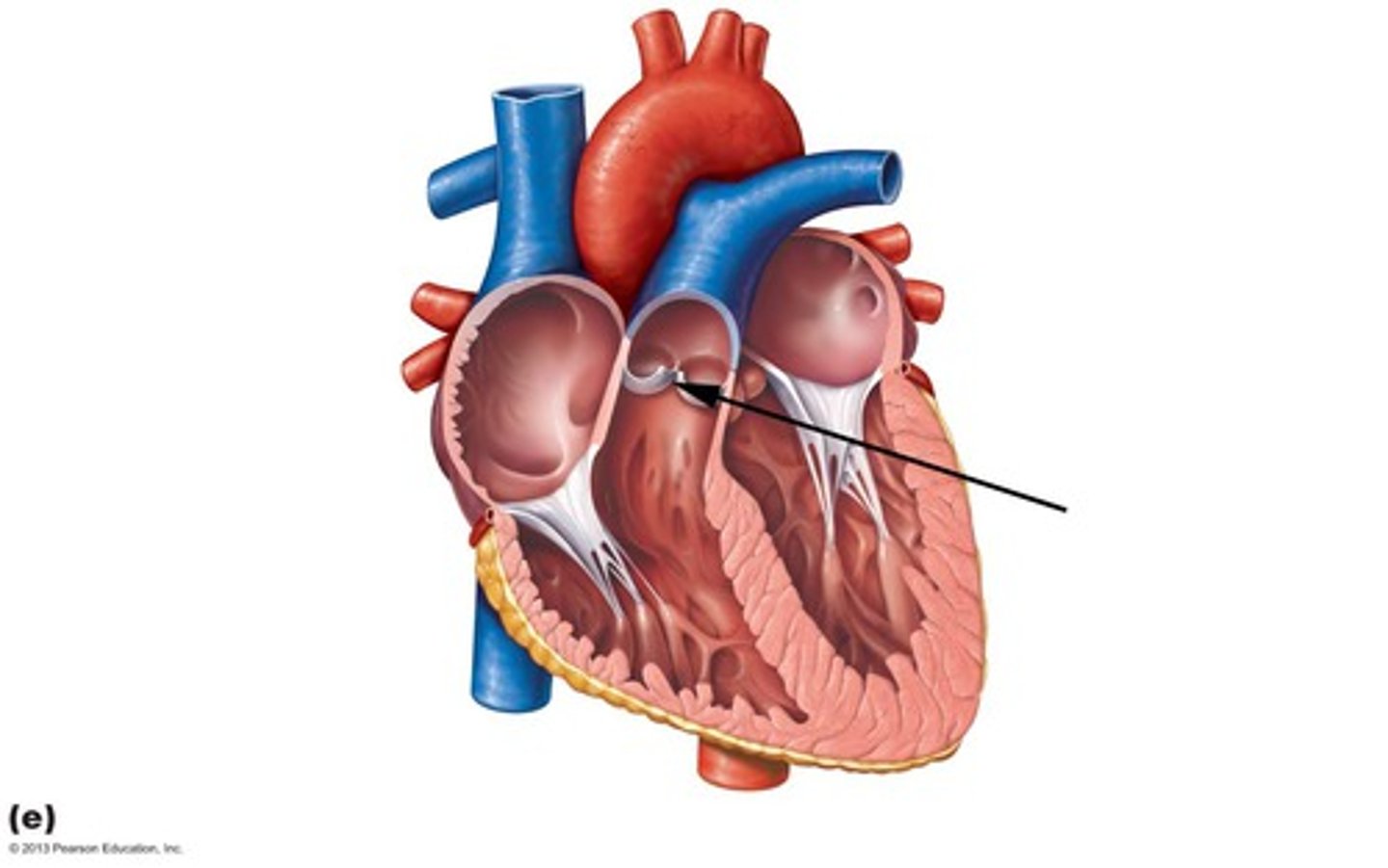
Pulmonary Semilunar Valve
Prevents backflow into the RIGHT ventricle; located between the R ventricle and Pulmonary Trunk
Aortic Semilunar Valve
Prevents backflow into the LEFT ventricle; located between the L ventricle and Aorta

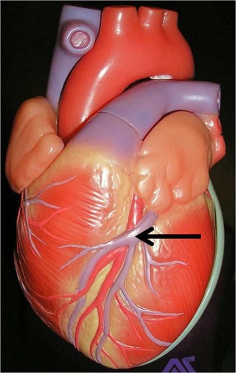
Anastomoses
Provide pathways for blood incase a pathway becomes blocked
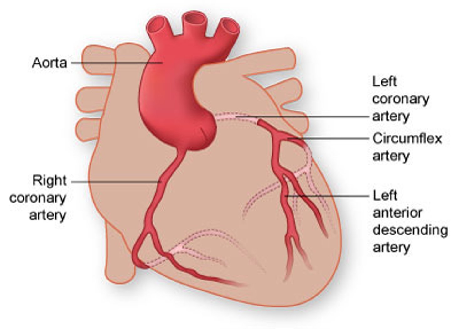
Cardiac Veins
Drain blood from the heart and carry it to the coronary sinus
Coronary Sinus
Big bulging vein where the blood drains to at the back of the heart
Systole
Contraction of the heart
Diastole
Relaxation of the heart
Functional Syncytium
A mass of cells that acts as a unit
Cardiac Conduction System
Muscle tissues that conducts impulses (electricity) throughout the myocardium
Sinoatrial (SA) Node
" Pacemaker " of the heart located in the right atrium. generated the impulses for heartbeat
Purkinje Fibers
Spread impulses to the ventricular wall and papillary muscles causing contraction
Electrocardiogram (ECG)
Records the electrical changes during the cardiac cycle
P Wave
Corresponds to the depolarization of the atria; this leads to the contraction of the atria

QRS Complex
Depolarization of ventricles, which leads to contraction of the ventricles; the repolarization of the atria occurs during the QRS complex, but is hidden behind the larger ventricular event.

T Wave
Corresponds to ventricular repolarization, and leads to ventricular relaxation

Lubb-Dupp
Lubb - ventricles contract and AV valves are closing
Dubb - ventricles relax and aortic/pulmonary valves are closing
Tachycardia
>100 bpm resting heart rate
Bradycardia
<60 resting heart rate
Cardiac Center of the Medulla Oblongata
Maintains balance btwn the sympathetic and parasympathetic divisions of the autonomic nervous system
Hyperkalemia
excessive K+ in the blood; decreases heart rate and force of contraction
Hypercalcemia
excessive calcium in the blood