cardiovascular systems
1/61
Earn XP
Description and Tags
blood & heart
Name | Mastery | Learn | Test | Matching | Spaced | Call with Kai |
|---|
No analytics yet
Send a link to your students to track their progress
62 Terms
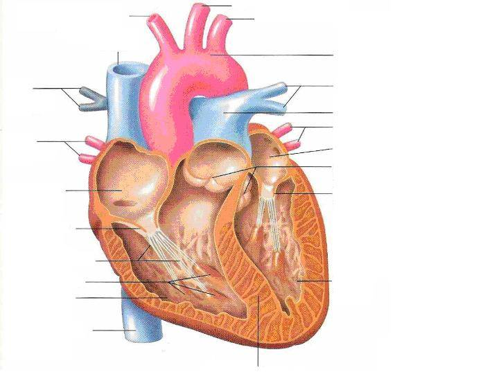
label (differentiate b/w semilunar valves)
semilunar valves —> right (side of heart): pulmonary valve, left: aortic valve
components that make up blood (& %)
plasma (55%)
buffy coat AKA white blood cells & platelets (1%)
red blood cells (44%)
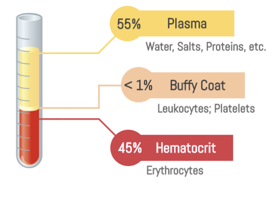
plasma
made of 90% water
transports blood cells & chemicals (nutrients, hormones, and waste) throughout body
white blood cells
immune defense (produce antigens)
red blood cells
transport oxygen with its hemoglobin
what 2 things makes the sound “Lubb Dubb” in the heart?
“Lubb” ← tricuspid & bicuspid valve closing
“Dubb” ← semilunar valves (aortic & pulmonary) closing
Another name for red blood cells?
Erythrocytes
what are red blood cells made of?
hemoglobin (iron-rich protein that carries oxygen)
define hematopoiesis
the process of creating new blood cells—including red blood cells, white blood cells, & platelets—that primarily occurs in red bone marrow
define leukocytes
aka white blood cells, protects the body from infection, 1% of blood
define lymphocytes
a major type of white blood cell that regulate immune cell function, attack infected cells/tumors, and produce antibodies
what blood type is the universal donor?
O- (b/c it lacks A, B, and Rh antigens)
what blood type is the universal recipient?
AB+ (b/c lacks A, B, and Rh antibodies)
what is a sphygmomanometer?
medical instrument used to measure blood pressure
systolic vs. diastolic blood pressure
measures force of circulating blood in arteries pumped by the heart
Systolic —> when heart contracts (max pressure during a beat)
Diastolic —> when heart relaxes (lowest pressure b/w beats)
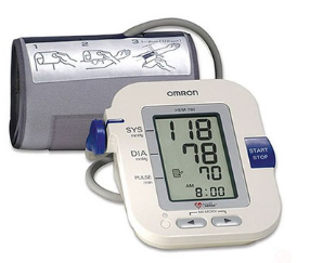
what blood pressure is considered normal?
120/80 mm Hg
tachycardia
fast heart rate (100+)
bradycardia
slow heart rate (x<60)
define EKG
measures electrical activity of heart (records heartbeats, rate, & rhythm)
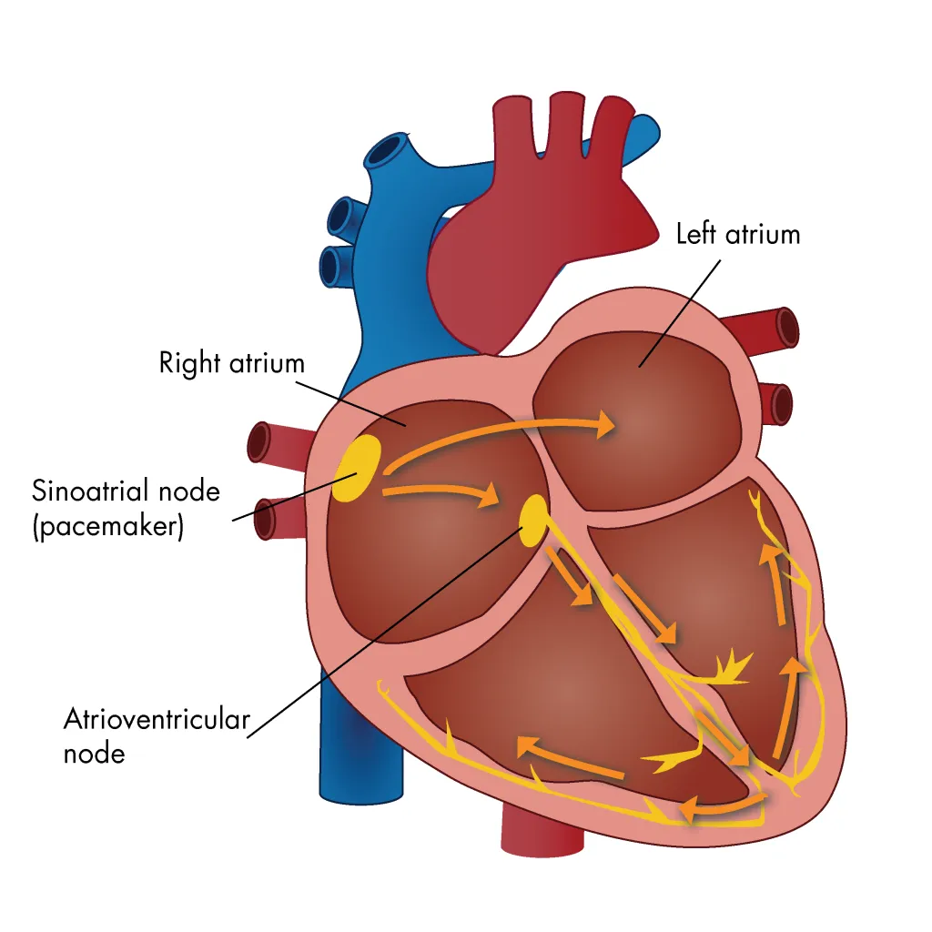
SA vs. AV node
SA: initiates impulses to contract the heart (controls heart rate)
AV: receives signal and delays it (so blood can contract)
3 functions of blood
transports nutrients, nutrients, hormones, & waste
regulates pH, temp, & pressure
clots & fights infection
Hemostasis
aka blood clotting process
vascular spasm (constriction)
primary hemostasis (platelet form a temp plug)
secondary hemostasis (body amps blood-clotting effects & fibrin creates mesh)
3 stages of hemostasis
vascular spasm — injured blood vessel constricts to minimize blood loss
primary hemostasis — platelet gather to blood vessel & forms a temp plug
secondary hemostasis — fibrin creates mesh that traps blood cells from leaving, surrounds and stabilizes the clot for wound healing
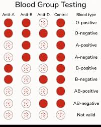
how is blood typing performed?
mixing a blood sample with antibodies (anti-A, anti-B, and anti-Rh) to detect antigen clumping.
Yes clumps —> does have that antibody
no clumps (solid color) —> doesn’t have that antibody
main components of cardiovascular system
heart, blood vessels, and blood
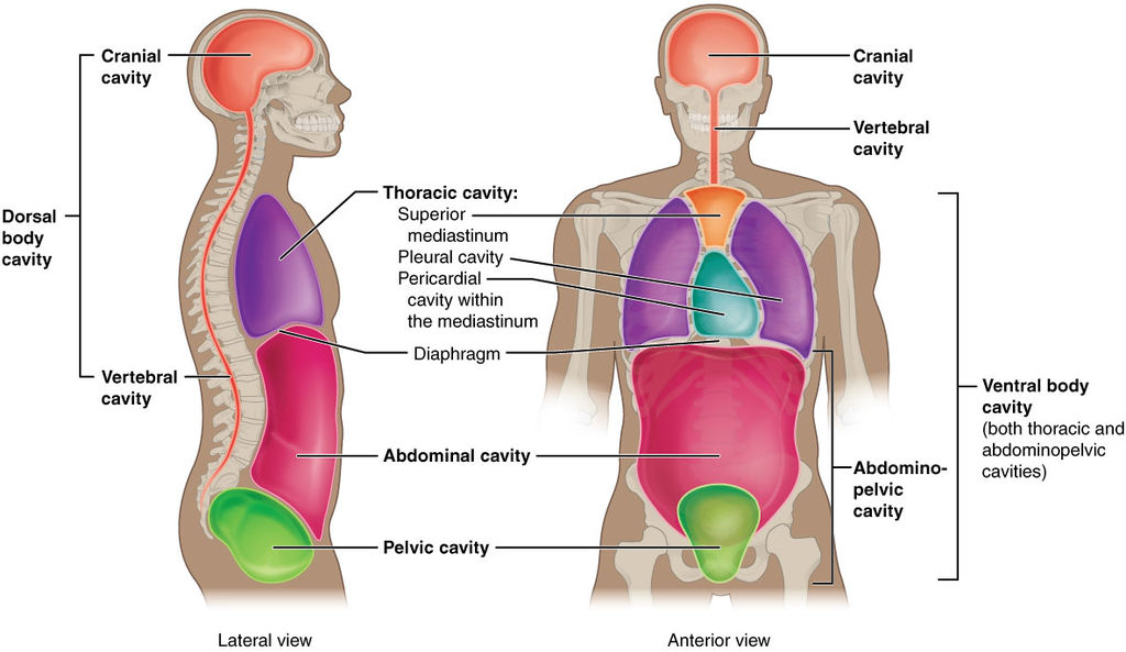
what cavity is the heart located in?
mediastinum cavity
list & describe the 3 layers of the heart
outermost —> innermost
epicardium — thin layer w/ connective tissue, fat, & coronary blood vessels
also part of pericardium
myocardium — thick layer made of cardiac muscle cells; responsible for contracting/pumping
endocardium — line chambers & cover valves; acts as protective barrier b/w blood & heart (myocardium)
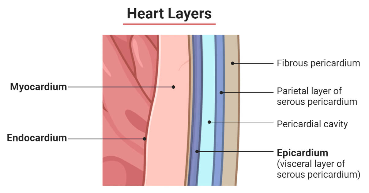
pericardium
protective, double-layered membrane that surrounds the outside of the heart

pulmonary circuit
system that transports blood from the heart to the lungs, becomes oxygenated, & back
systemic circuit
system that transports oxygenated blood from the heart to the rest of the body & back
difference b/w pulmonary circuit vs systemic circuit
P: (deoxygenated) blood goes from heart to lungs & back
S: (oxygenated) blood goes from heart to rest of body & back
what do heart valves do?
control blood flow, ensuring it moves in one direction, & prevents backward flow
electrical pathway of the heart (list stages/flow)
SA node — “pacemaker” initiates heartbeat by sending electrical impulses
AV node — delays signal to the ventricle slightly until the atria are empty (to ensure the blood fills the ventricles before contraction starts)
AV bundle (bundle of His) — carries signal to Purkinje fibers through bundle branches
Purkinje fibers — distribute electrical signal to ventricles —> contracts & delivers blood out to body
trace pathway of blood flow throughout the heart
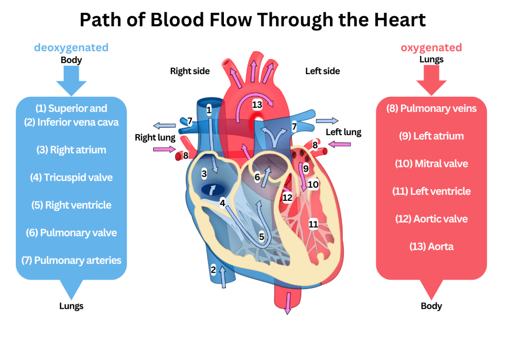
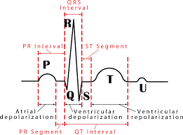
what occurs at each letter? (excluding s)
P —> SA node sends electrical impulse, so atria contracts
PR segment —> delay caused by AV node
QRS —> ventricles contract, atria relaxes
T —> ventricles relax
U —> sign of low potassium
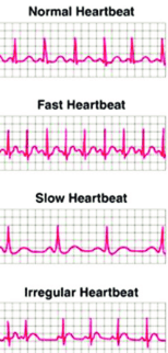
4 primary sinus rhythms
normal: 60-100 bpm
tachycardia: more than 100 bpm
bradycardia: less than 60 bpm
arrhythmia: irregular
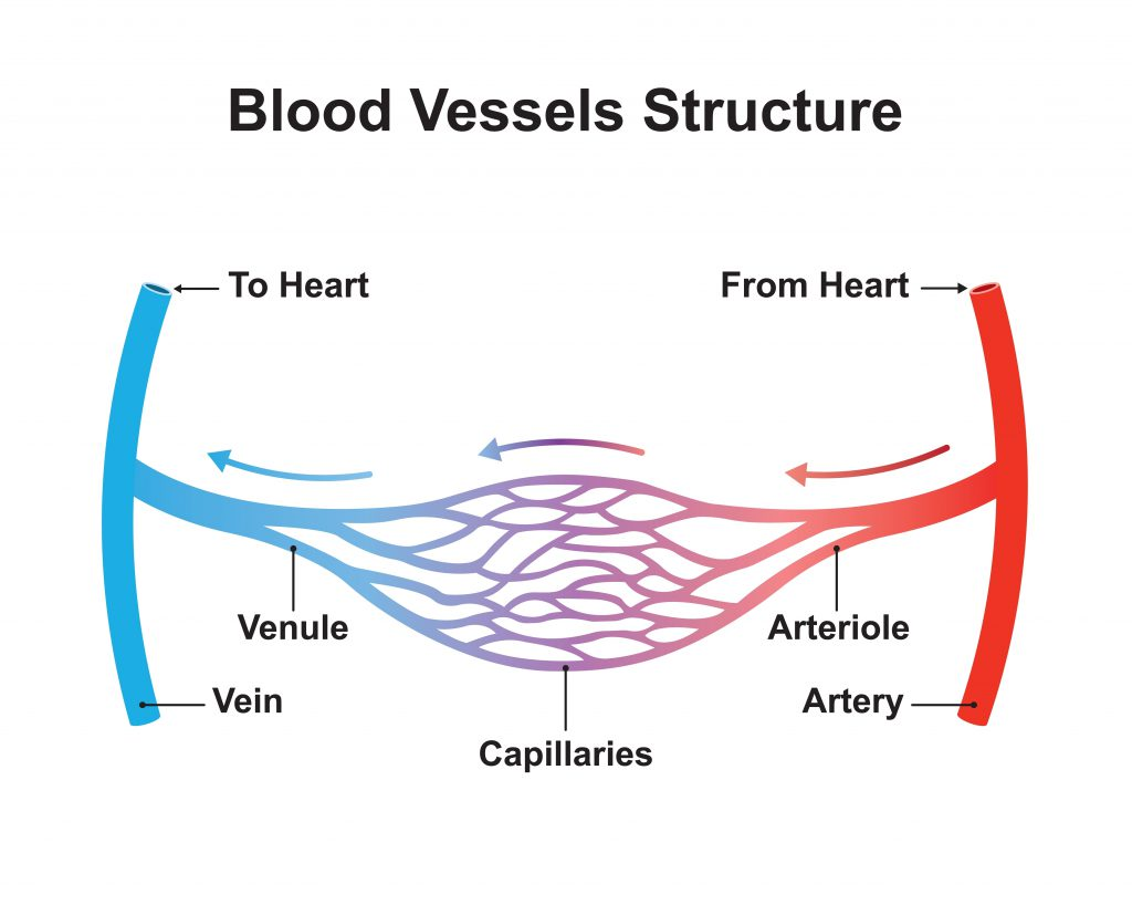
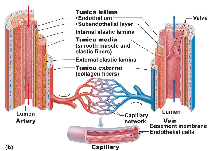
arteries
moves blood away from the heart
thicker, rigid walls
high pressure
“art” —> renaissance art w/ high society & thick paint
capillaries
1-cell thick walls
exchanges gases (O² & CO²) b/w blood & tissue cells
veins
brings blood toward heart
thinner, flexible walls
low pressure
large lumen (central, hollow channel allowing blood flow)
contains valves
3 layers of blood vessels & their functions
outermost —> innermost
tunica externa: fibrous connective tissue (protects)
tunica media: smooth muscle & elastic tissue (dilates & constricts)
tunica intima: lined w/ endothelium aka endothelial cells (decreases friction as blood flows)
what is a pulse & where to find one?
expansion & recoil of arteries close to the skin where blood pumps through the body from the heart; indicates BPM; some locations are
wrist (radial artery)
elbow (brachial artery)
neck (carotid artery)
what is resting heart rate?
70-76 bpm
what is considered to be high blood pressure?
140+/90+
hypertension
high blood pressure
circulatory shock
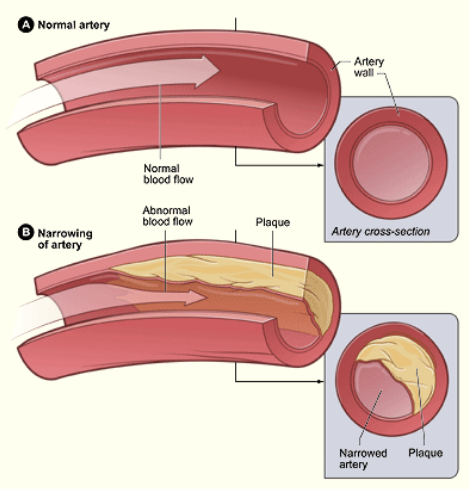
severe hypotension (low blood pressure) from inadequate blood flow & oxygen delivery caused by atherosclerosis (plaque buildup in arteries)
hypotension
low blood pressure
atherosclerosis
the thickening of artery walls due to the buildup of fatty deposits (plaques) from dmg to endothelial lining
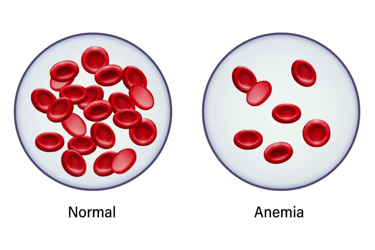
anemia
insufficient amount of healthy red blood cells or hemoglobin to carry oxygen to the body’s tissues
caused by low iron, blood, or nutrient
symptoms: fatigue, dizziness, pale skin
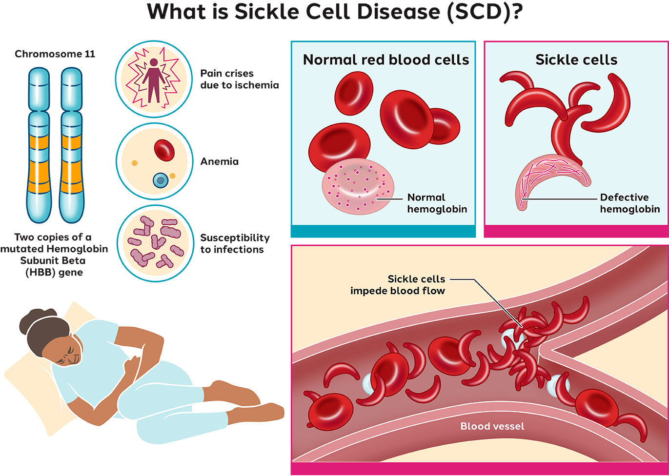
sickle-cell disease
genetic blood disorder where red blood cells turn sickle-shaped, blocking blood flow
causes chronic anemia, severe pain, & organ dmg
leukemia
cancer of blood-forming tissues, producing an excessive, abnormal white blood cells.
embolus
blood clot that is a detached, traveling mass that moves through the bloodstream until it lodges somewhere
hemophilia
blood doesn’t clot properly; primarily affects males
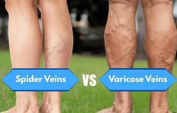
varicose veins
swollen, twisted veins (typically in legs) caused by weakened valves (blood flows backward & collects in the veins)
angina pectoris
chest pain caused by reduced blood flow & oxygen to heart muscle
myocardial infarction
aka heart attack as of result of when blood flow is blocked or greatly reduced
ischemia
serious condition caused by reduced blood flow to organs or tissues, depriving them of necessary oxygen & nutrients —> tissue dmg or death
fibrillation
arrhythmia (irregular heartbeat) causing risk of stroke, blood clots, and heart failure
heart murmur
extra or unusual “whooshing” sound heard during a heartbeat made by
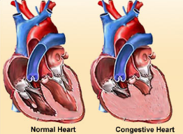
congestive heart failure
progressive weakening of heart b/c inadequate circulation to tissue; caused by
coronary atherosclerosis
persistent high blood pressure
multiple heart attacks
A-Fib vs V-Fib
irregular heartbeats
A: chronic, manageable condition in upper heart chambers
V: fatal immediate emergency in lower heart chambers
arterioles
small, branching vessels from arteries connecting to capillaries

venules
small, thin blood vessels leading away from capillaries to veins