Chapter 6: Skeletal System Lab
1/199
There's no tags or description
Looks like no tags are added yet.
Name | Mastery | Learn | Test | Matching | Spaced | Call with Kai |
|---|
No analytics yet
Send a link to your students to track their progress
200 Terms
SKELETAL
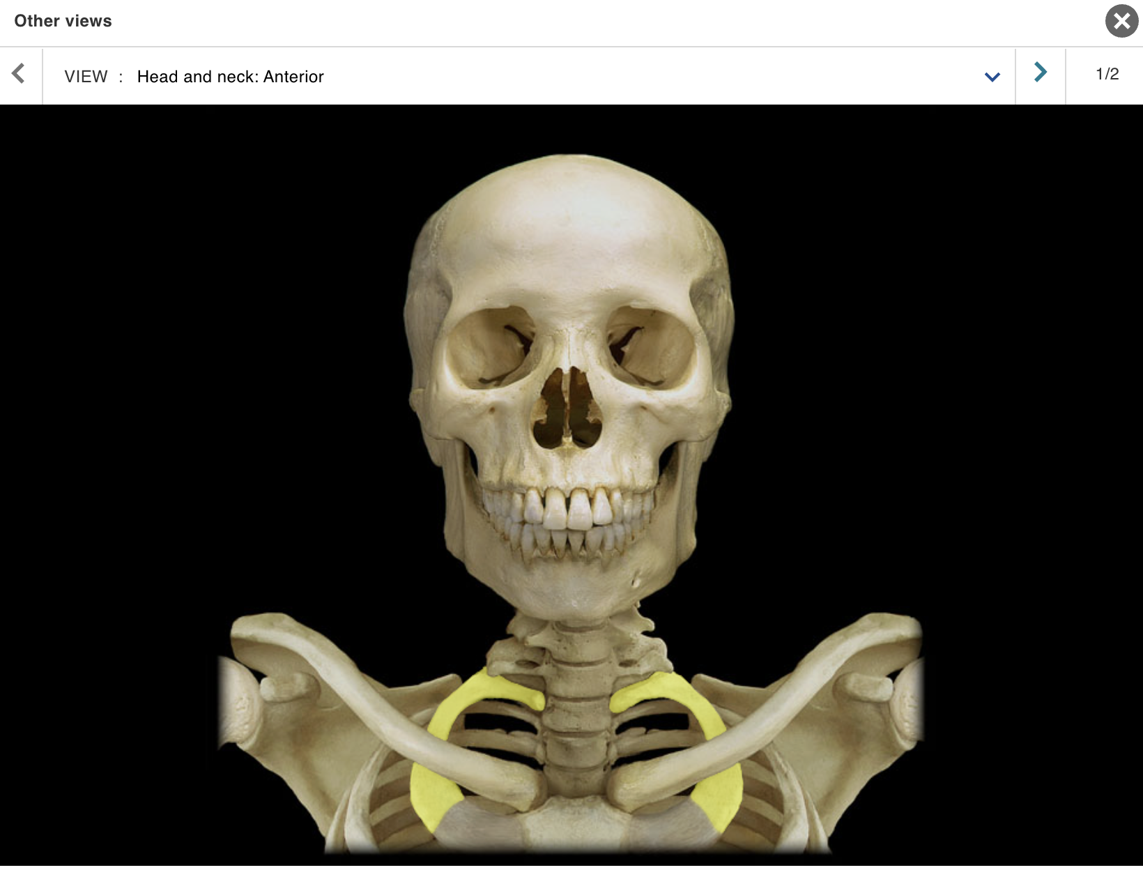
TOPIC: HEAD / NECK / VIEW : ANTERIOR
Cervical vertebra
Clavicle
Frontal bone
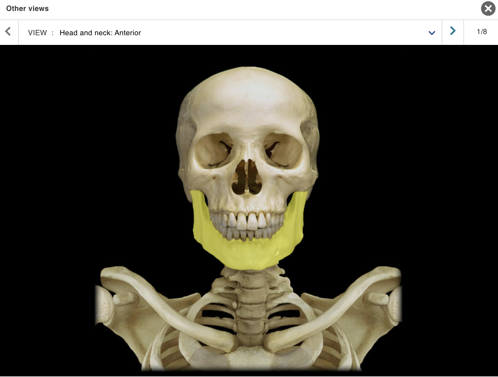
Mandible
Manubrium
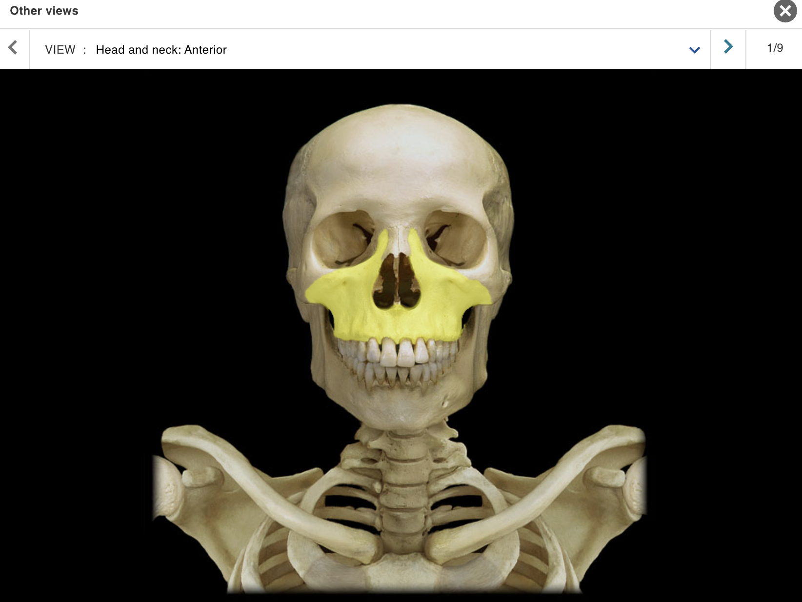
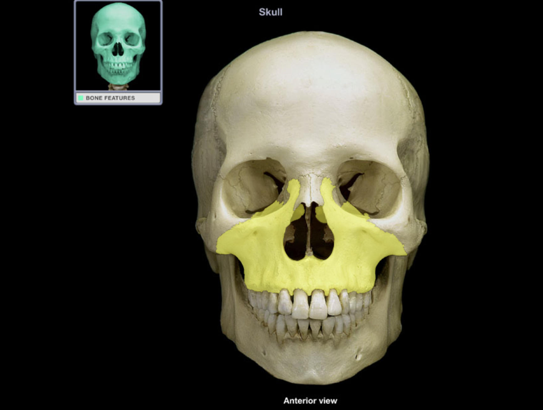
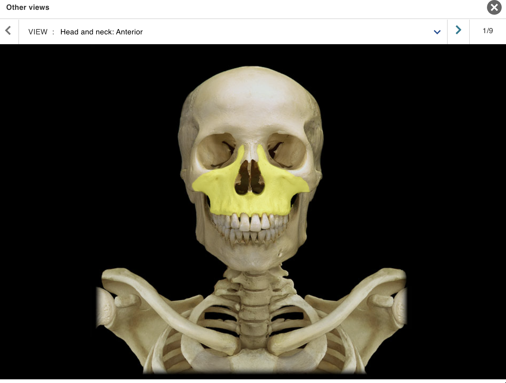
Maxilla
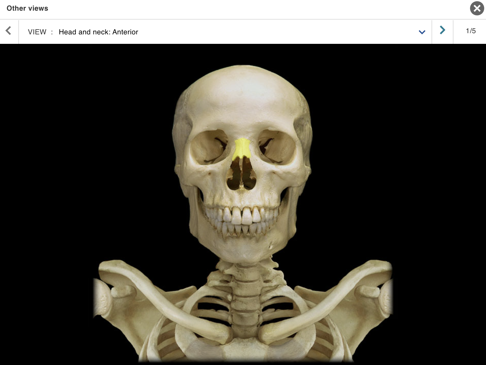
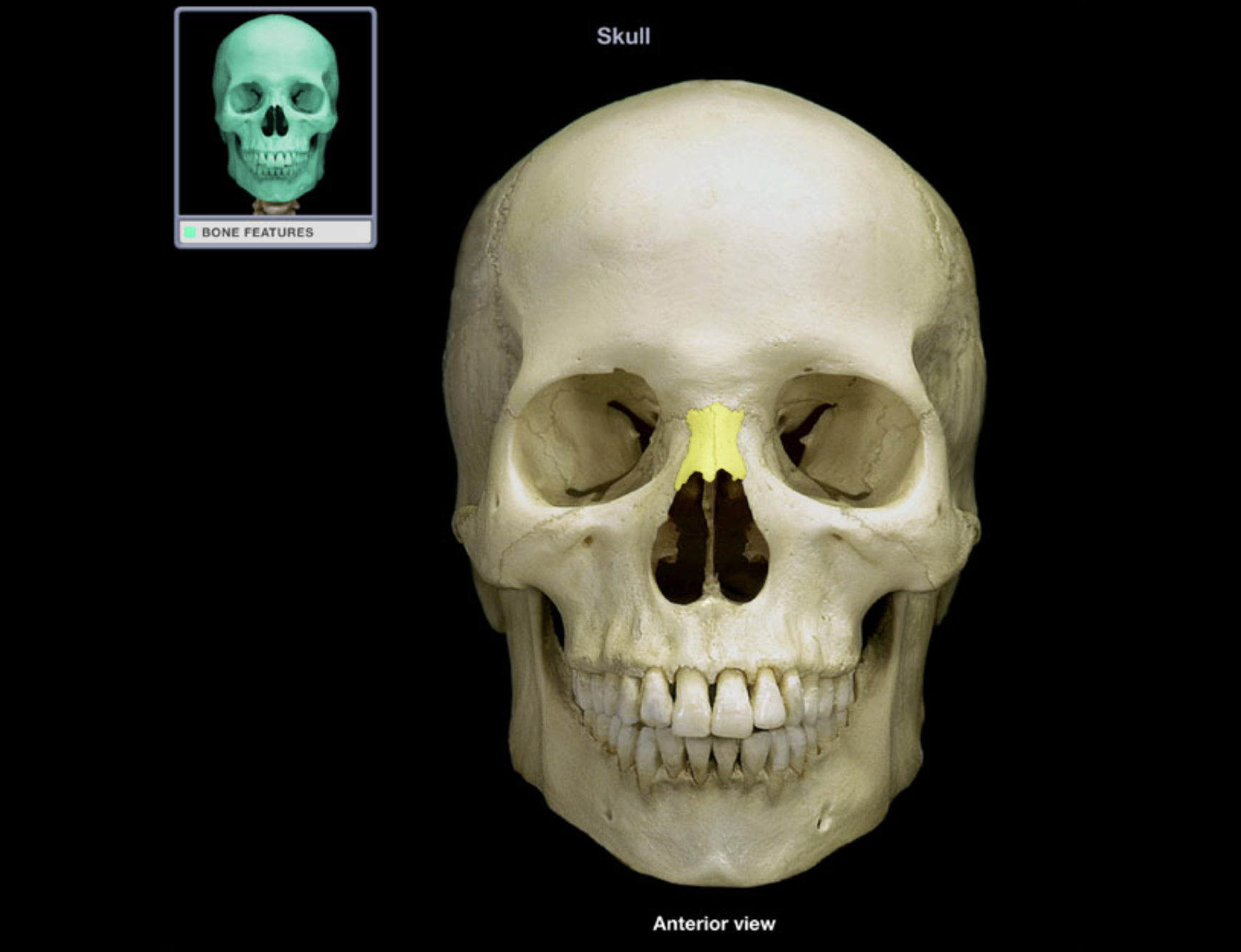
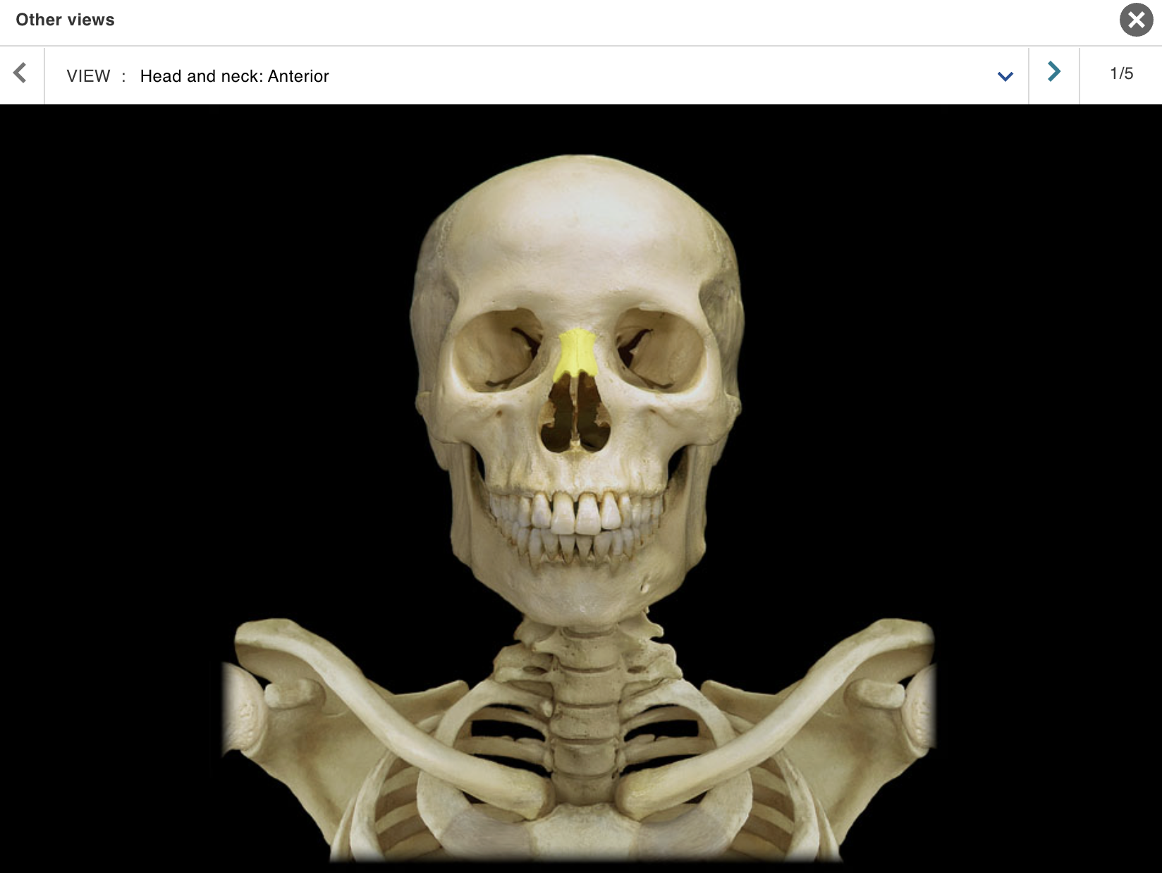
Nasal bone
Orbit
Rib 1
Scapula
Skull
Sphenoid bone
Thoracic vertebra
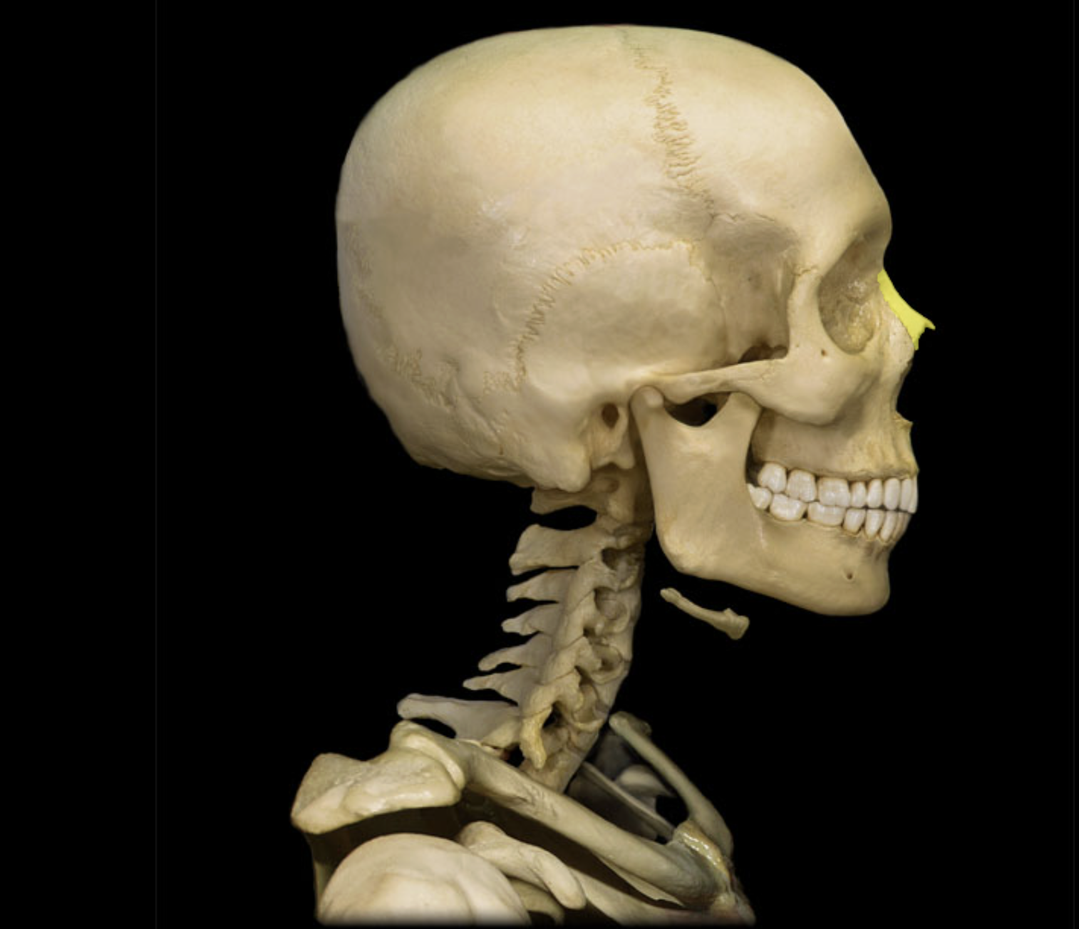
Zygomatic bone
Cervical vertebra
Location:
Neck
Between occipital bone and T1 vertebra
Description:
Seven individual vertebrae
Characteristic features include transverse foramen and bifid (split) spinous process on C3-C6
Comment:
• Atlas (C1 vertebra) articulates with skull
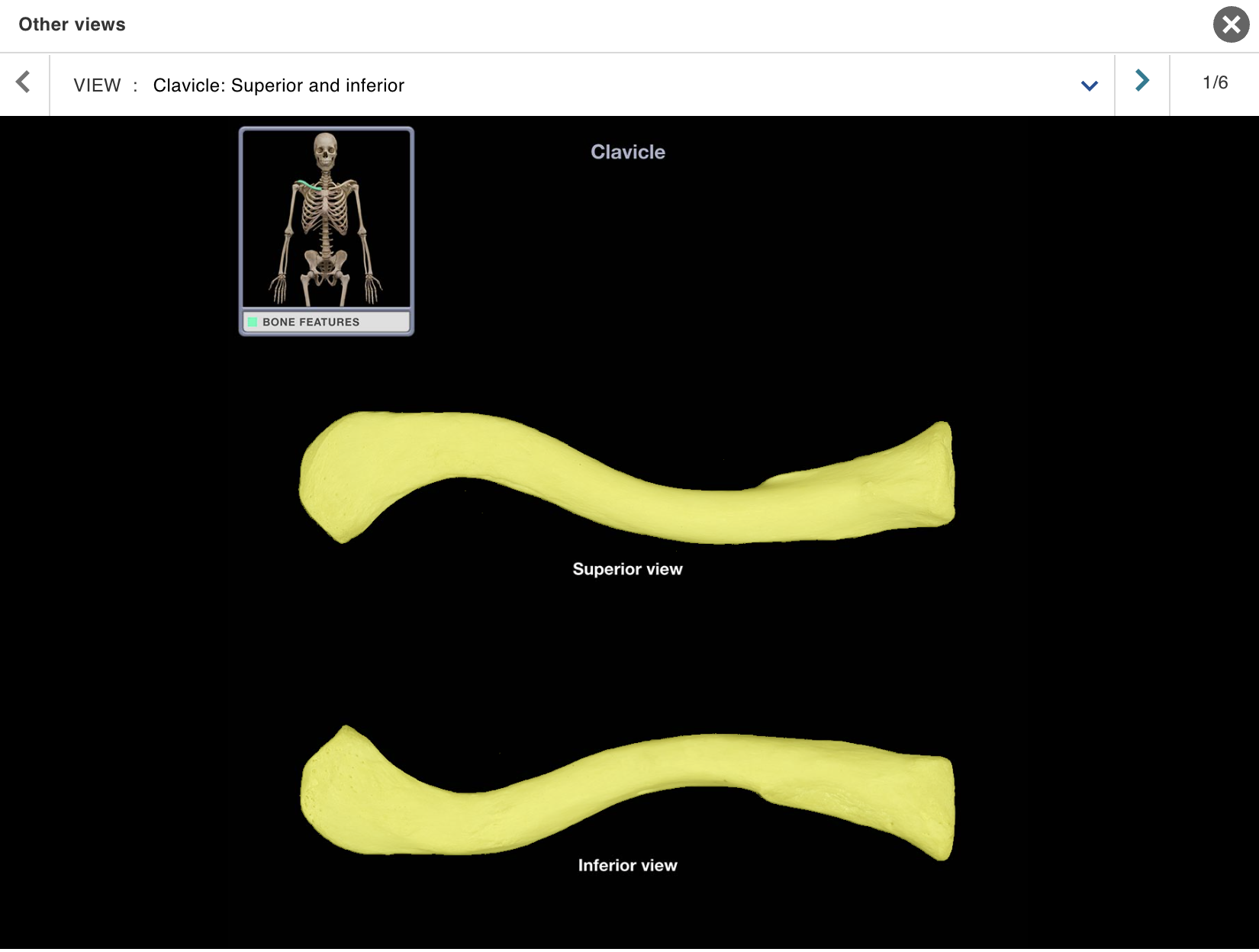
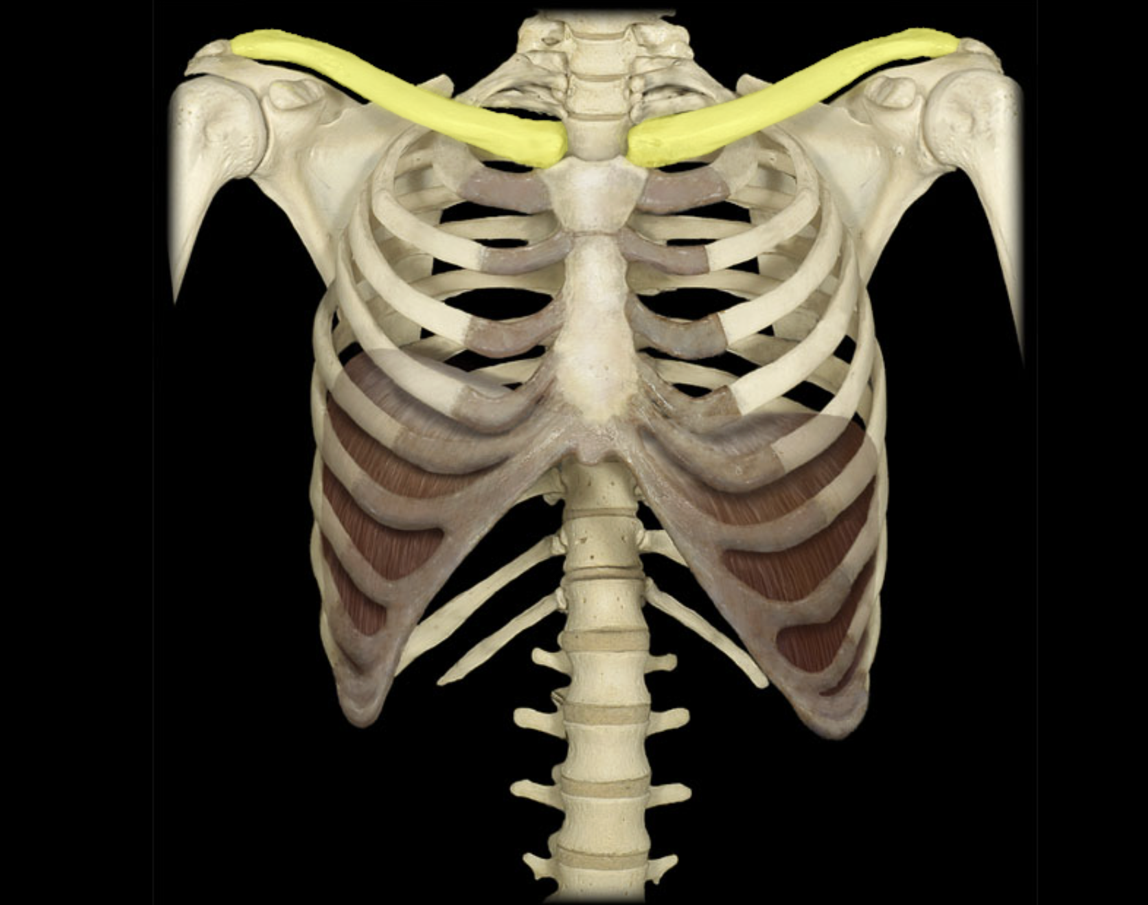
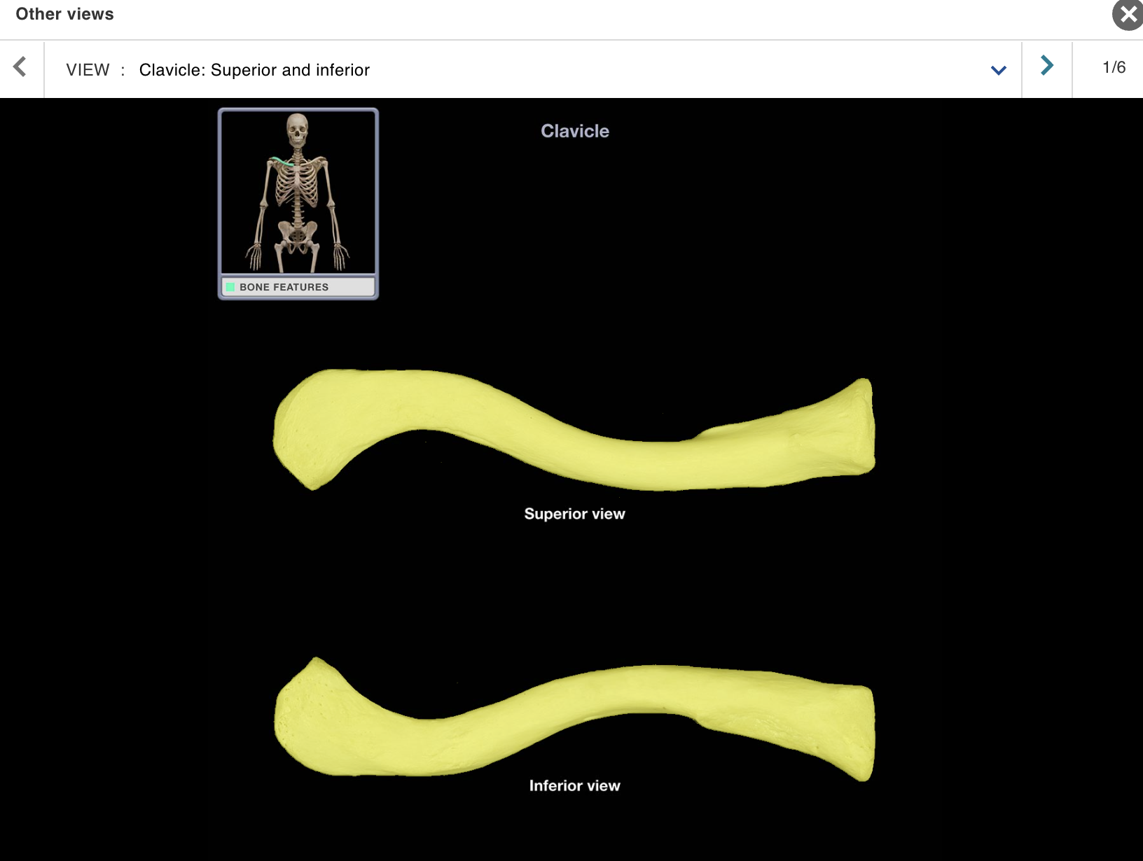
Clavicle
Location:
• Junction of neck and anterior thorax
Description:
Subcutaneous, S-shaped bone
Medial end articulates with sternum at sternoclavicular joint
Lateral end articulates with acromion of scapula at acromioclavicular joint
Also known as:
• "Collar bone"
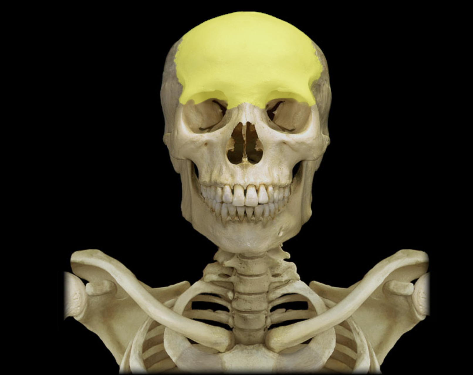
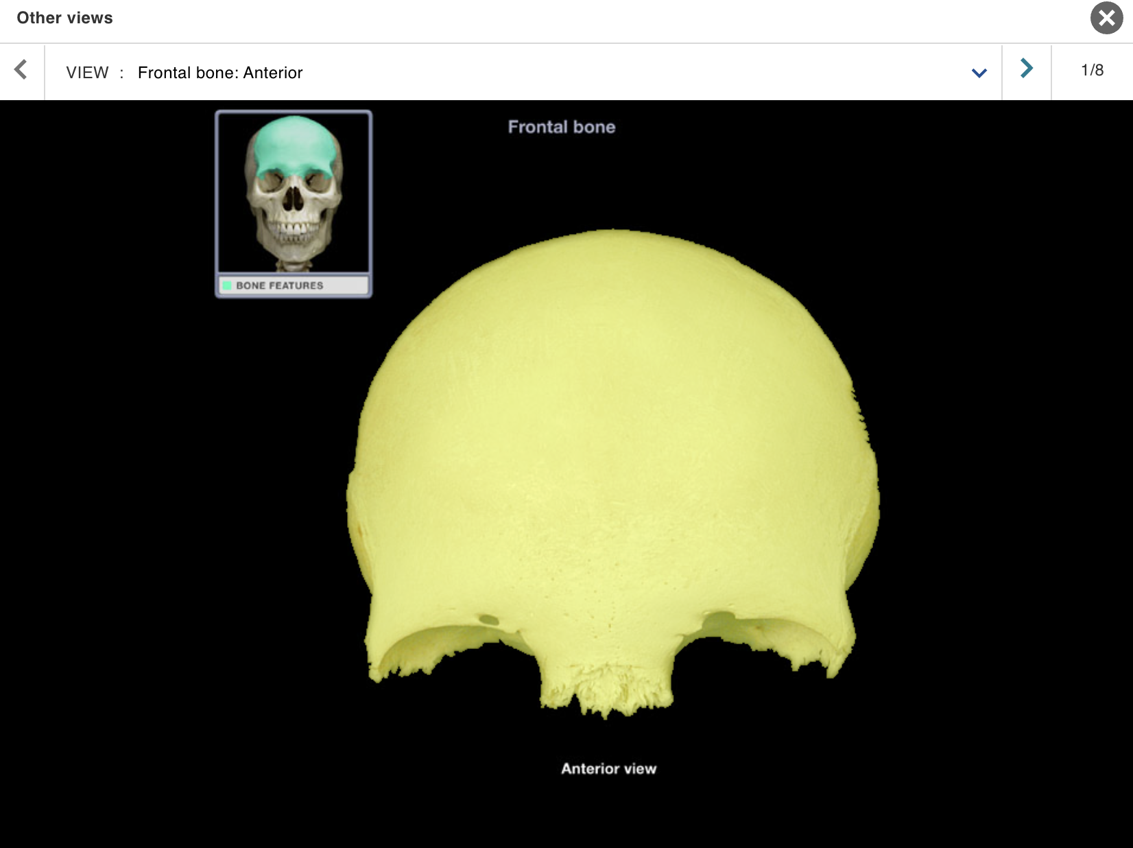
Frontal bone
Location:
• Skull (anterior superior part)
Description:
Unpaired, irregular-shaped, flat bone
Forms forehead, roof of orbits, and most of anterior cranial fossa
Contains frontal air sinuses
Comment:
• Articulates with parietal bone at coronal suture and sphenoid bone at sphenosquamosal suture
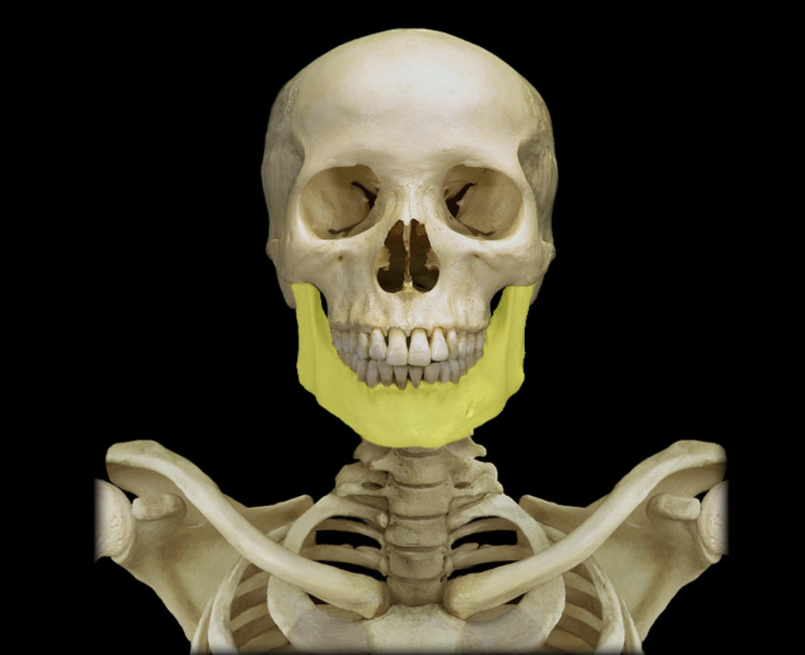
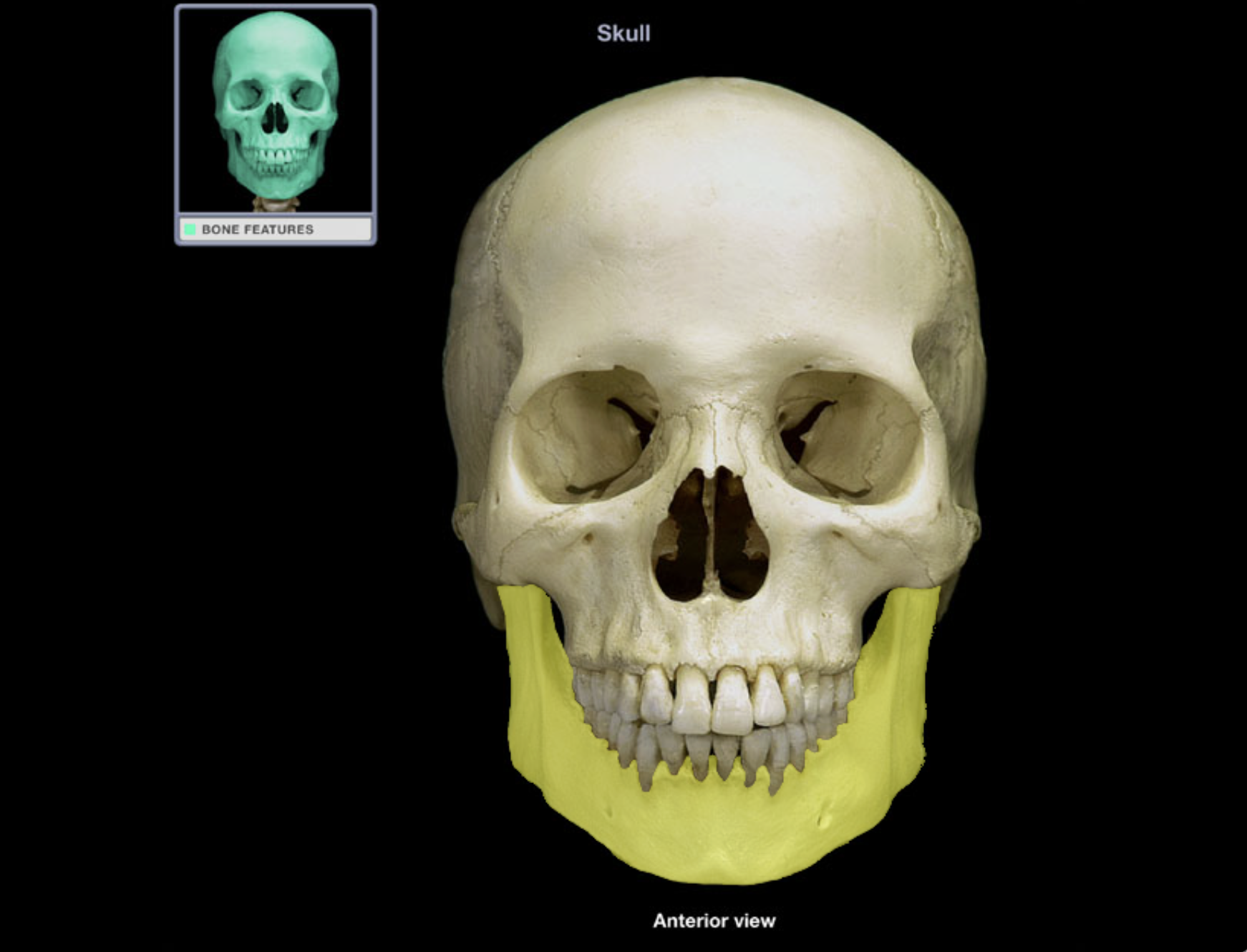
Mandible
Location:
• Skull (anterior)
Description:
U-shaped bone
Each side consists of body (horizontal) and ramus (vertical) with coronoid and condylar processes
Mental protuberance forms point of chin
Contains alveoli ("sockets") for teeth
Also known as:
• "Lower jaw"
Comment:
• Contributes to temporomandibular joint (TMJ
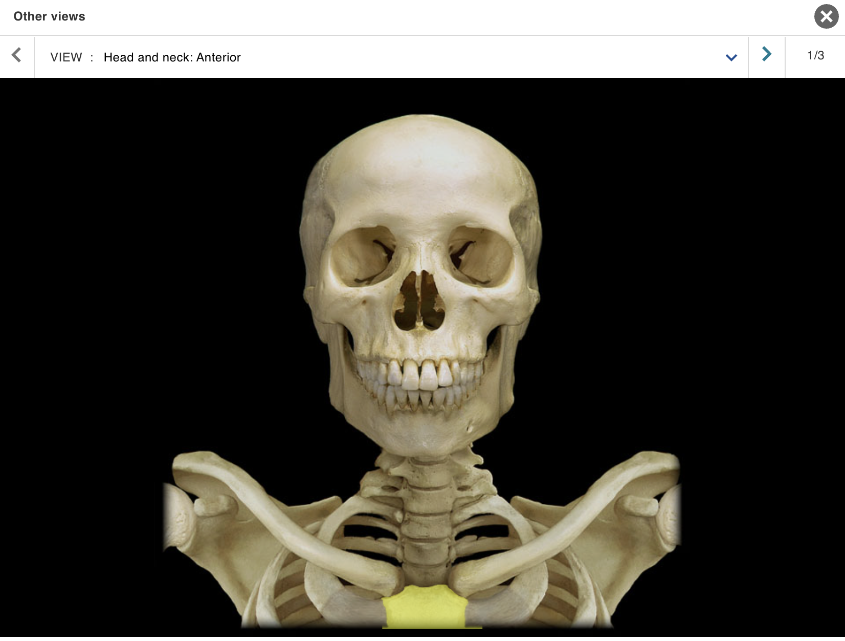
Manubrium
Location:
• Sternum
Description:
Triangular shape
Superior part of sternum
Articulates with costal cartilages of ribs 1-2 and clavicles
Comment:
• Provides attachment for sternocleidomastoid and pectoralis major muscles
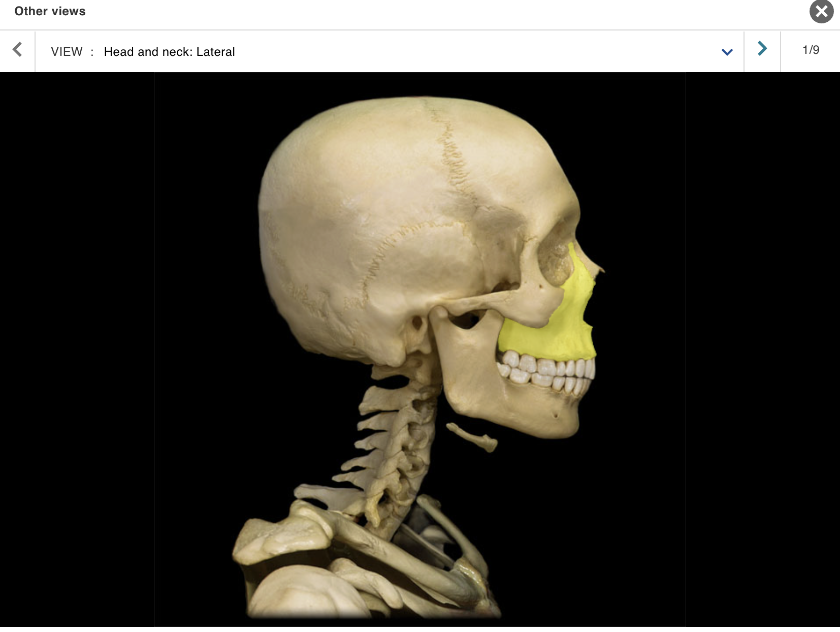
Maxilla
Location:
• Skull (anterior)
Description:
Paired, irregular-shaped bone
Left and right maxillae unite to form "upper jaw"
Contains alveoli ("sockets") for teeth
Contains maxillary air sinus on each side
Also known as:
• "Upper jaw"
Comment:
Forms part of floor of orbit and anterior part of hard palate
Contributes to upper face
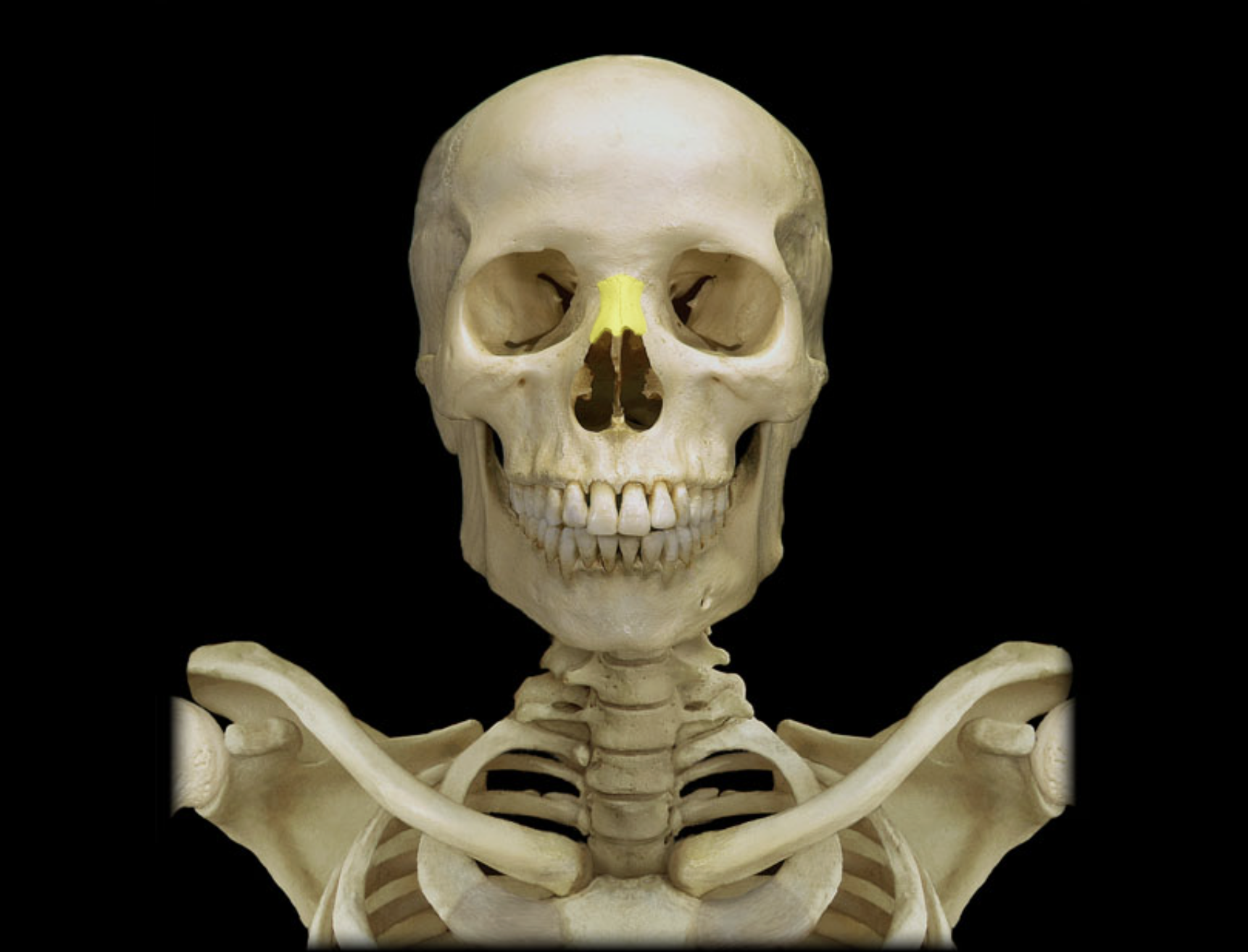
Nasal bone
Location:
• Skull (anterior)
Description:
Small, paired bone
Articulates at midline with nasal bone from opposite side
Also known as:
• "Bridge of nose"
Comment:
• Forms bony part of nose
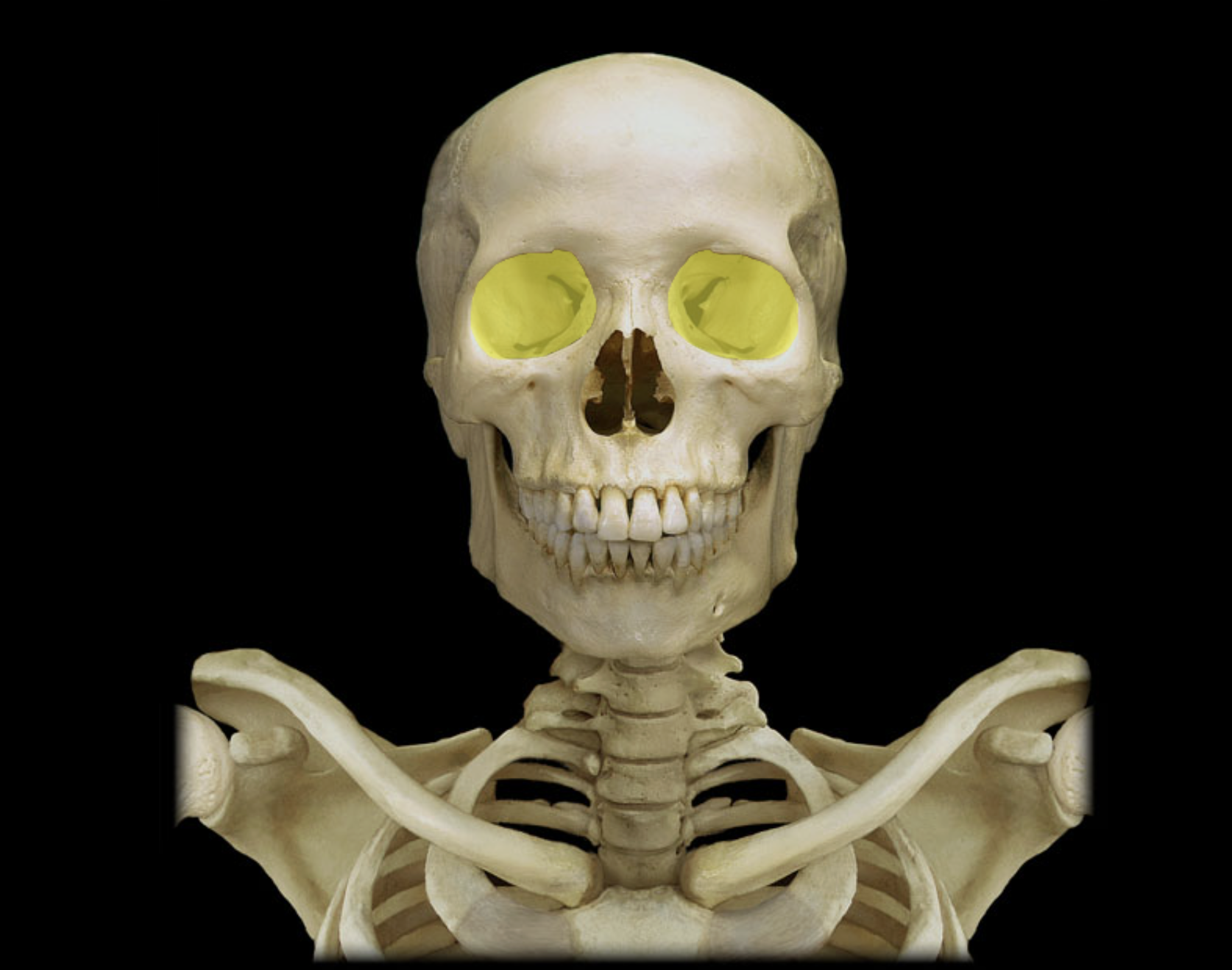
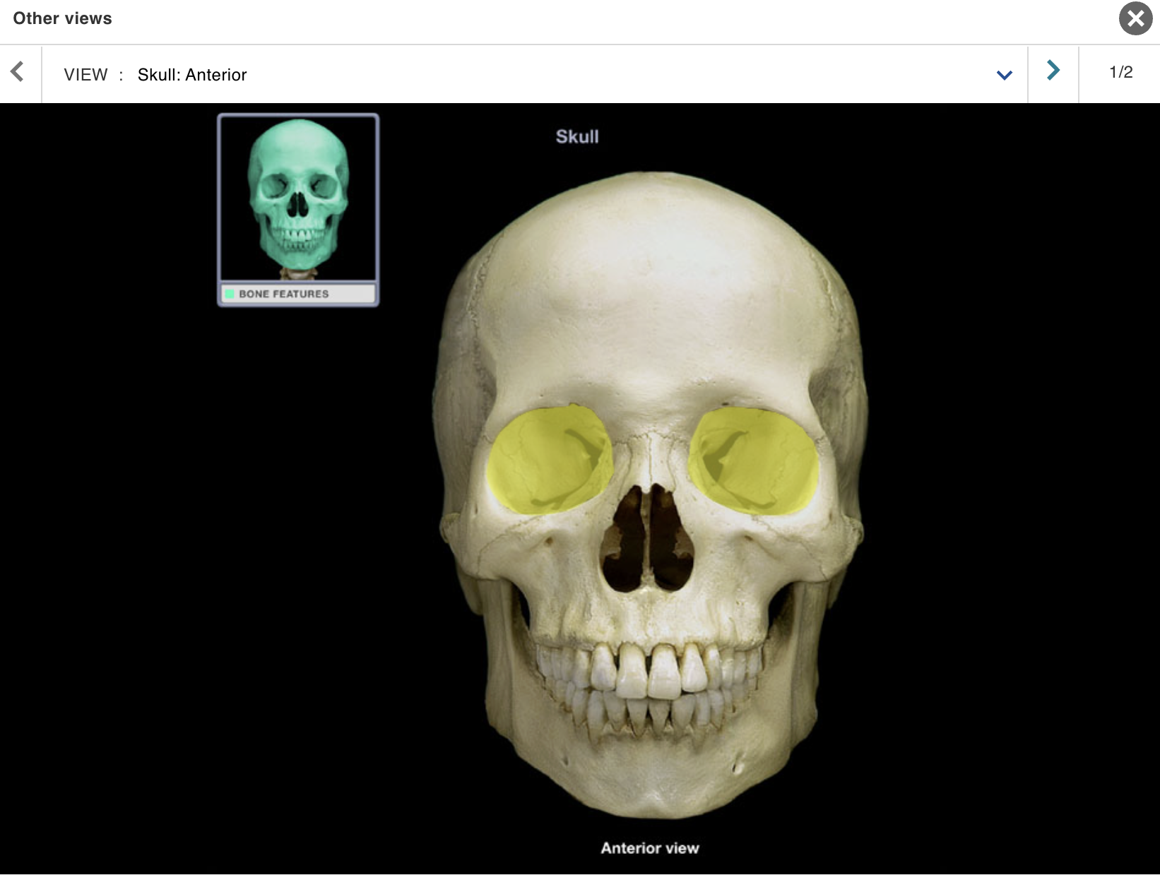
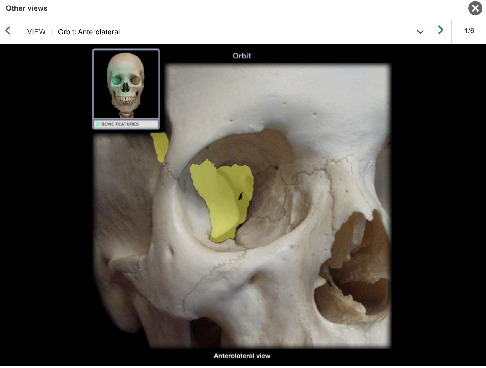
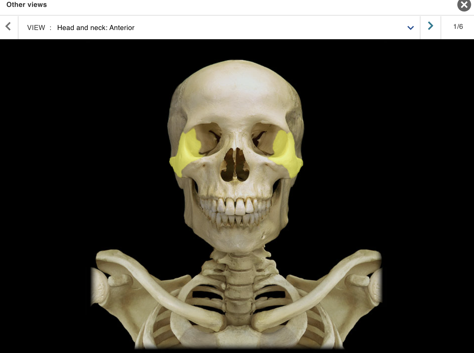
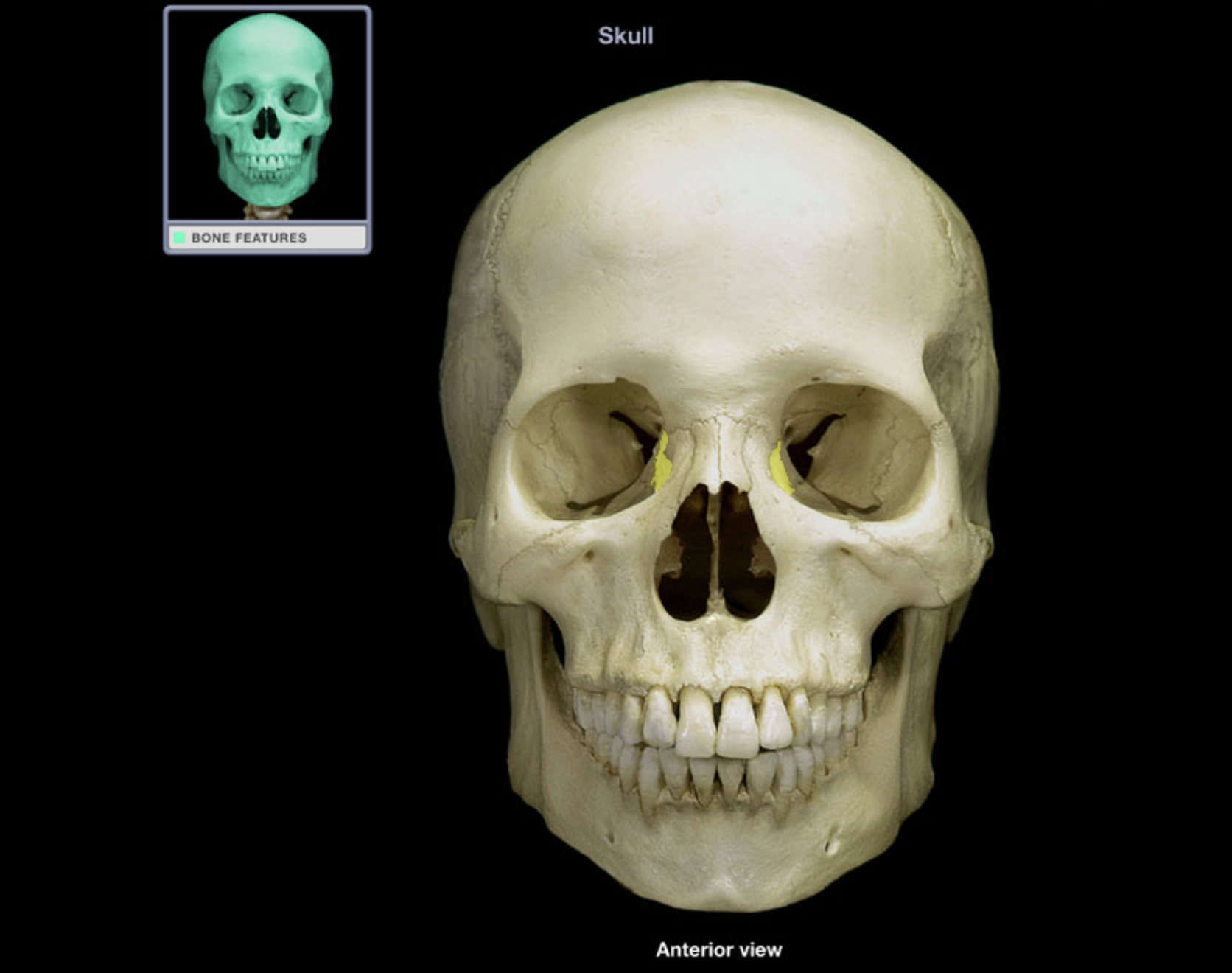
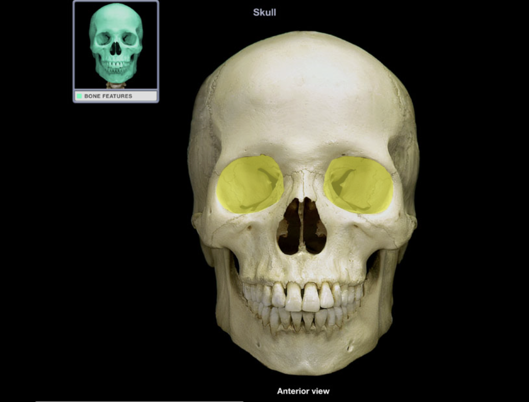
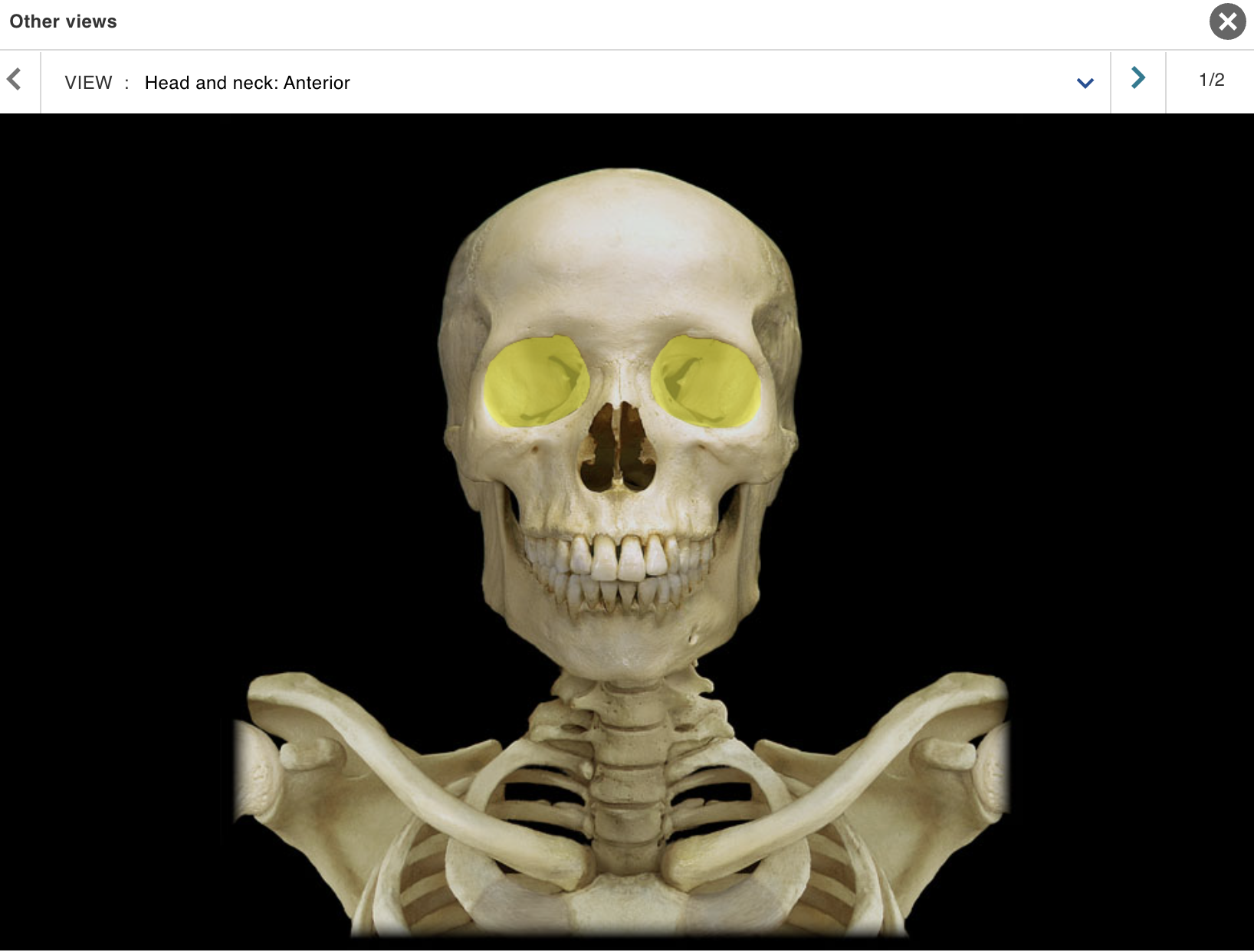
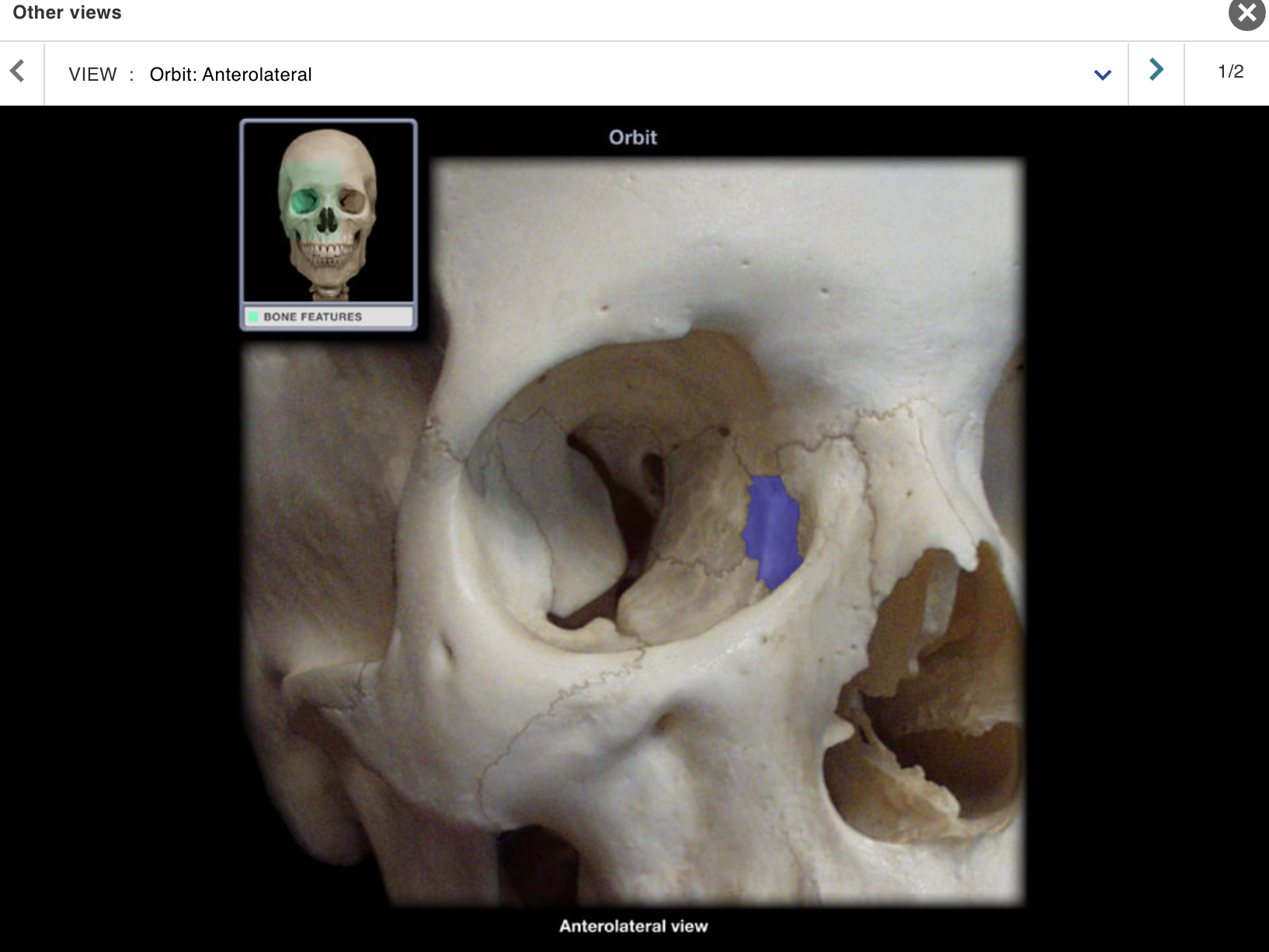
Orbit
Location:
Skull (anterior)
Lateral to nasal cavity
Description:
Pyramidal space with roof, floor, and medial and lateral walls
Roof: frontal and sphenoid bones
Floor: palatine and zygomatic bones, and maxilla
Lateral wall: zygomatic, sphenoid, and frontal bones
Medial wall: ethmoid and lacrimal bones, and maxilla
Openings include superior and inferior orbital fissures, and optic canal
Also known as:
• Eye socket
Comment:
• Contains eyeball and its muscles; cranial nerves II, III, IV, V1, and VI; vasculature; lacrimal apparatus; and fat
Rib 1
Location:
• Thorax
Description:
A true rib (shortest)
Articulates with T1 vertebra and, via its costal cartilage, with manubrium of sternum
Subclavian artery and vein groove superior surface
Comment:
All ribs: articulate with thoracic vertebra
True ribs (ribs 1-7): attached directly to sternum by costal cartilages
False ribs (ribs 8-10): attach indirectly to sternum via shared costal cartilages
Floating ribs (ribs 11-12): not attached to sternum
Alternate definition: some include the floating ribs (11-12) as a subcategory of false ribs
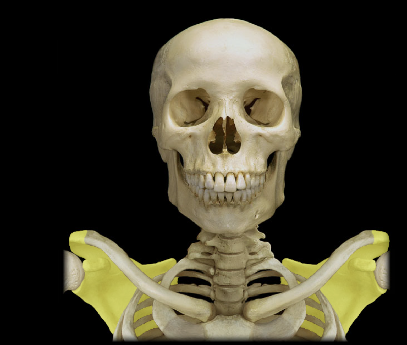
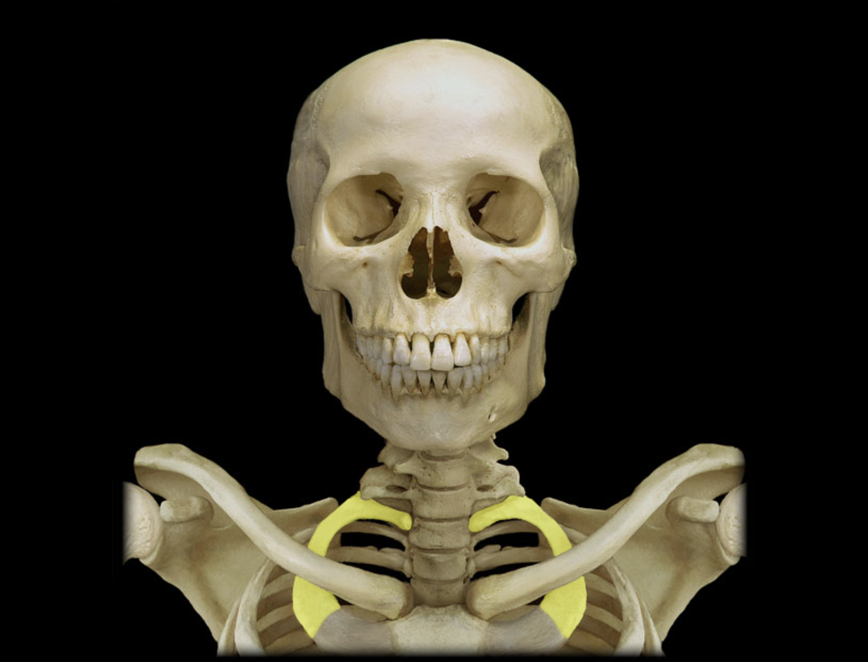
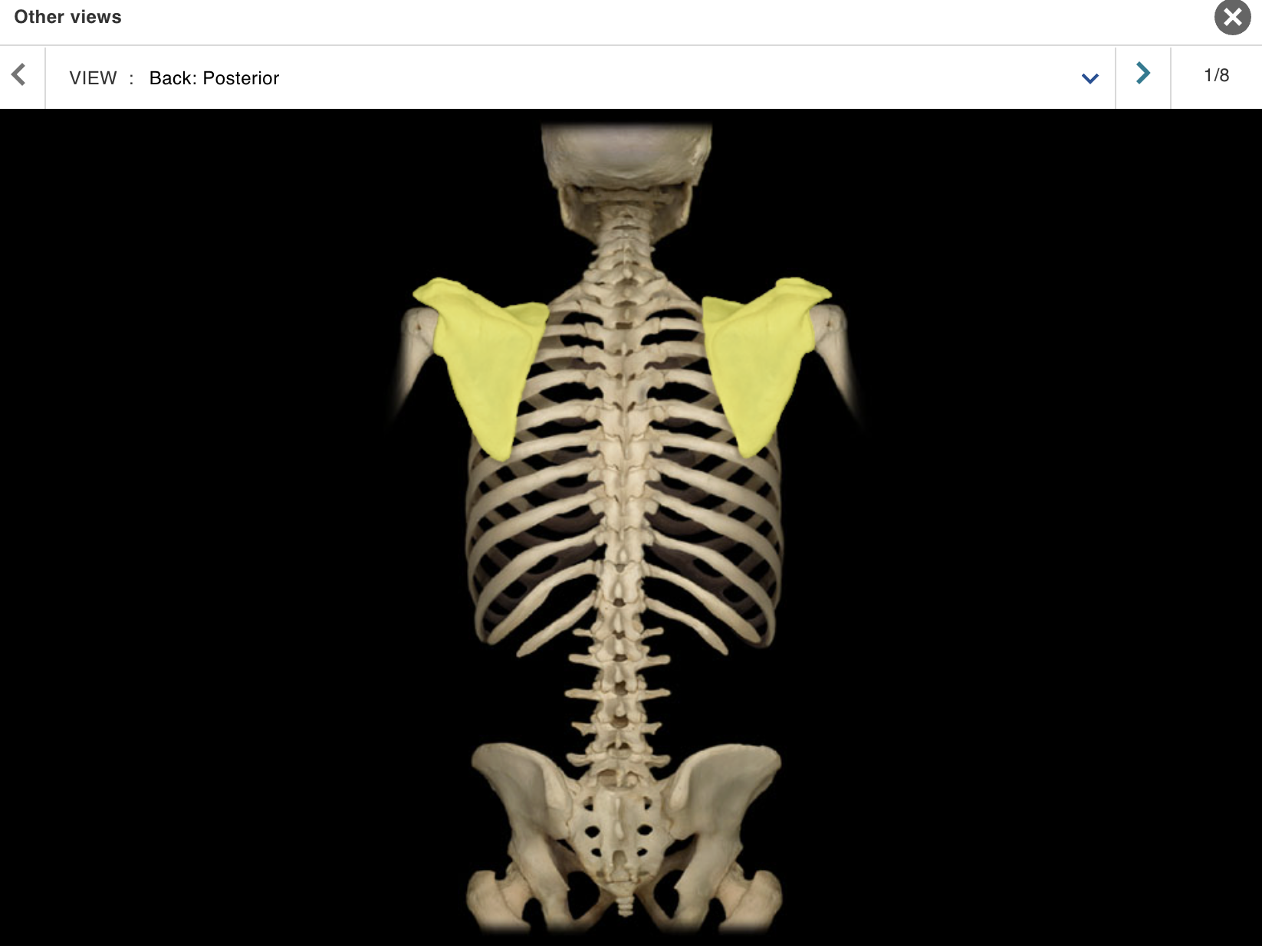
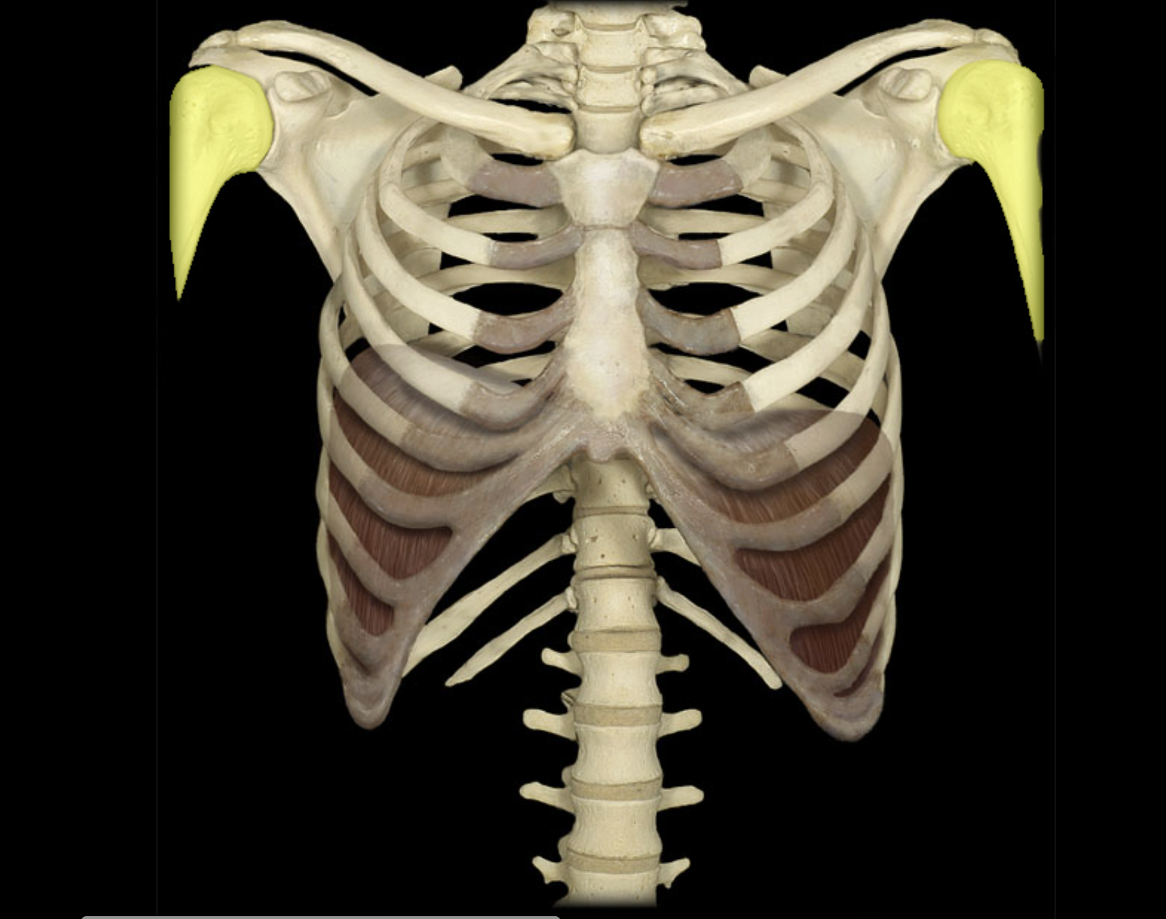
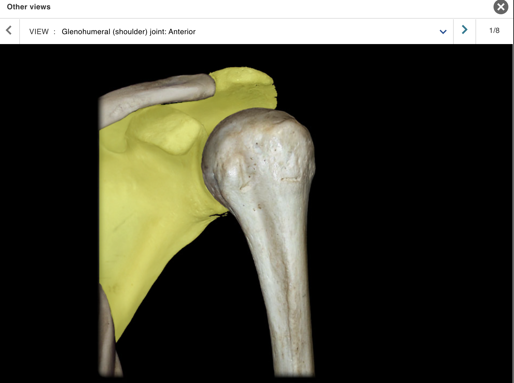
Scapula
Location:
Posterior thorax
Overlies ribs 2-7
Description:
Large, triangular, flat bone
Characteristic features include spine, acromion, coracoid process, and glenoid cavity
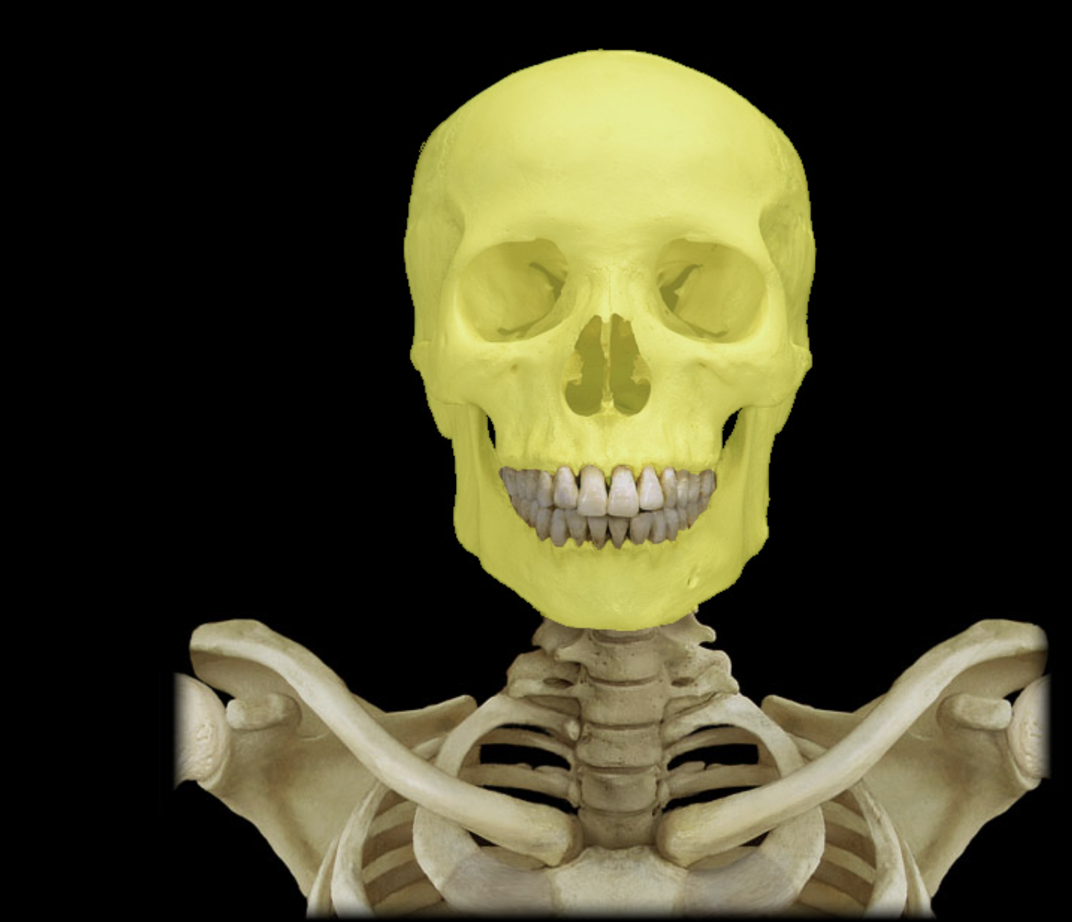
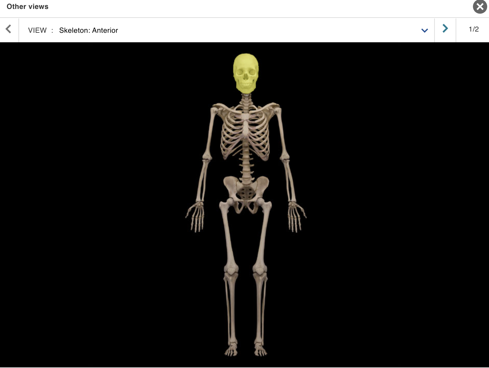
Skull
Location:
• Head
Description:
Paired and unpaired bones divided into cranial and facial
groupsCranial group: frontal, sphenoid, ethmoid, parietal (2), occipital, and temporal bones (2)
Facial group: maxilla, mandible, vomer, nasal (2), lacrimal (2), and zygomatic bones (2)
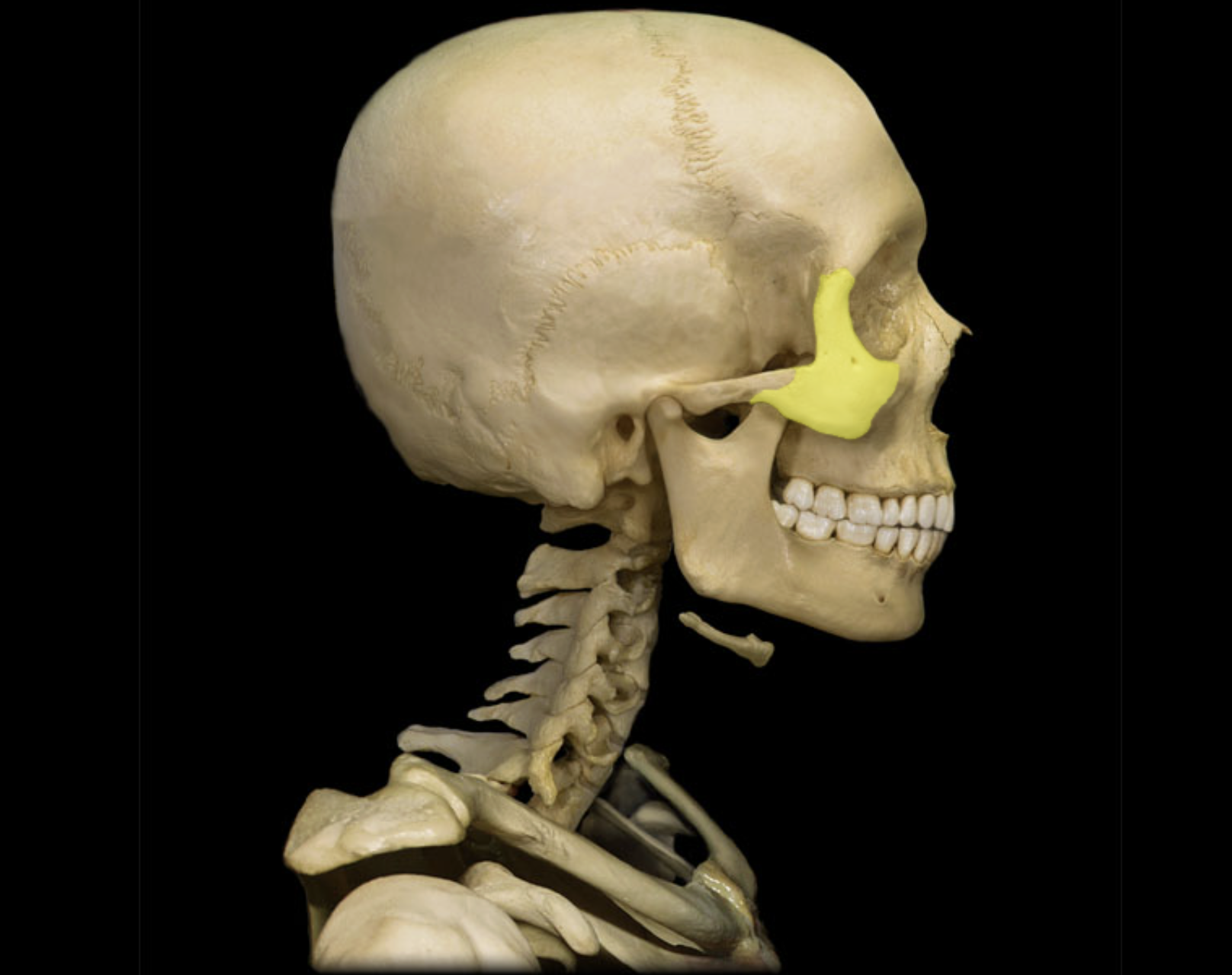
Sphenoid bone
Location:
• Skull
Description:
Unpaired, irregular-shaped bone
Shape resembles a butterfly
Consists of body, and greater and lesser wings
Comment:
Contributes to middle cranial fossa, nasal cavities, orbits, and lateral skull (temples)
Contains sella turcica ("Turkish saddle"), sphenoidal air sinus, and many foramina

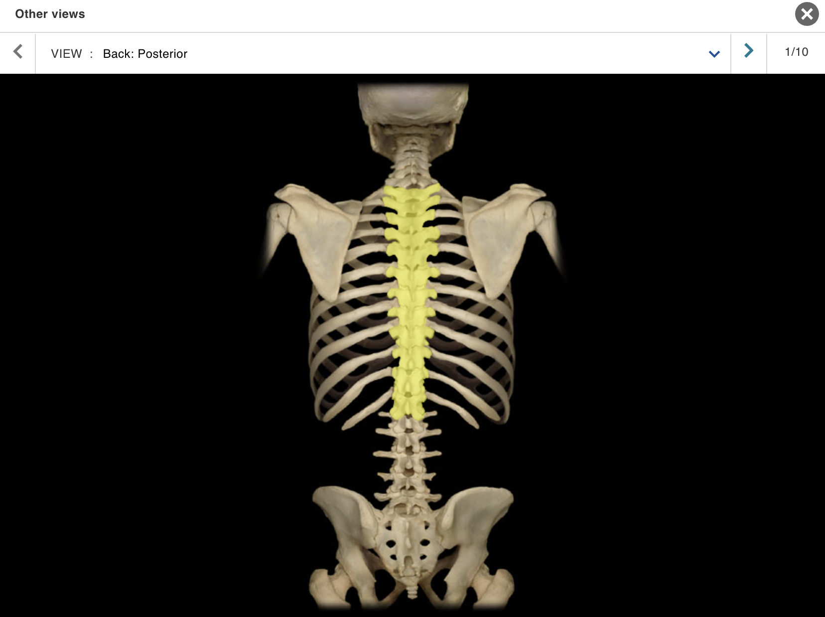
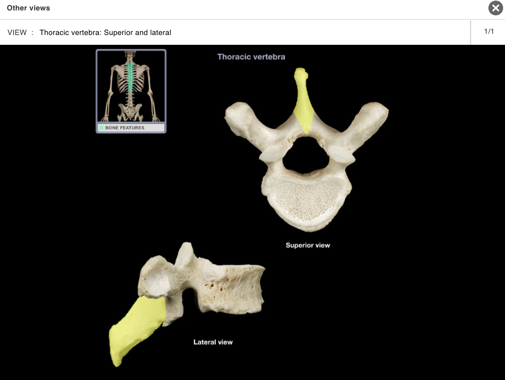
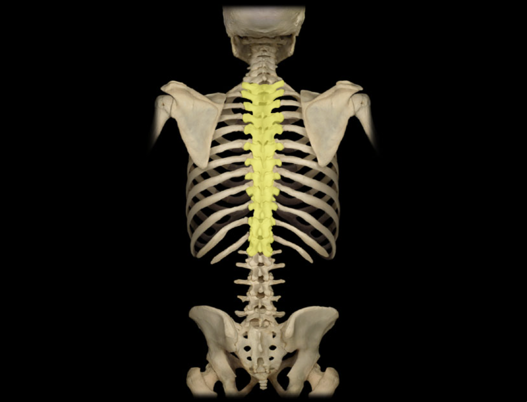
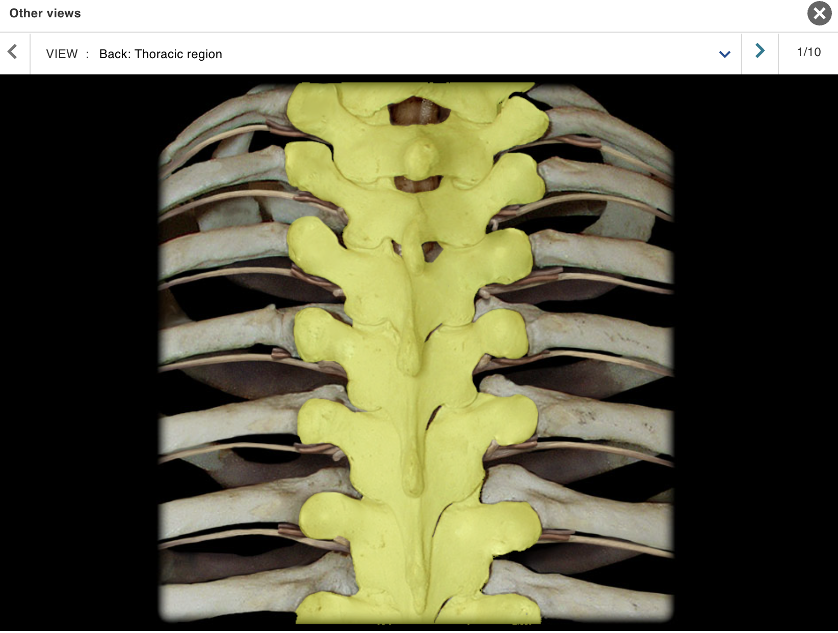
Thoracic vertebra
Location:
Trunk
Between C7 and L1 vertebrae
Description:
12 individual vertebrae
Characteristic features include costal demifacets (or facets) for articulation with head of rib, spinous process slopes inferiorly, and heart-shaped vertebral body
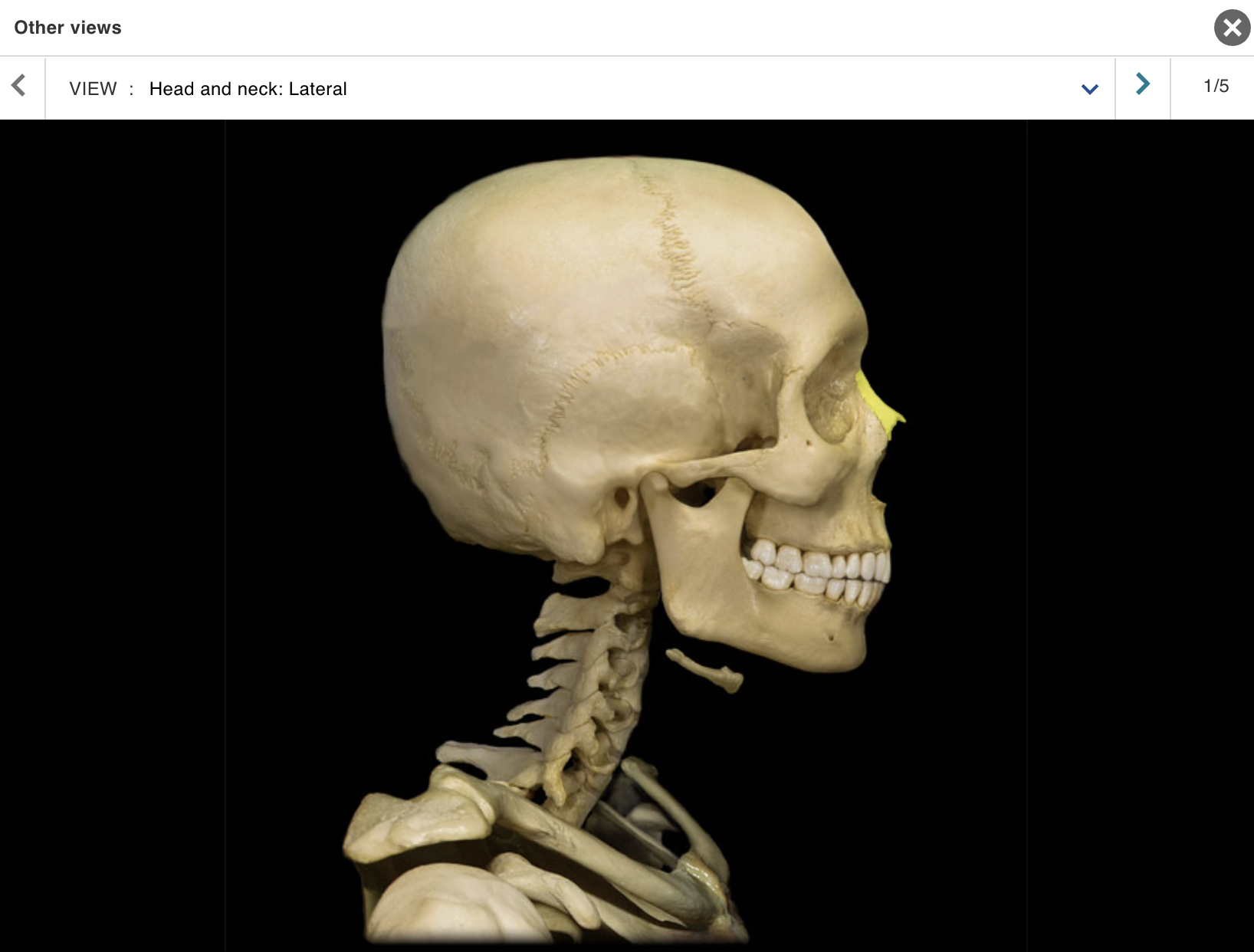
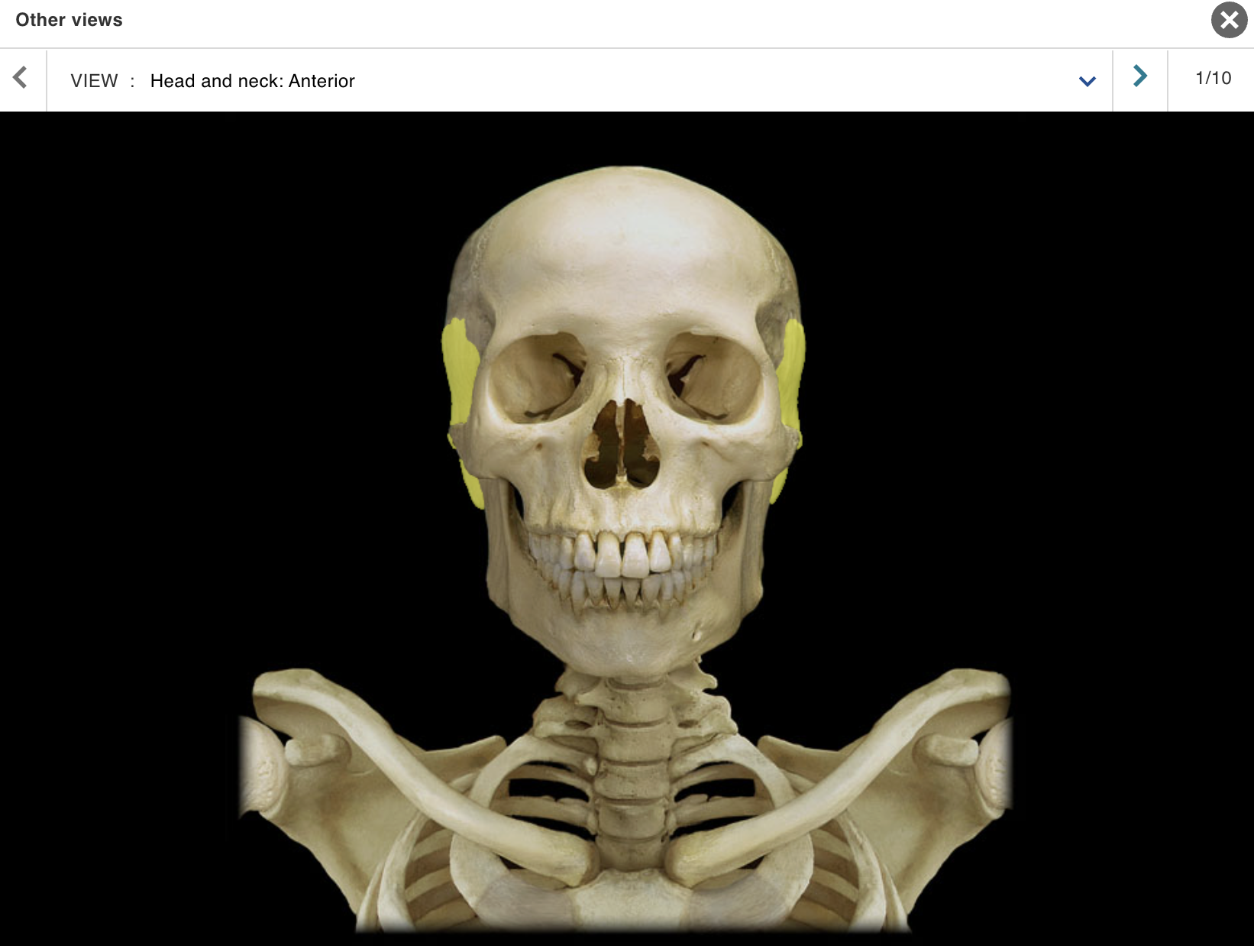
Zygomatic bone
Location:
• Skull (anterior and lateral)
Description:
Paired, irregular-shaped bone
Temporal process contributes to zvaomatic arch
Also known as:
• "Cheekbone"
Comment:
• Forms part of floor and lateral wall of orbit
SKELETAL
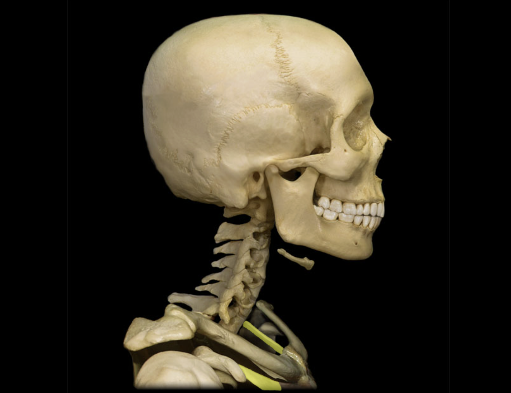
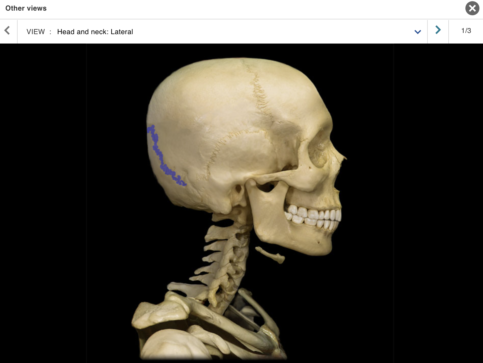
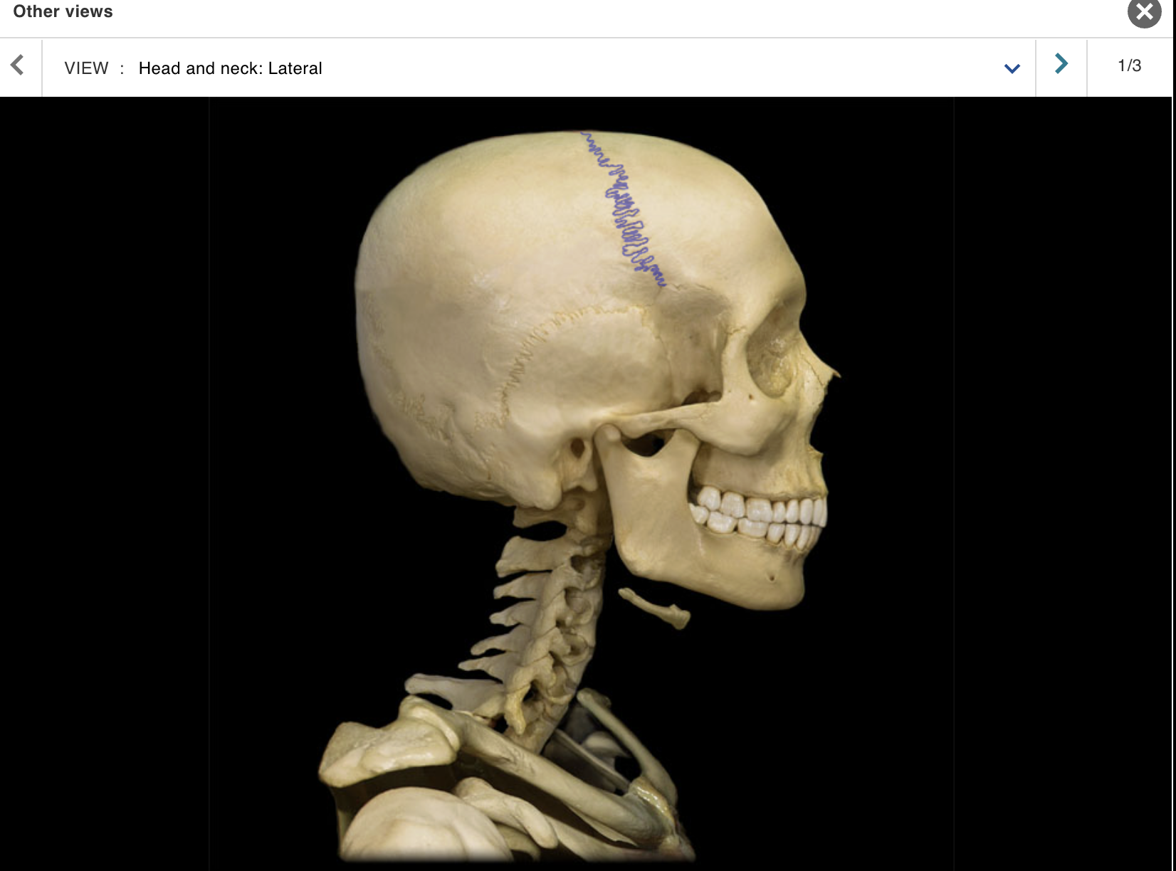
TOPIC: HEAD / NECK / VIEW : LATERAL
Cervical vertebra
Clavicle
Frontal bone
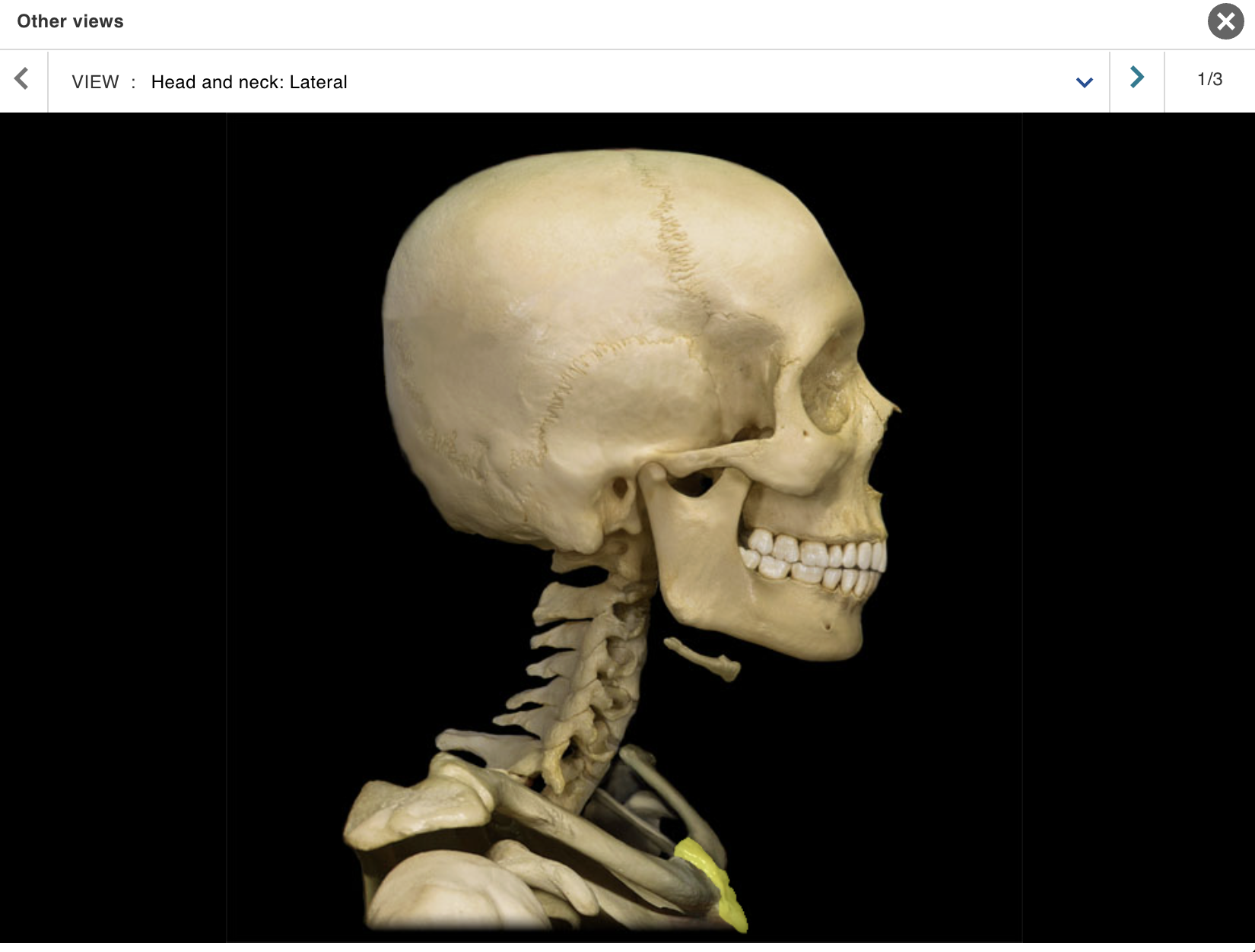
Hyoid bone
Intervertebral disc
Mandible
Manubrium
Maxilla
Nasal bone
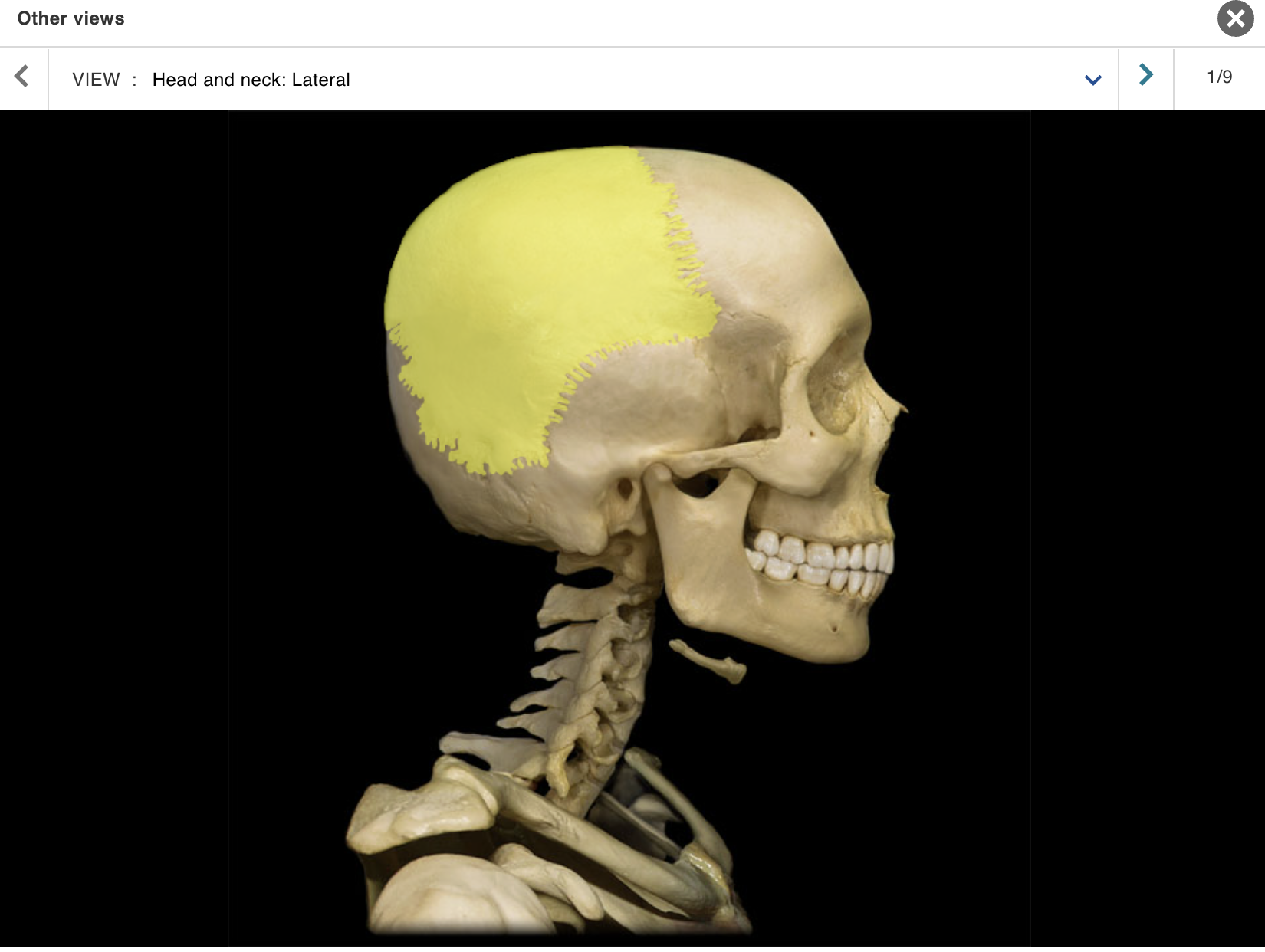
Parietal bone
Rib 1
Scapula
Temporal bone
Zygomatic bone
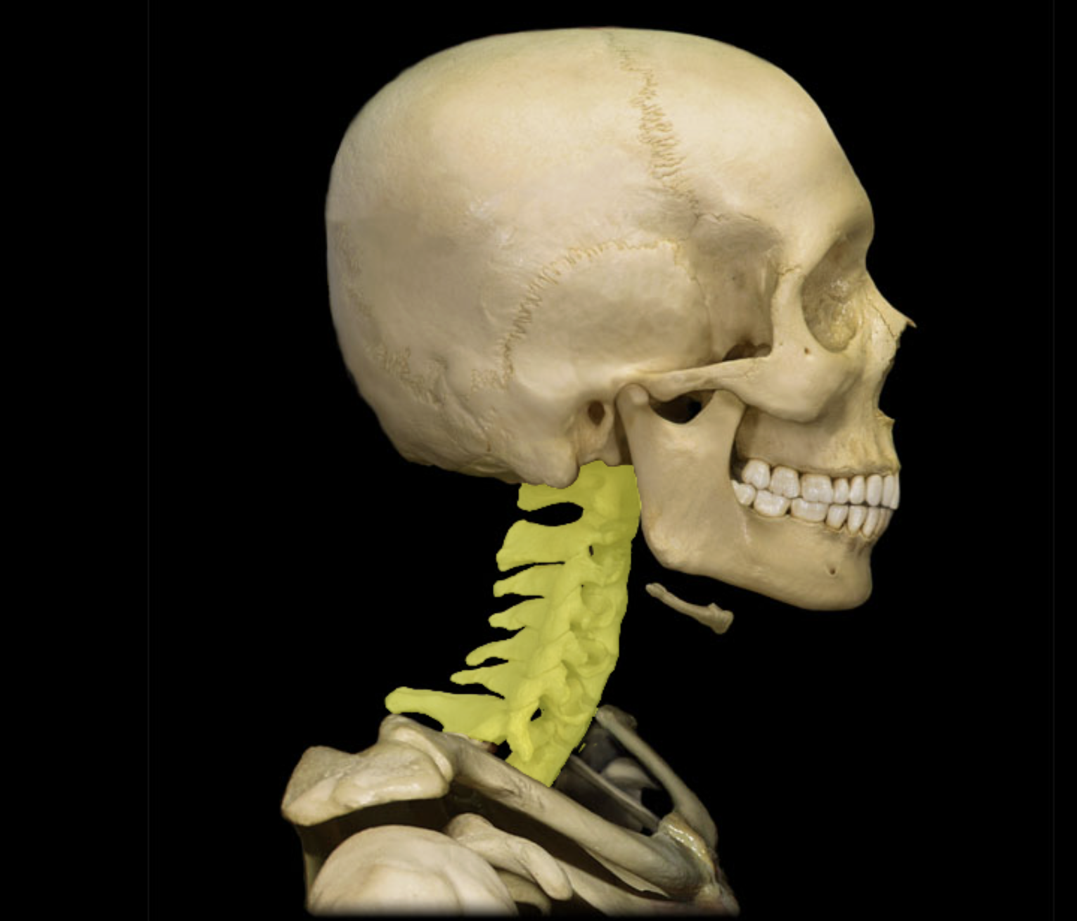
Cervical vertebra
Location:
Neck
Between occipital bone and T1 vertebra
Description:
Seven individual vertebrae
Characteristic features include transverse foramen and bifid (split) spinous process on C3-C6
Comment:
• Atlas (C1 vertebra) articulates with skull
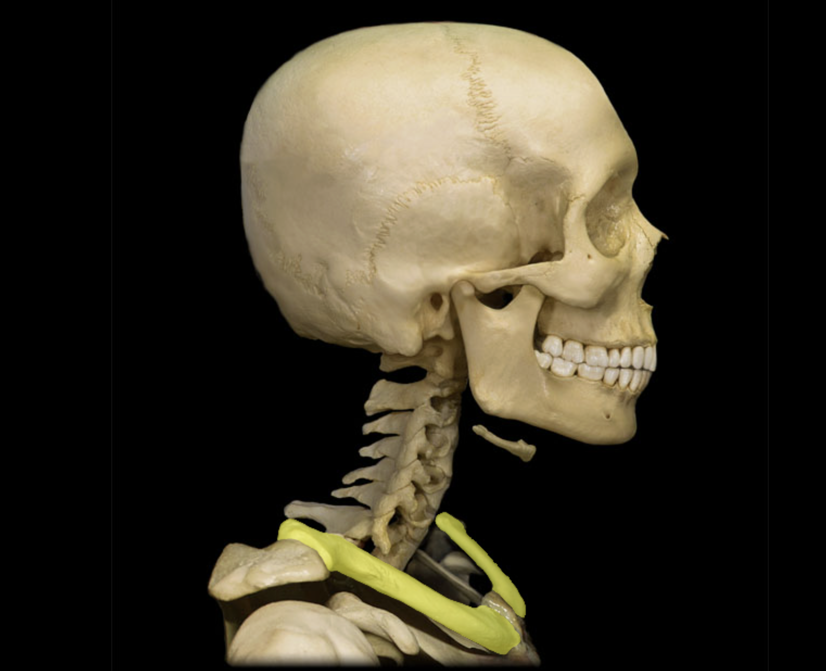
Clavicle
Location:
• Junction of neck and anterior thorax
Description:
Subcutaneous, S-shaped bone
Medial end articulates with sternum at sternoclavicular joint
Lateral end articulates with acromion of scapula at acromioclavicular joint
Also known as:
• "Collar bone"

Frontal bone
Location:
• Skull (anterior superior part)
Description:
Unpaired, irregular-shaped, flat bone
Forms forehead, roof of orbits, and most of anterior cranial fossa
Contains frontal air sinuses
Comment:
• Articulates with parietal bone at coronal suture and sphenoid bone at sphenosquamosal suture

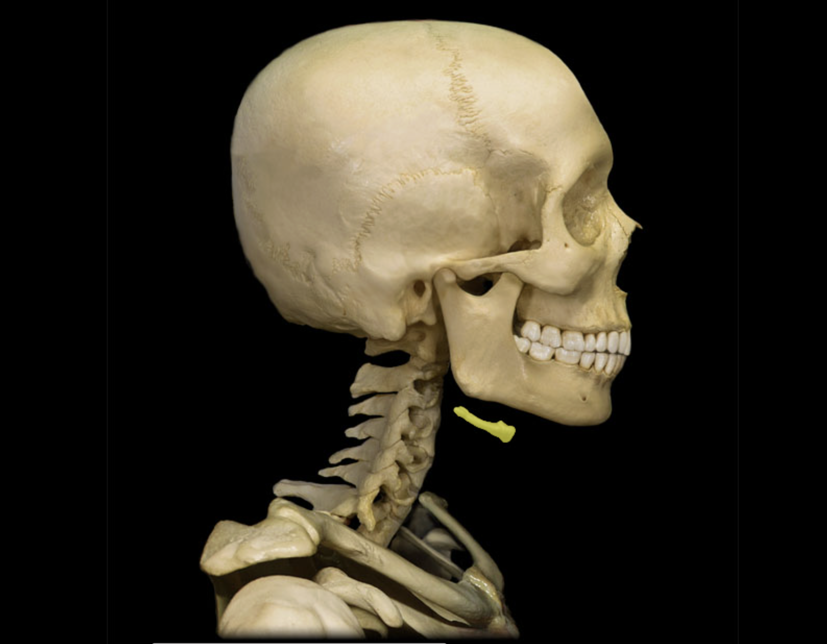
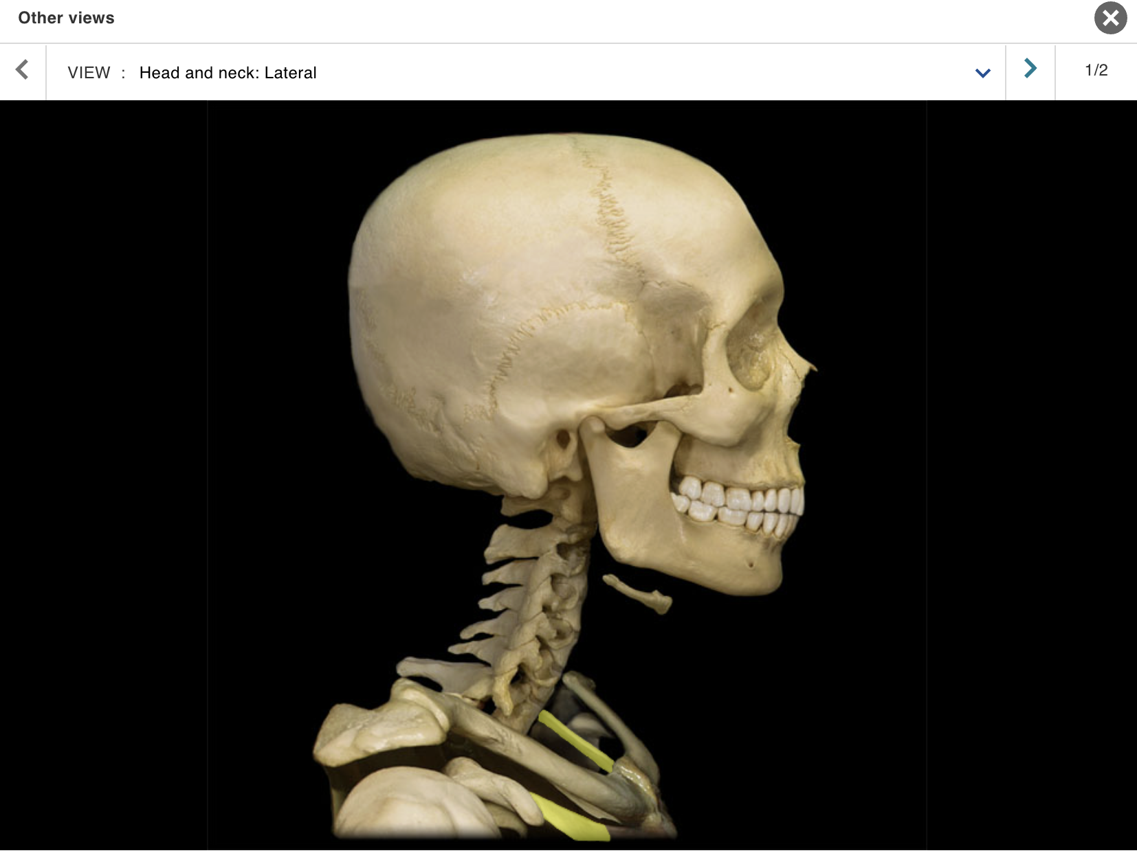
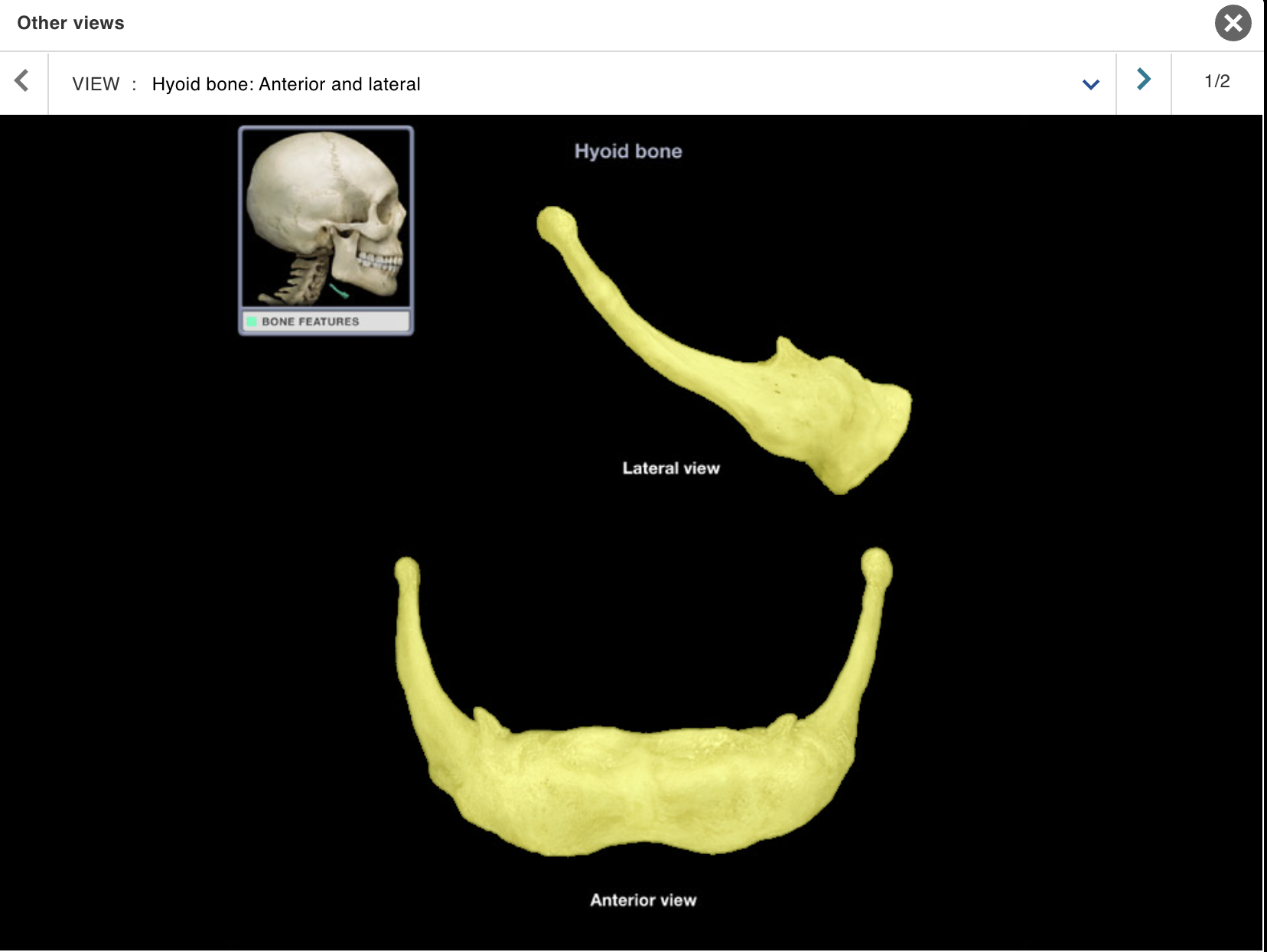
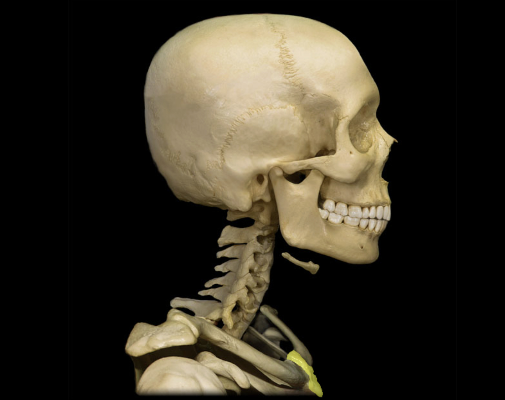
Hyoid bone
Location:
Anterior neck
Between thyroid cartilage and mandible
Description:
U-shaped bone
Paired projections (greater and lesser horns) on each side
Comment:
Attached structures include stylohyoid ligament, suprahyoid and infrahyoid muscles, and extrinsic tongue muscles
Does not articulate with any other bone
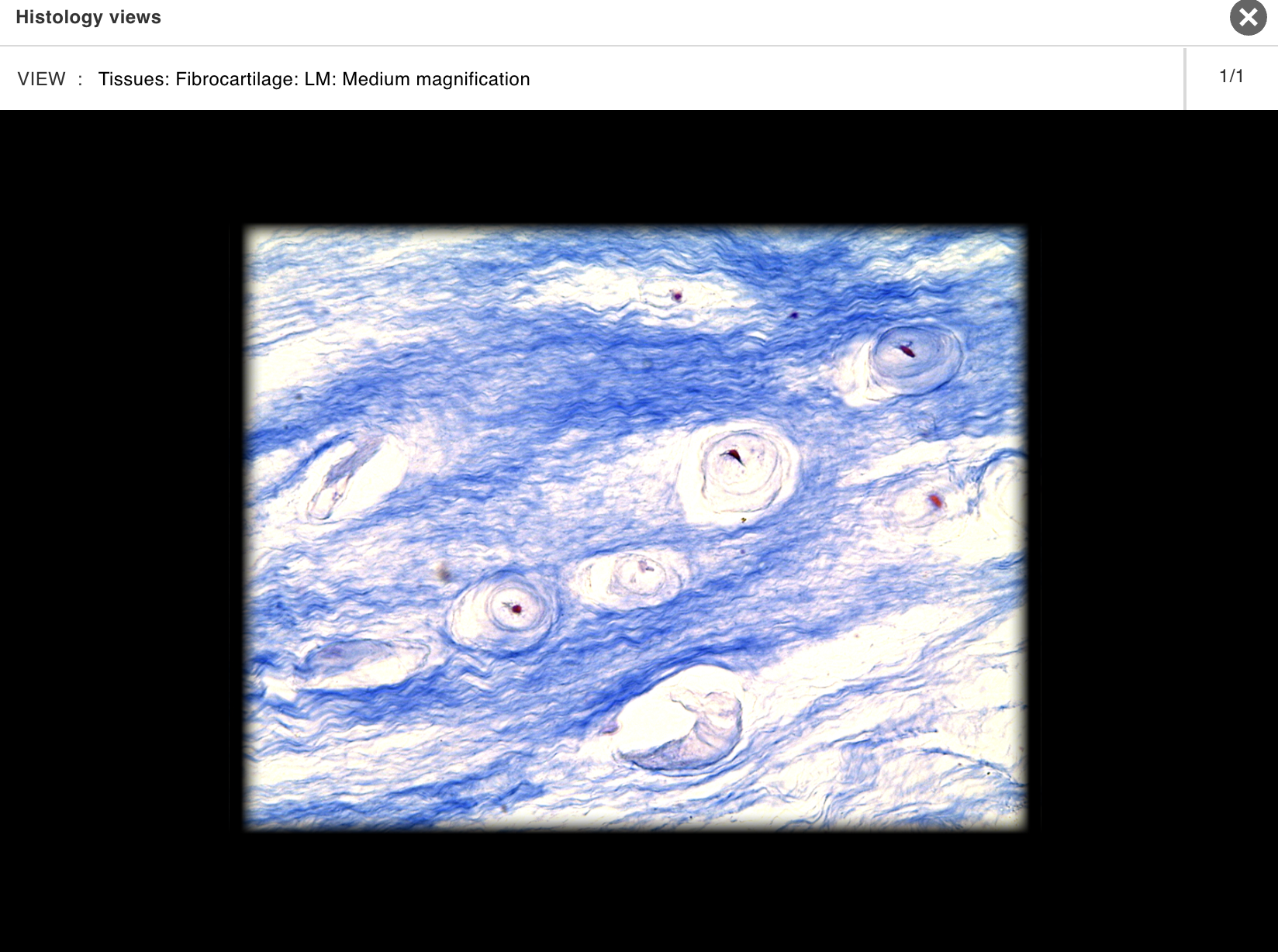
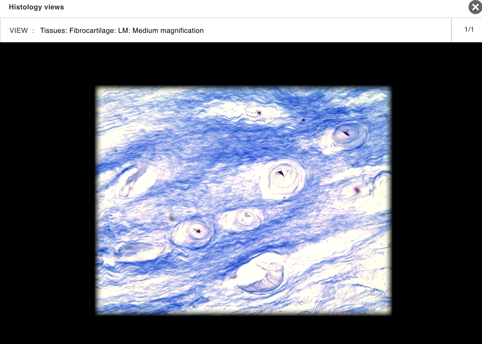
Intervertebral disc
Location:
• Between vertebral bodies (C2 to S1)
Description:
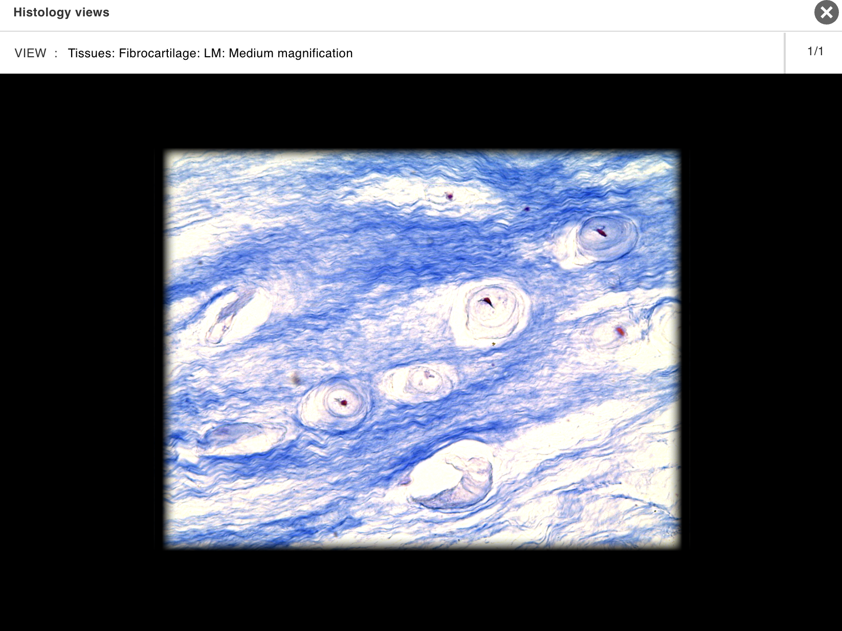
Outer part: anulus fibrous consisting of a fibrocartilage ring
Inner part: nucleus pulposus composed of mucoid material
Comment:
• No disk between atlas (C1 vertebra) and axis (C2 vertebra)
Mandible
Location:
• Skull (anterior)
Description:
U-shaped bone
Each side consists of body (horizontal) and ramus (vertical) with coronoid and condylar processes
Mental protuberance forms point of chin
Contains alveoli ("sockets") for teeth
Also known as:
• "Lower jaw"
Comment:
• Contributes to temporomandibular joint (TMJ
Manubrium
Location:
• Sternum
Description:
Triangular shape
Superior part of sternum
Articulates with costal cartilages of ribs 1-2 and clavicles
Comment:
• Provides attachment for sternocleidomastoid and pectoralis major muscles
Maxilla
Location:
• Skull (anterior)
Description:
Paired, irregular-shaped bone
Left and right maxillae unite to form "upper jaw"
Contains alveoli ("sockets") for teeth
Contains maxillary air sinus on each side
Also known as:
• "Upper jaw"
Comment:
Forms part of floor of orbit and anterior part of hard palate
Contributes to upper face
Nasal bone
Location:
• Skull (anterior)
Description:
Small, paired bone
Articulates at midline with nasal bone from opposite side
Also known as:
• "Bridge of nose"
Comment:
• Forms bony part of nose
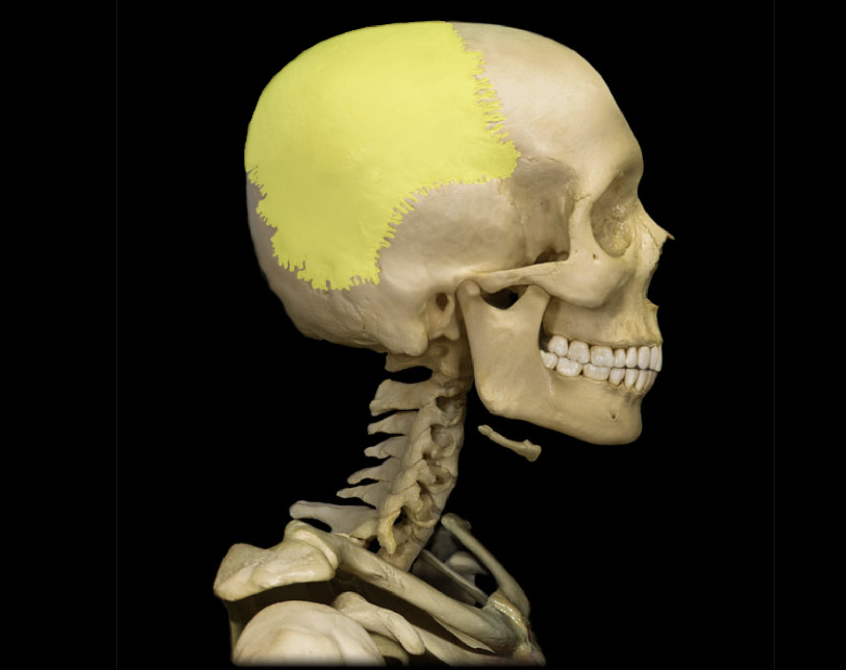
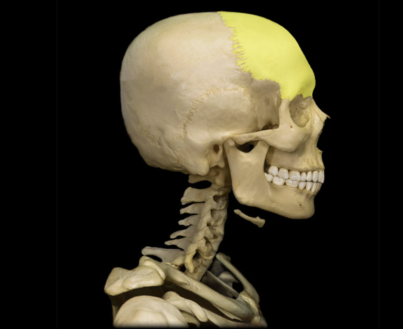
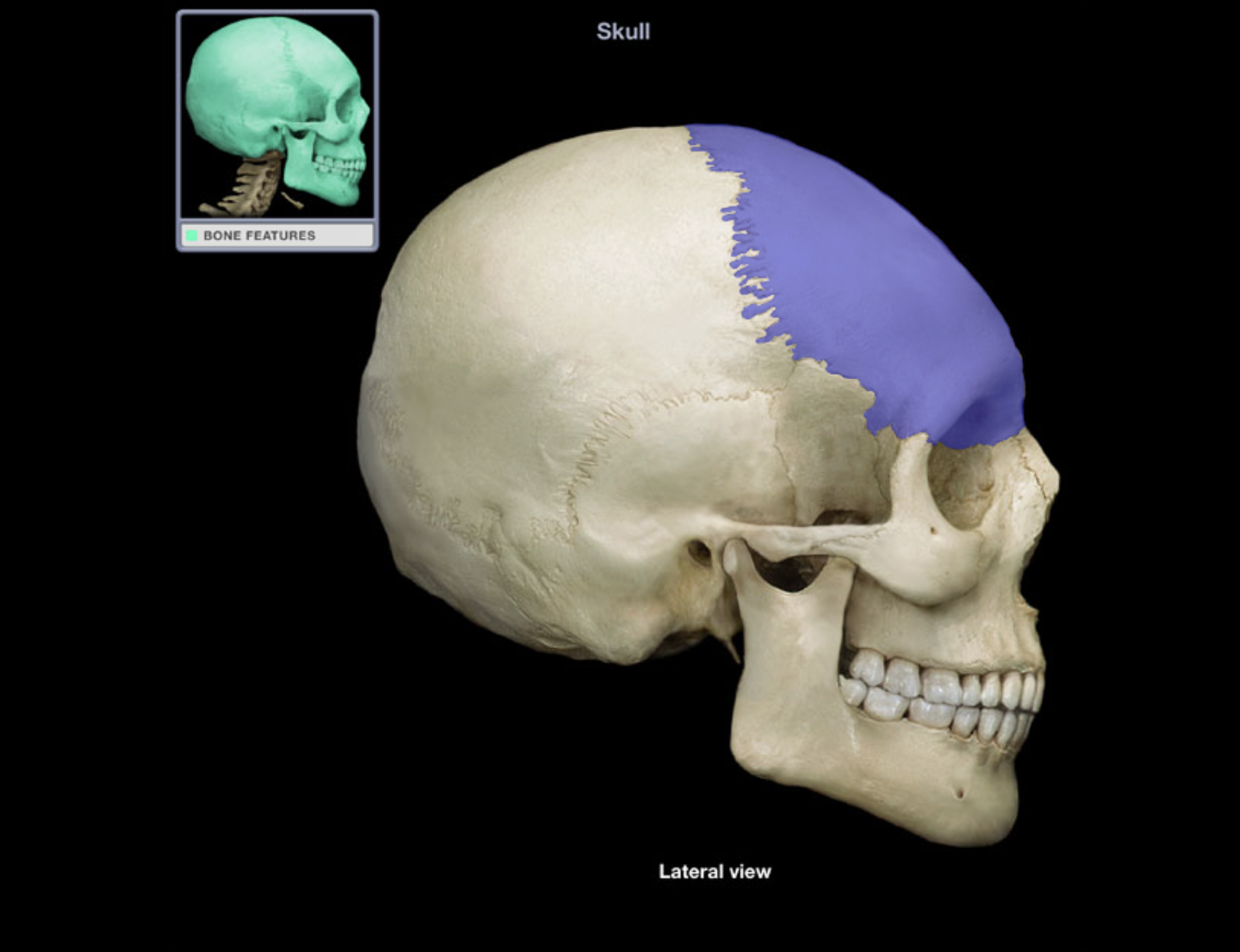
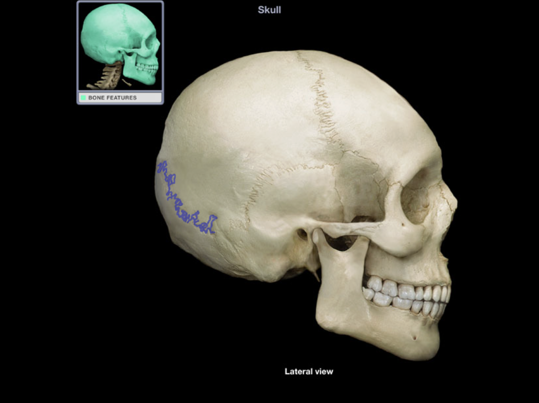
Parietal bone
Location:
• Skull (lateral)
Description:
Paired, flat bone
Forms most of lateral skull
Comment:
Parietal bones articulate at sagittal suture (midline)
Articulates with frontal bone at coronal suture, occipital bone at lambdoid suture, and temporal bone at squamosal suture

Rib 1
Location:
• Thorax
Description:
A true rib (shortest)
Articulates with T1 vertebra and, via its costal cartilage, with manubrium of sternum
Subclavian artery and vein groove superior surface
Comment:
All ribs: articulate with thoracic vertebra
True ribs (ribs 1-7): attached directly to sternum by costal cartilages
False ribs (ribs 8-10): attach indirectly to sternum via shared costal cartilages
Floating ribs (ribs 11-12): not attached to sternum
Alternate definition: some include the floating ribs (11-12) as a subcategory of false ribs
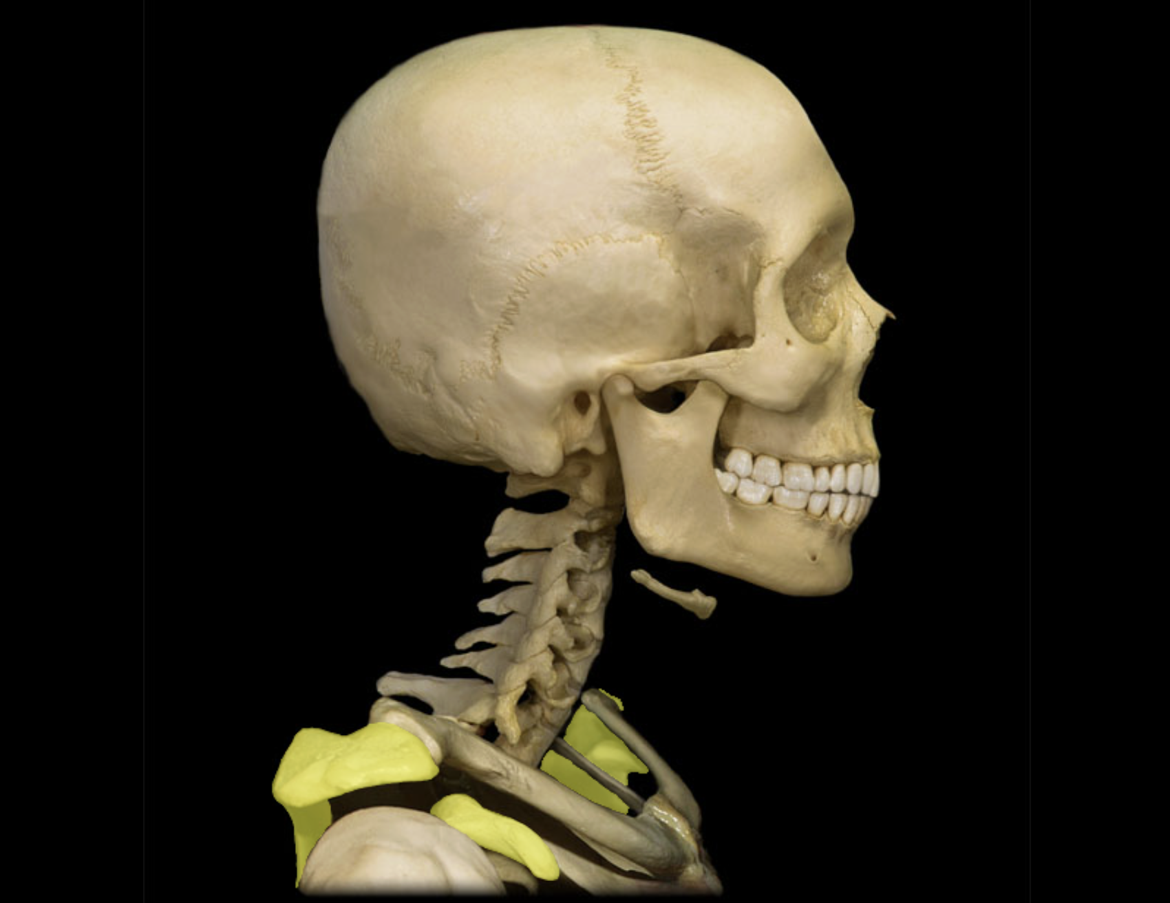
Scapula
Location:
Posterior thorax
Overlies ribs 2-7
Description:
Large, triangular, flat bone
Characteristic features include spine, acromion, coracoid process, and glenoid cavity


Temporal bone
Location:
• Skull (lateral and inferior)
Description:
Paired, irregular-shaped, flat bone
Consists of squamous, petrous, mastoid, and tympanic parts
Comment:
Forms part of lateral skull (temple)
Forms inferior and lateral walls of middle cranial fossa
Articulates with sphenoid bone at sphenosquamosal suture, occipital bone at lambdoid suture, and parietal bone at squamosal suture
Has mandibular fossa that forms temporomandibular joint with head of mandible
Forms zygomatic process that forms zygomatic arch with temporal process of zygomatic bone
Zygomatic bone
Location:
• Skull (anterior and lateral)
Description:
Paired, irregular-shaped bone
Temporal process contributes to zygomatic arch
Also known as:
• "Cheekbone"
Comment:
• Forms part of floor and lateral wall of orbit
SKELETAL
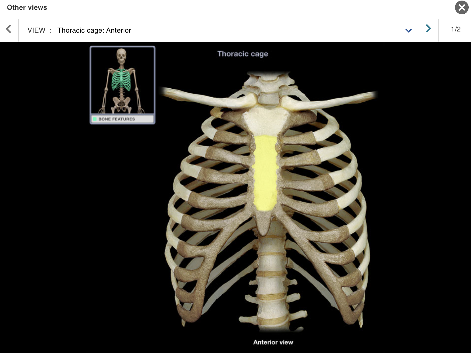
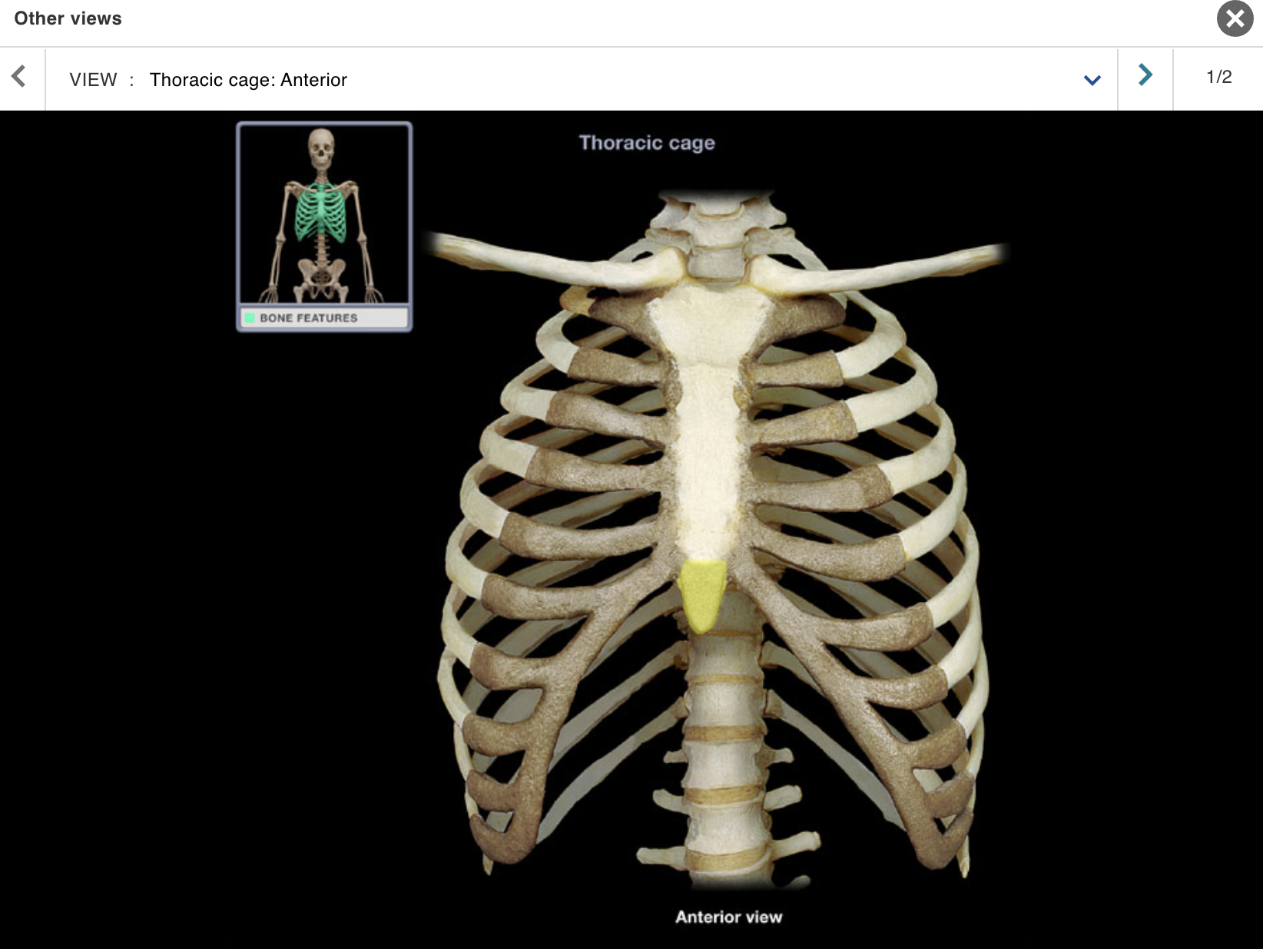
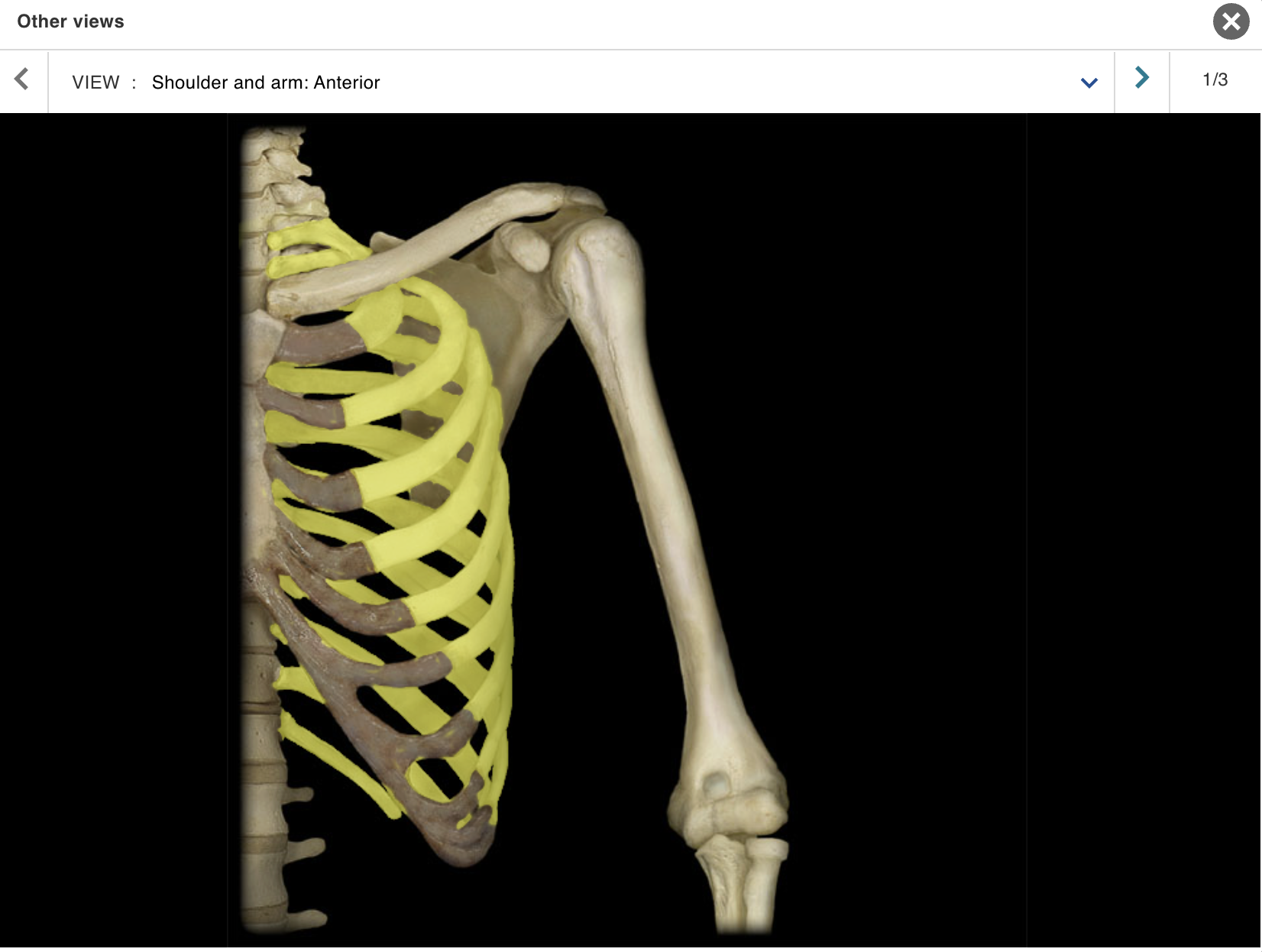
TOPIC: THORAX / VIEW : ANTERIOR
Cervical vertebra
Clavicle
Costal cartilages
Humerus
Intervertebral disc
Lumbar vertebra
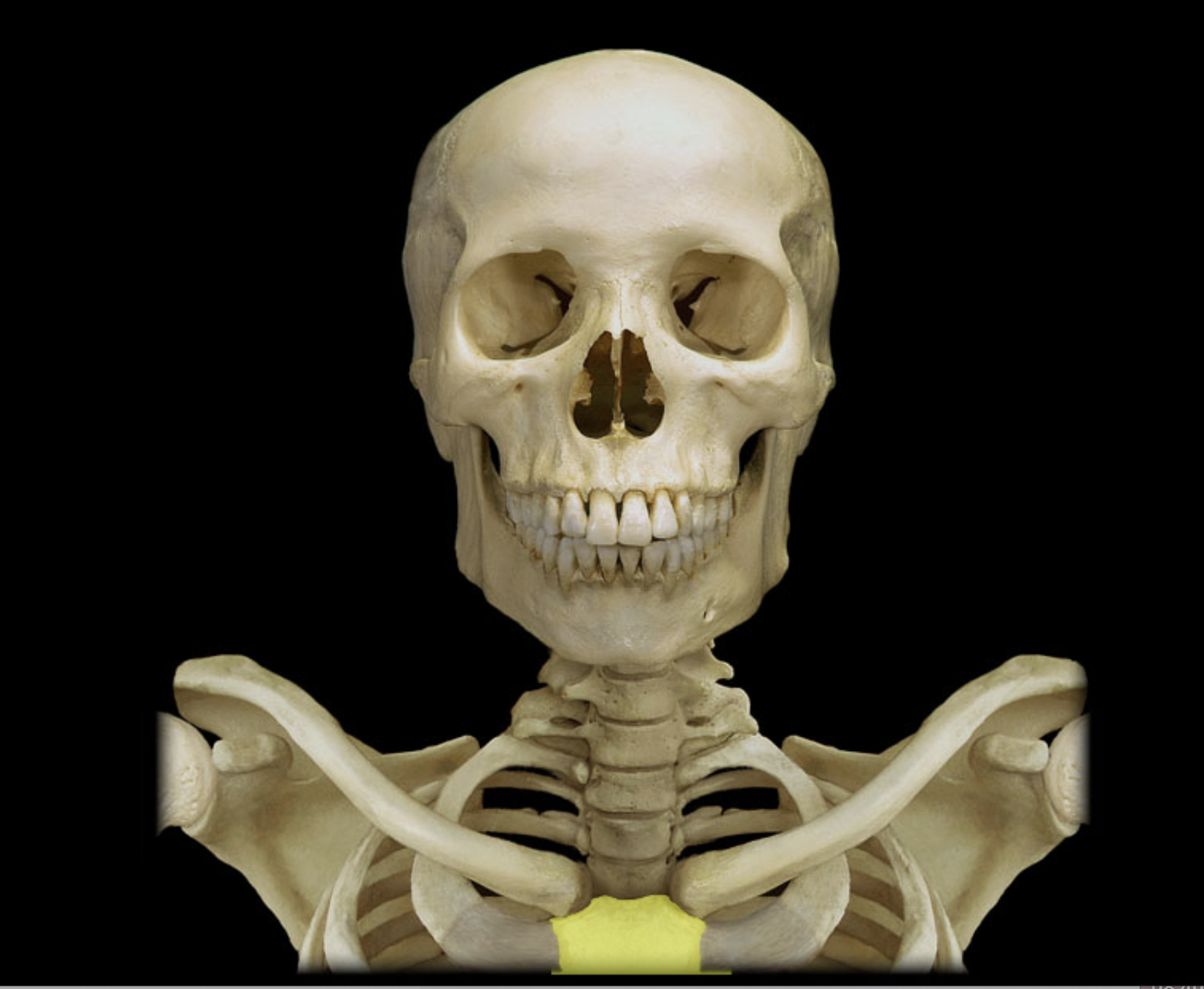
Manubrium
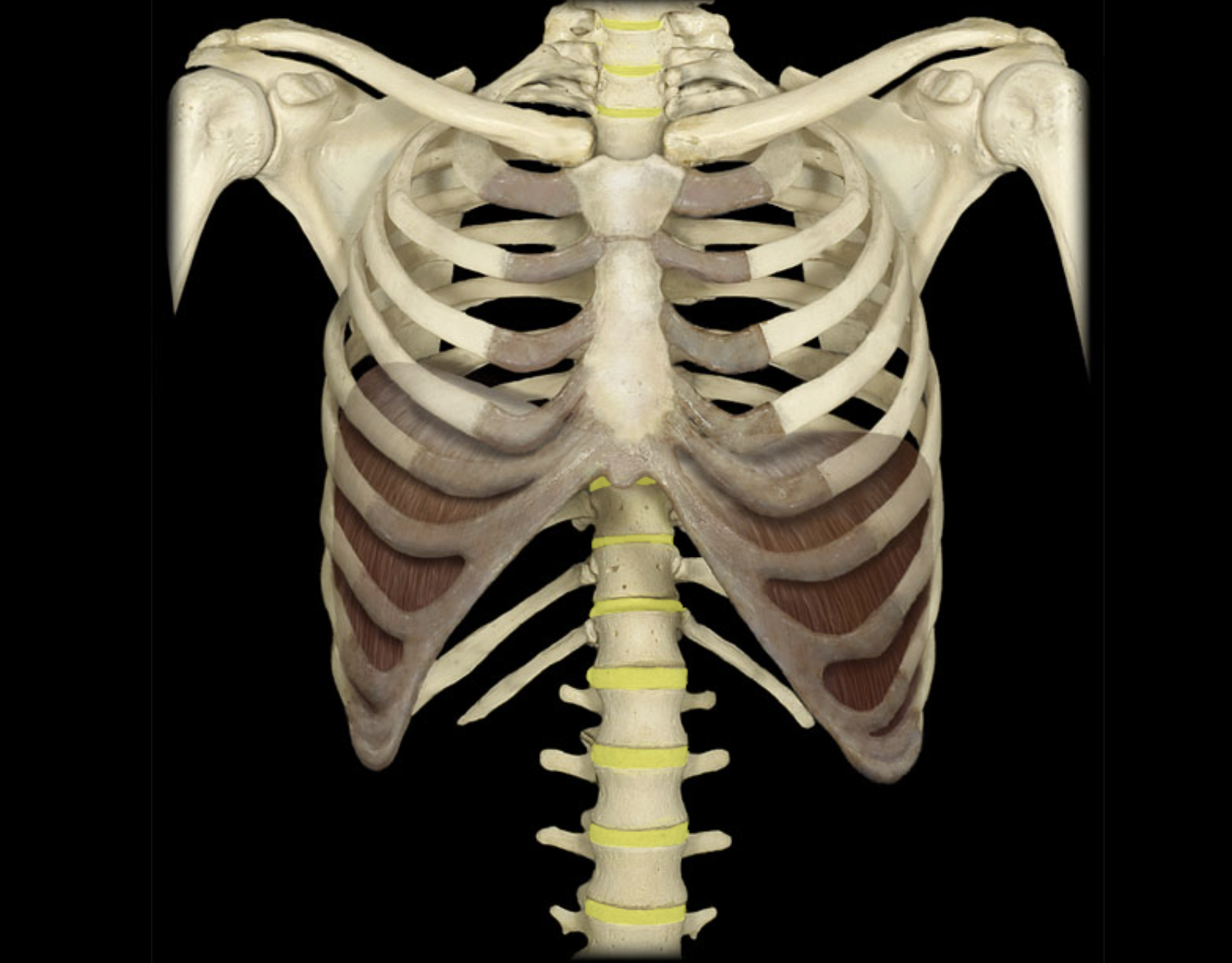
Ribs 1 - 12
Scapula
Sternum
Thoracic vertebra
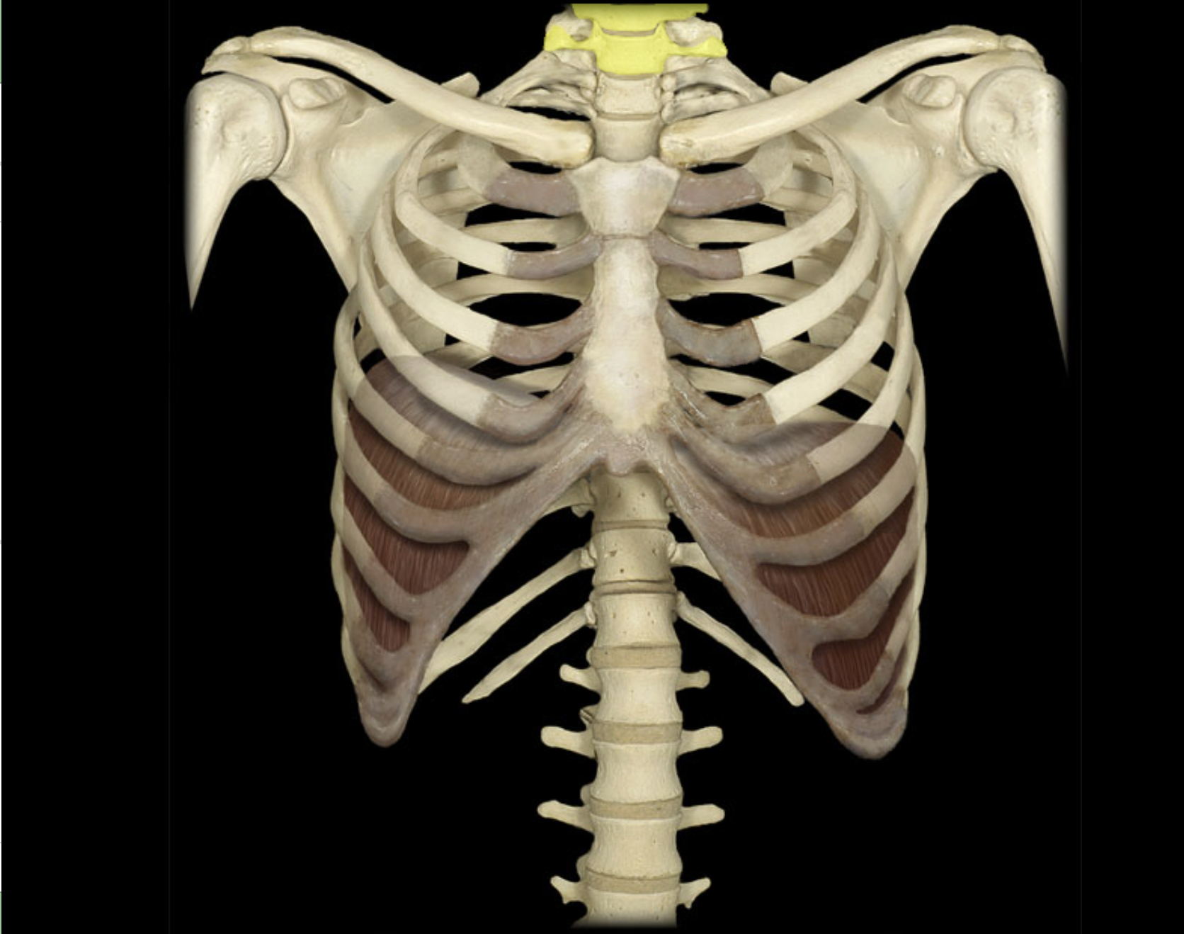
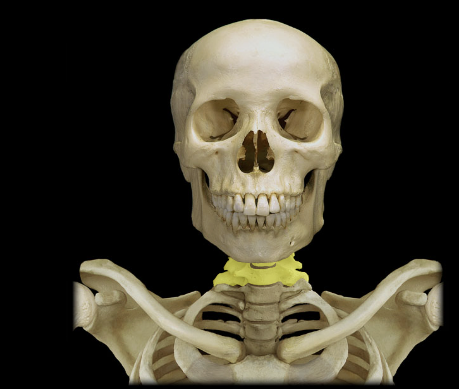
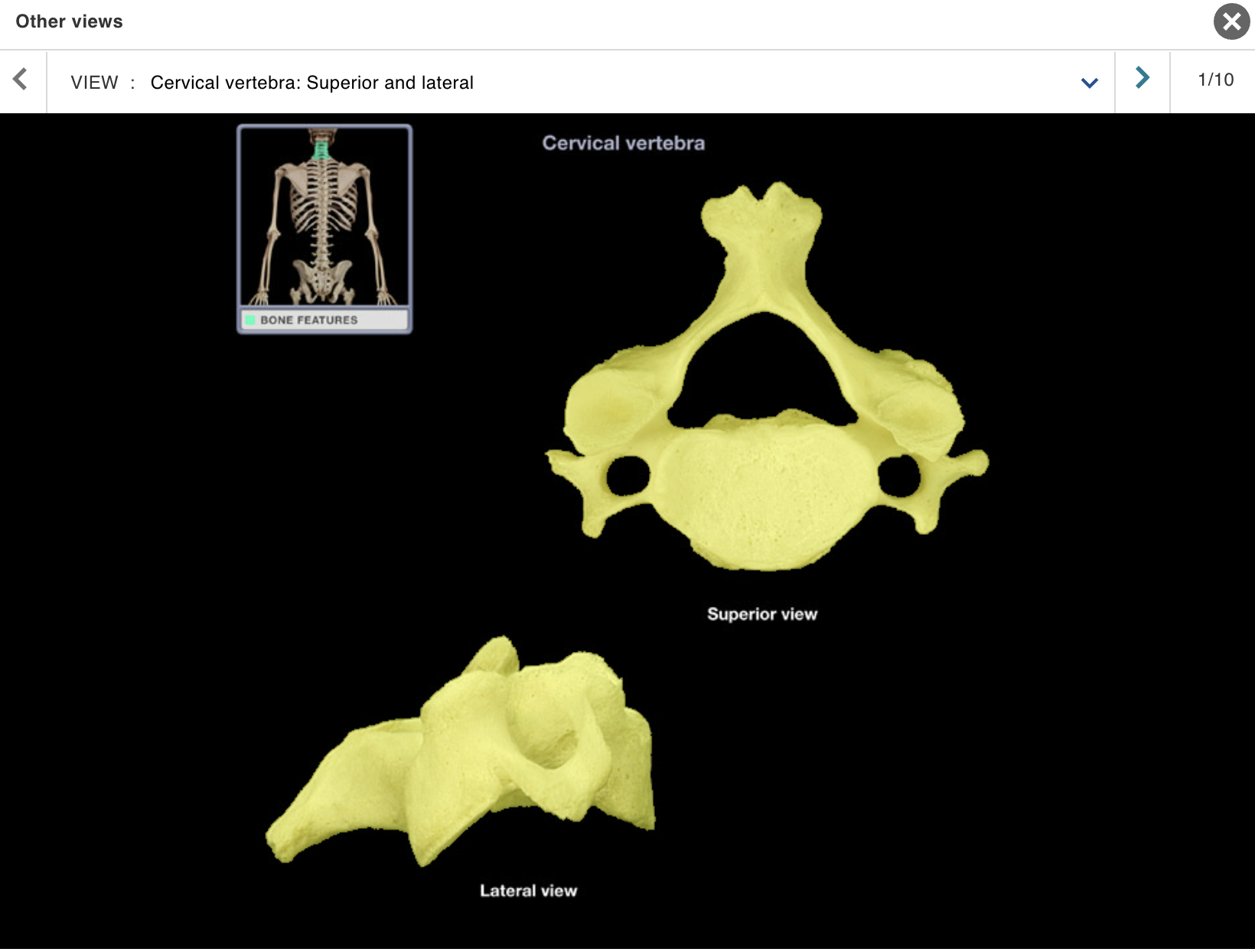
Cervical vertebra
Location:
Neck
Between occipital bone and T1 vertebra
Description:
Seven individual vertebrae
Characteristic features include transverse foramen and bifid
(split) spinous process on C3-C6
Comment:
• Atlas (C1 vertebra) articulates with skull
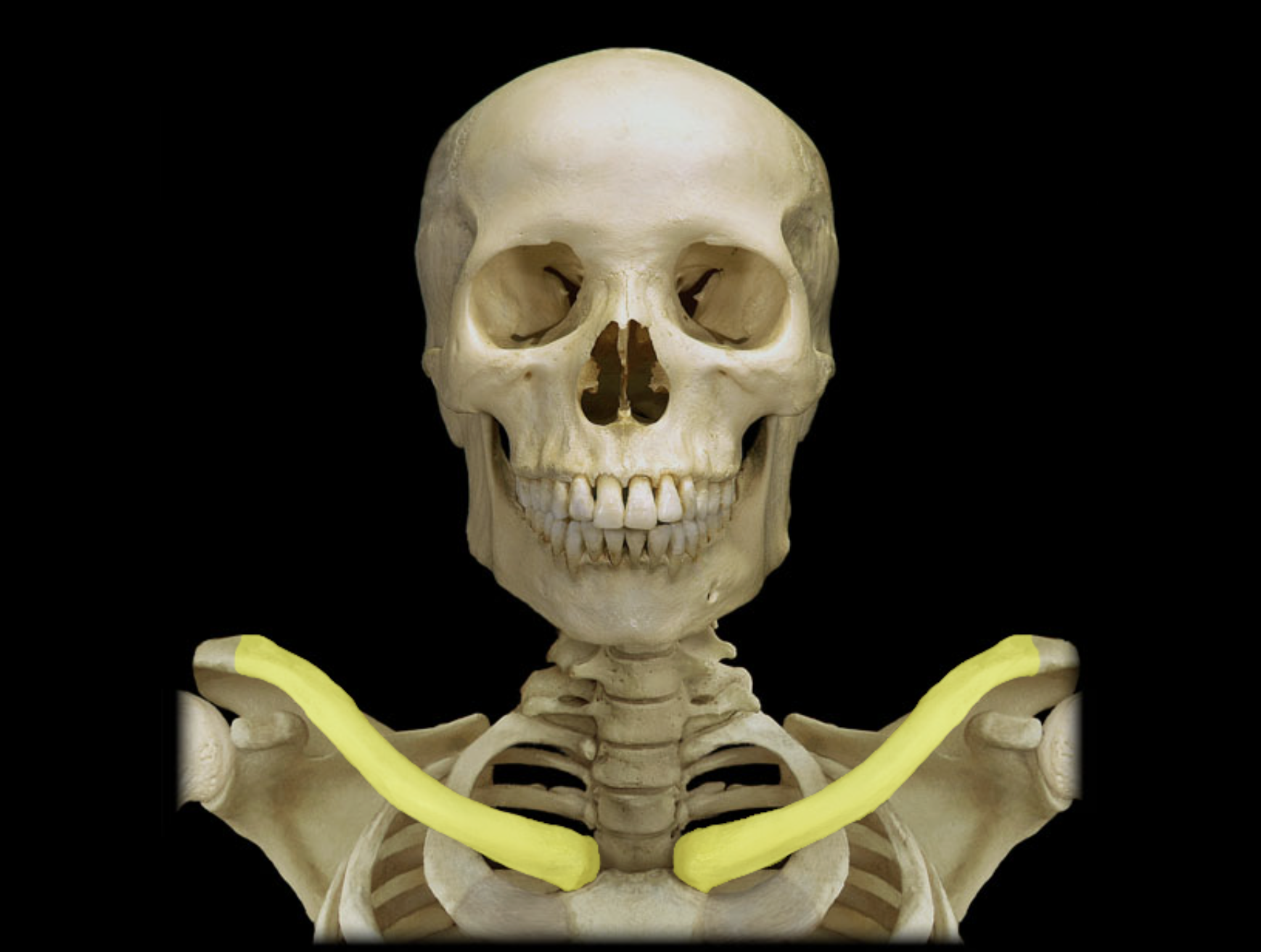
Clavicle
Location:
• Junction of neck and anterior thorax
Description:
Subcutaneous, S-shaped bone
Medial end articulates with sternum at sternoclavicular joint
Lateral end articulates with acromion of scapula at acromioclavicular joint
Also known as:
• "Collar bone"
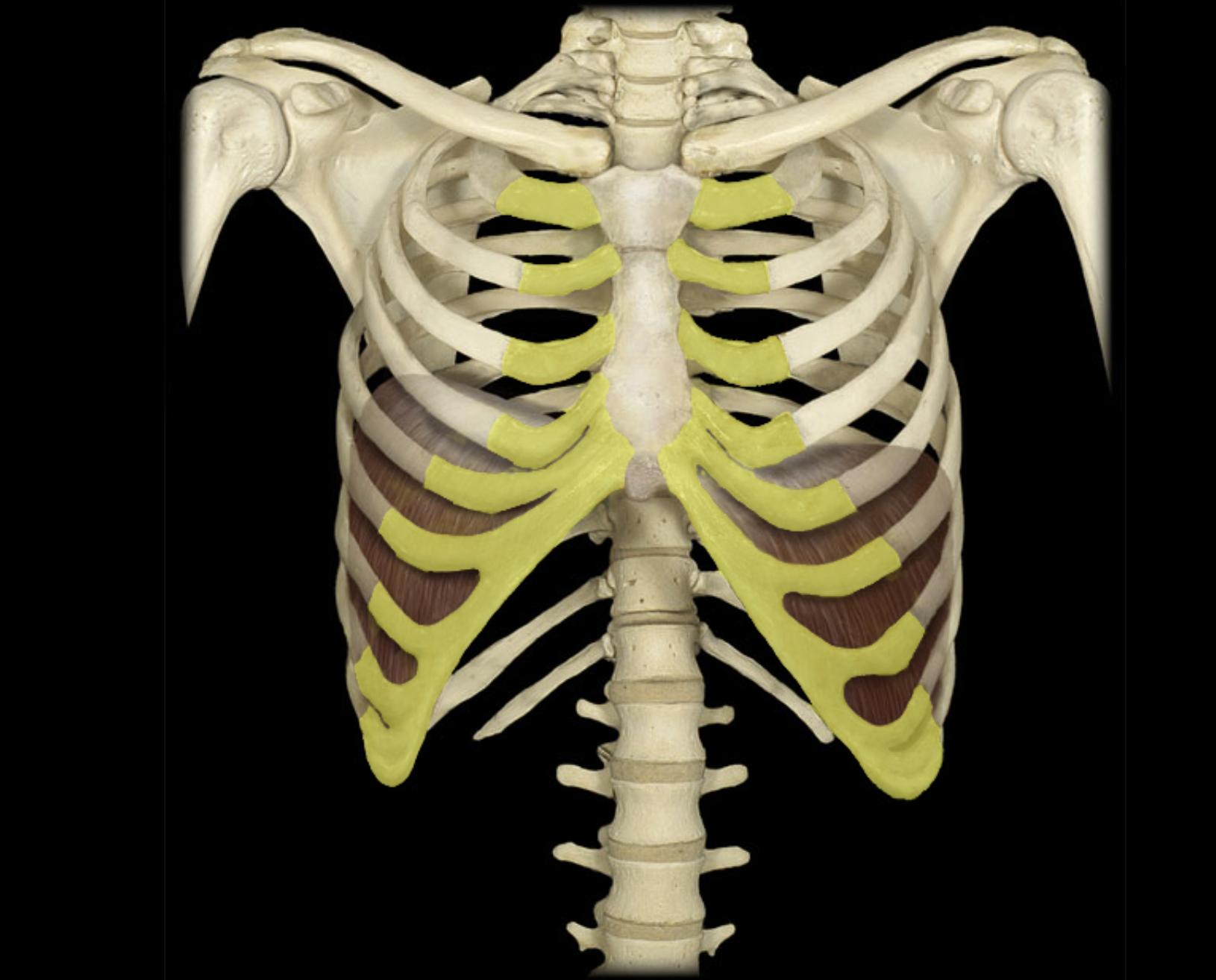
Costal cartilages
Location:
• Thorax
Description:
Attaches rib to sternum
True ribs (ribs 1-7): attached directly to sternum by costal cartilages
False ribs (ribs 8-10): attach indirectly to sternum via a shared costal cartilage
Costal margin created by costal cartilages of ribs 7-10
Comment:
All ribs: articulate with thoracic vertebrae
Floating ribs (ribs 11-12): not attached to sternum
Alternate definition: some include the floating ribs (11-12) as a subcategory of false ribs
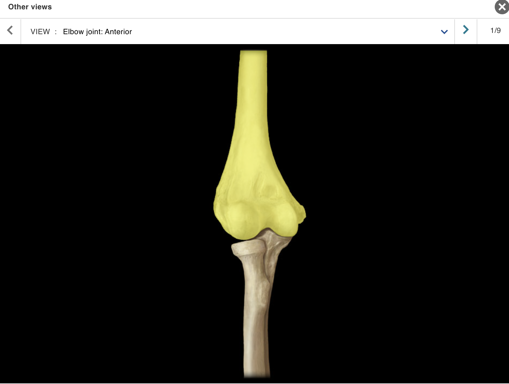
Humerus
Location:
• Arm
Description:
Long bone
Characteristic features include head, neck, greater and lesser tubercles, shaft, medial and lateral epicondyles, capitulum, and trochlea
Comment:
• Largest bone of upper limb
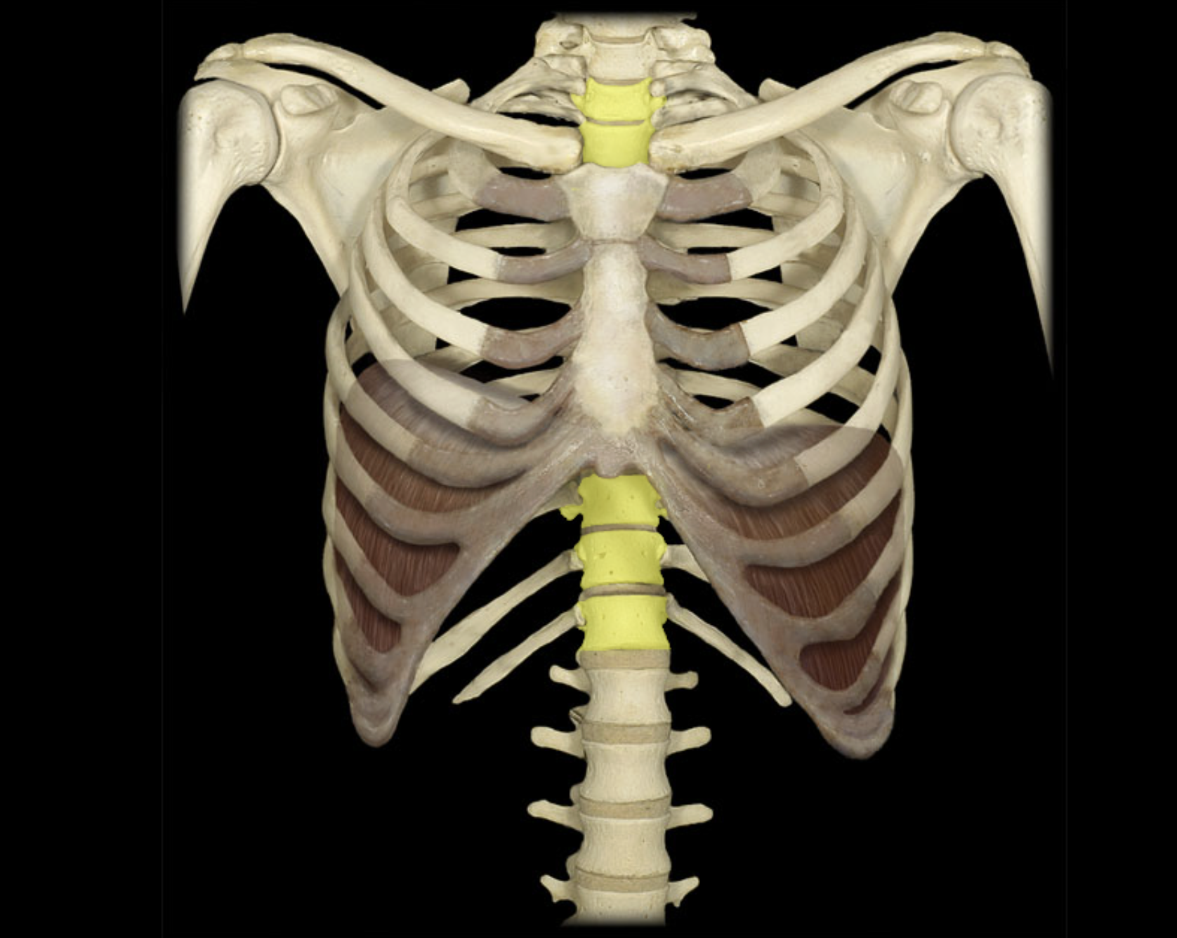
Intervertebral disc
Location:
• Between vertebral bodies (C2 to S1)
Description:
Outer part: anulus fibrosus consisting of a fibrocartilage ring
Inner part: nucleus pulposus composed of mucoid material
Comment:
• No disk between atlas (C1 vertebra) and axis (C2 vertebra)
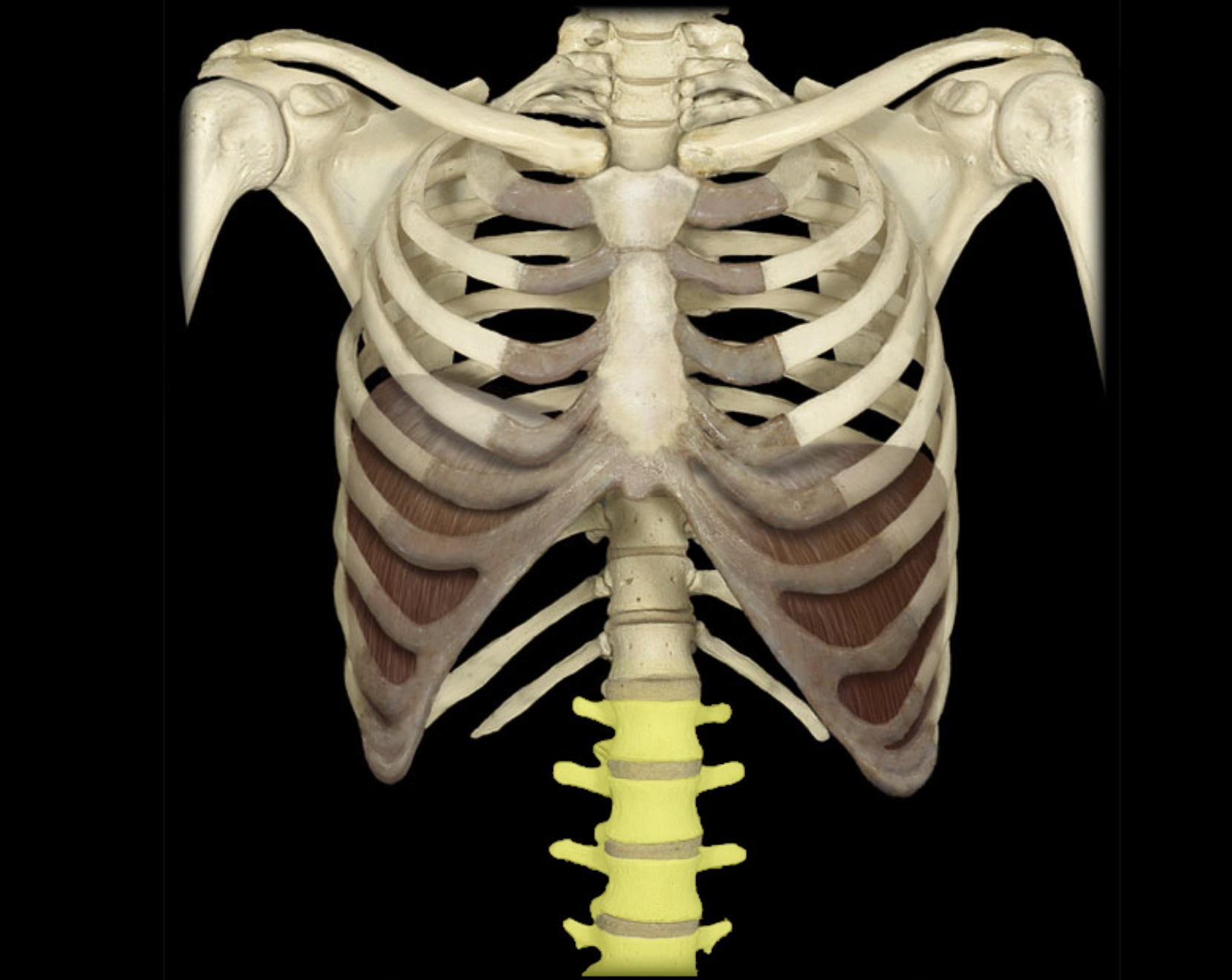
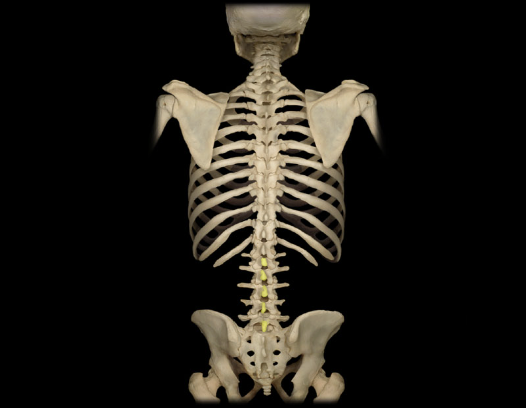
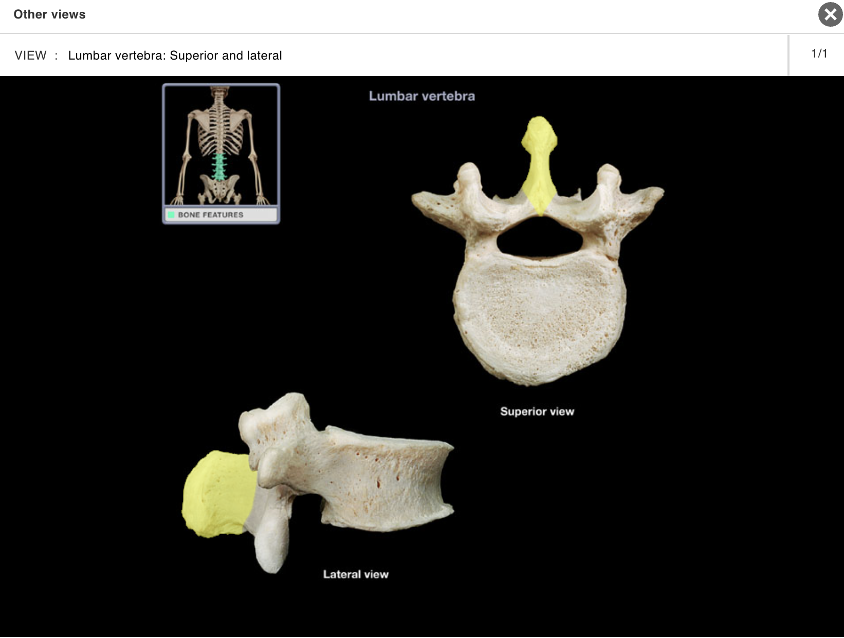
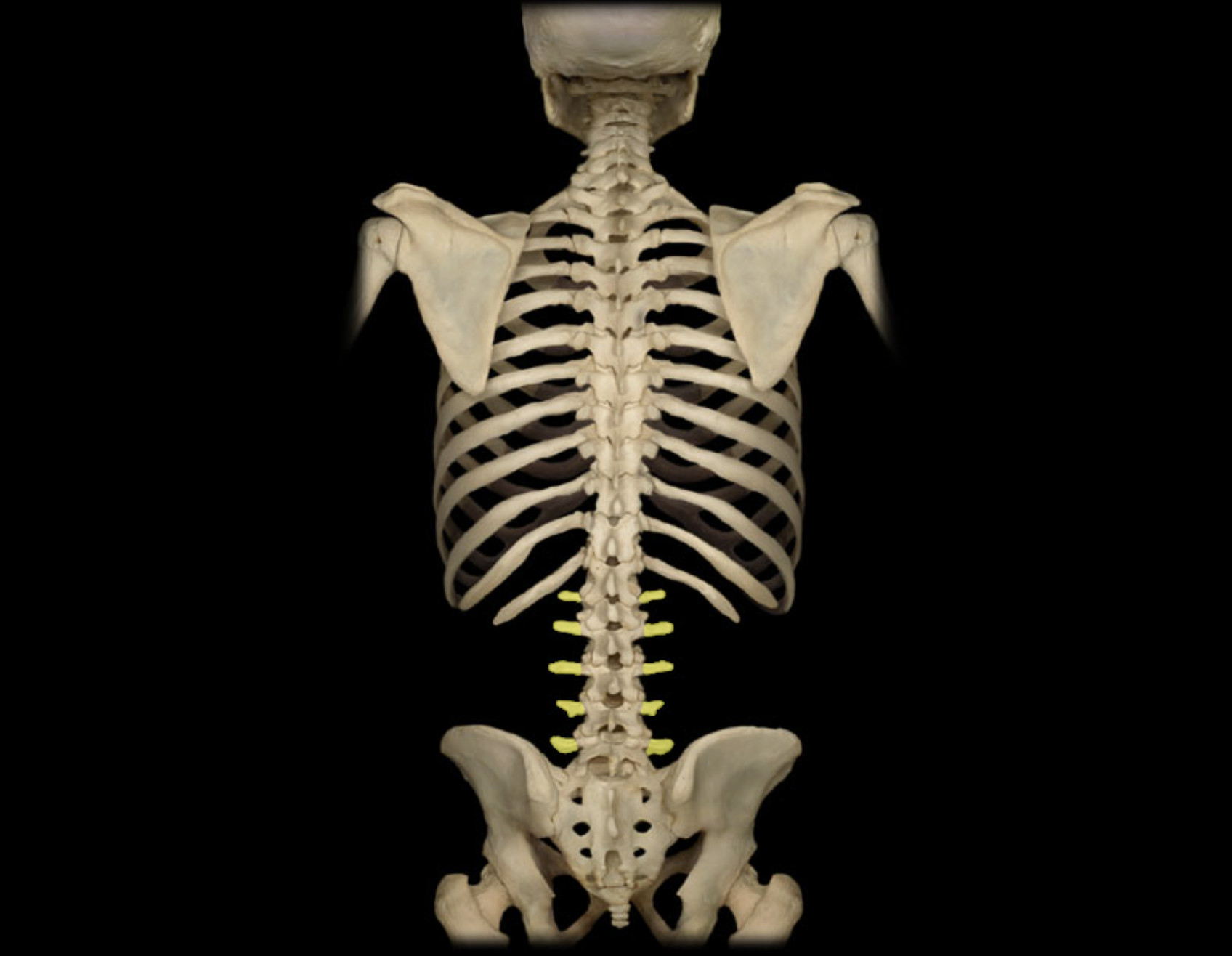
Lumbar vertebra
Location:
Lower back
Between T12 and S1 vertebrae
Description:
Five individual vertebrae
Characteristic features include large size, kidney bean-shaped body, and a thick, blunt spinous process
Comment:
Bodies arranged to form prominent anterior convexity (lumbar curvature; also Known as lumbar lordosis, which can be accentuated pathologically)
Intervertebral discs between lumbar vertebrae most commonly herniate ("slipped-disk")
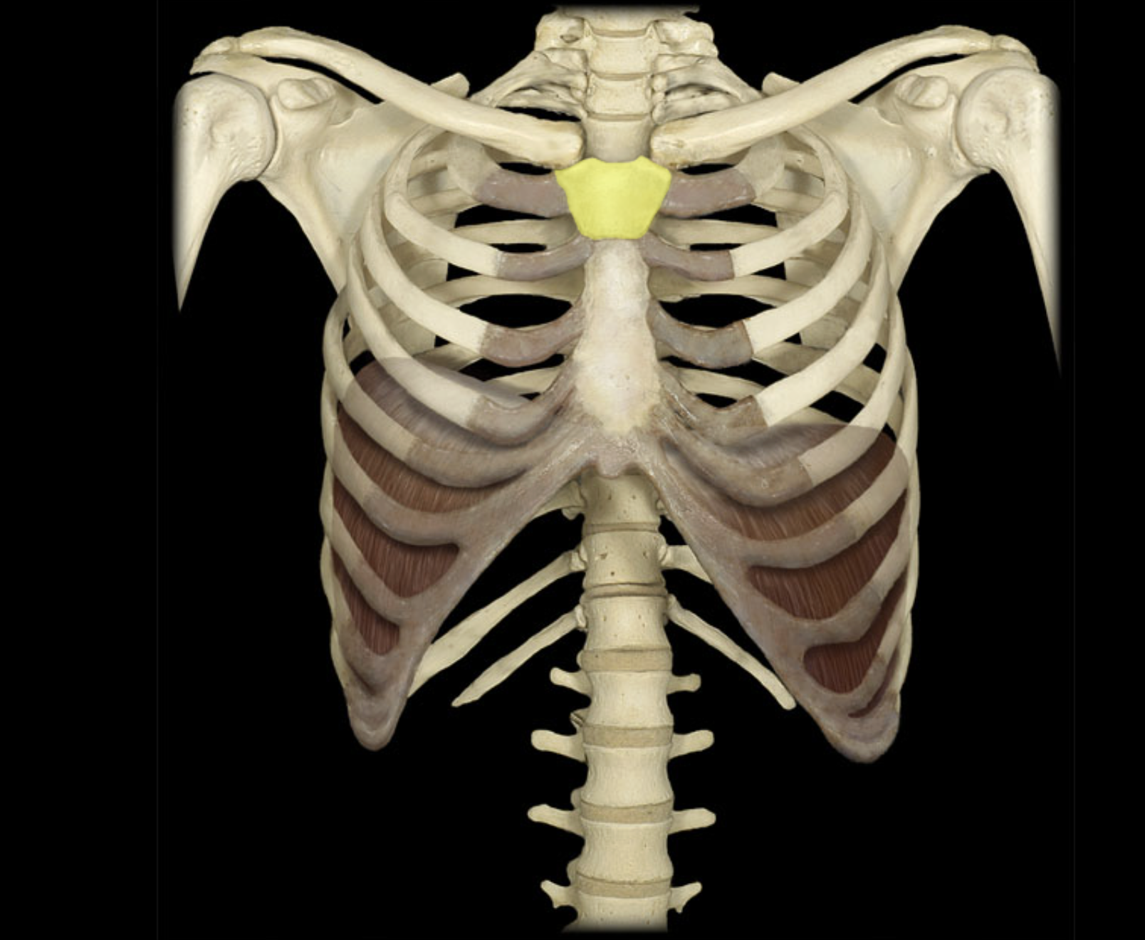
Manubrium
Location:
• Sternum
Description:
Triangular shape
Superior part of sternum
Articulates with costal cartilages of ribs 1-2 and clavicles
Comment:
• Provides attachment for sternocleidomastoid and pectoralis major muscles
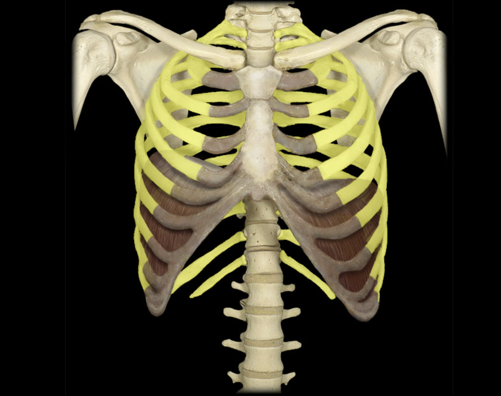
Ribs 1 - 12
Location:
• Thorax
Description:
Twelve pairs of curved, flat bones
All ribs: articulate with thoracic vertebrae
True ribs (ribs 1-7): attached directly to sternum by costal cartilages
False ribs (ribs 8-10): attach indirectly to sternum via shared costal cartilages
Floating ribs (ribs 11-12): not attached to sternum
Comment:
• Alternate definition: some include the floating ribs (11-12) as a subcategory of false ribs
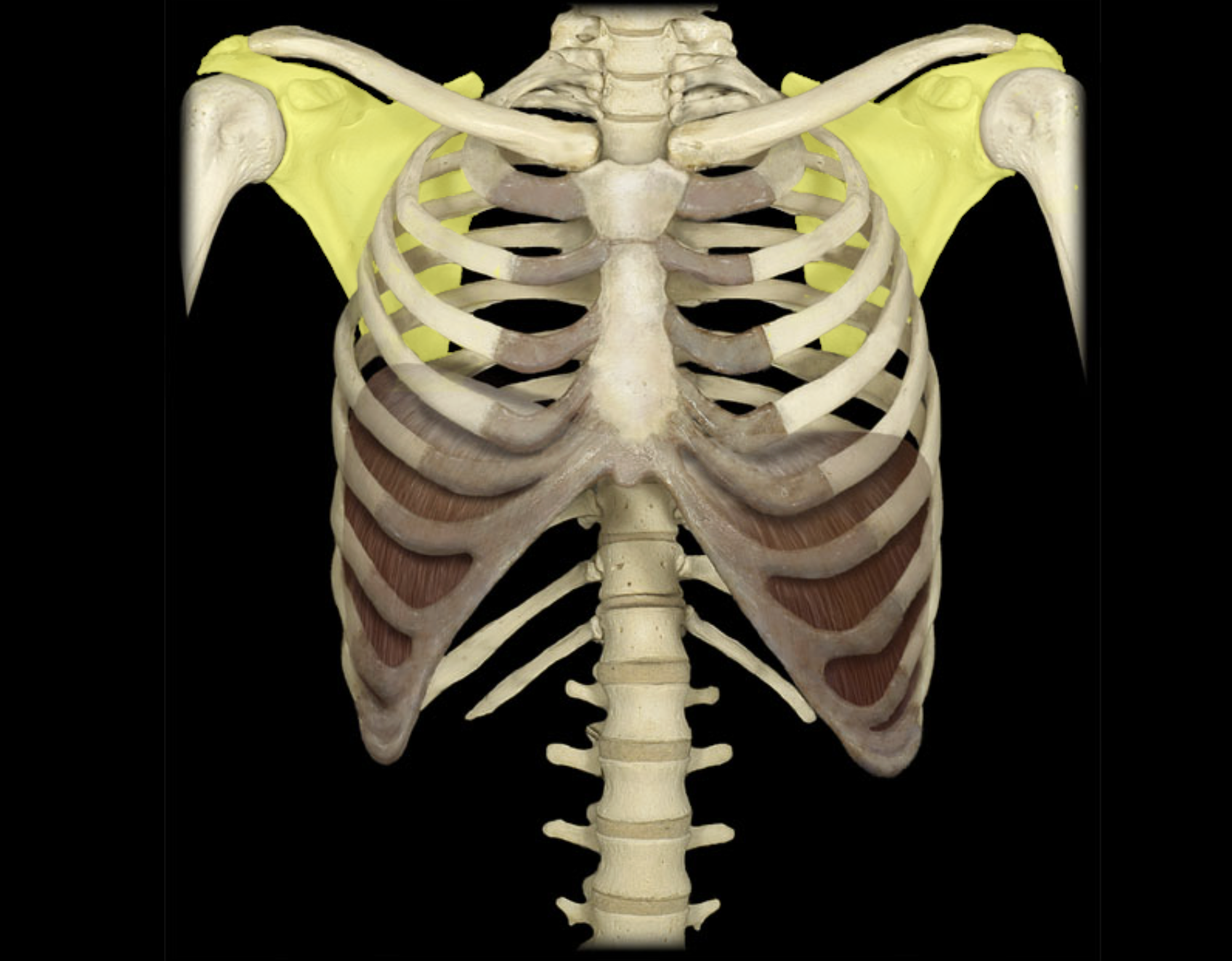
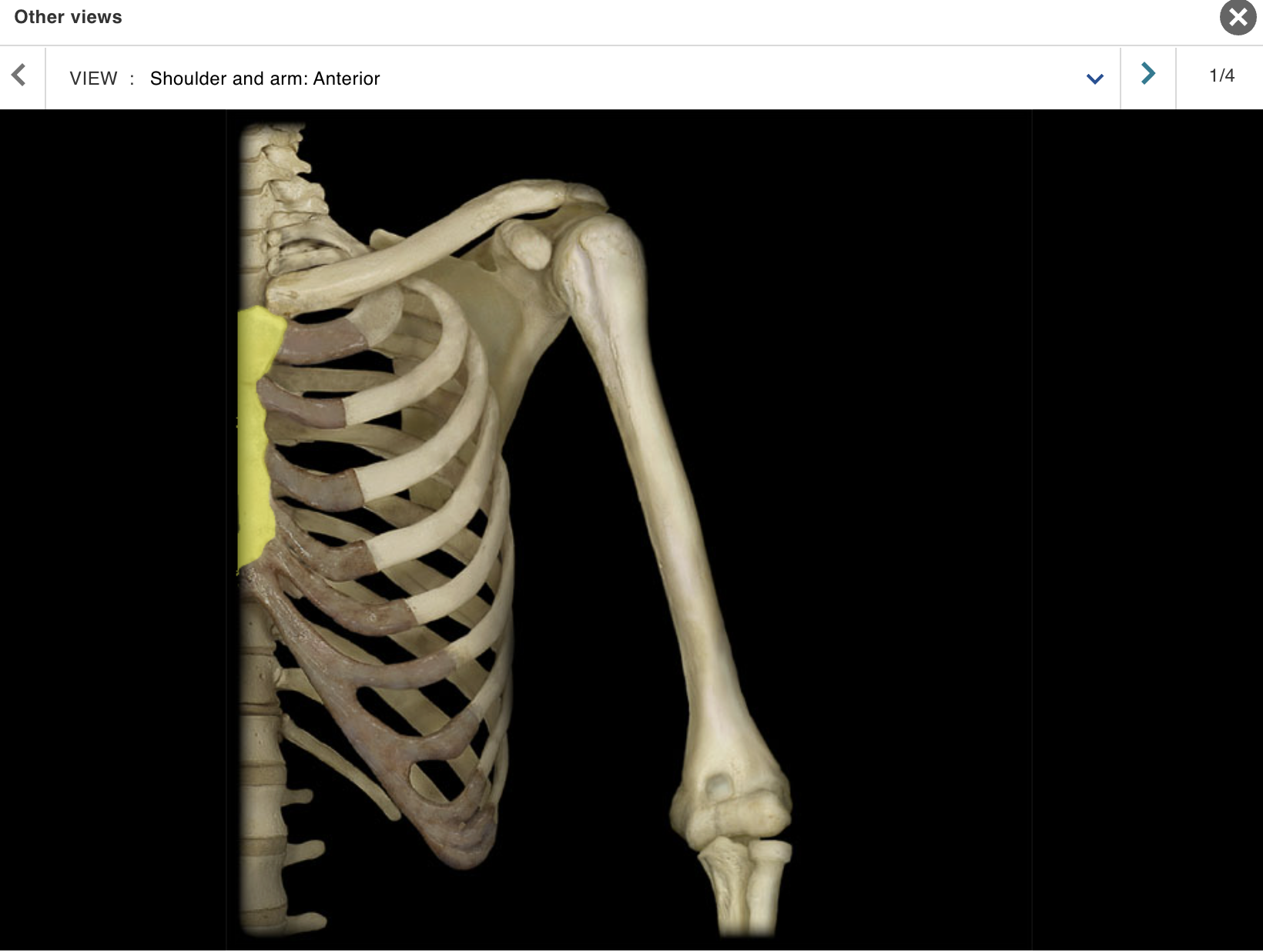
Scapula
Location:
Posterior thorax
Overlies ribs 2-7
Description:
Large, triangular, flat bone
Characteristic features include spine, acromion, coracoid process, and glenoid cavity
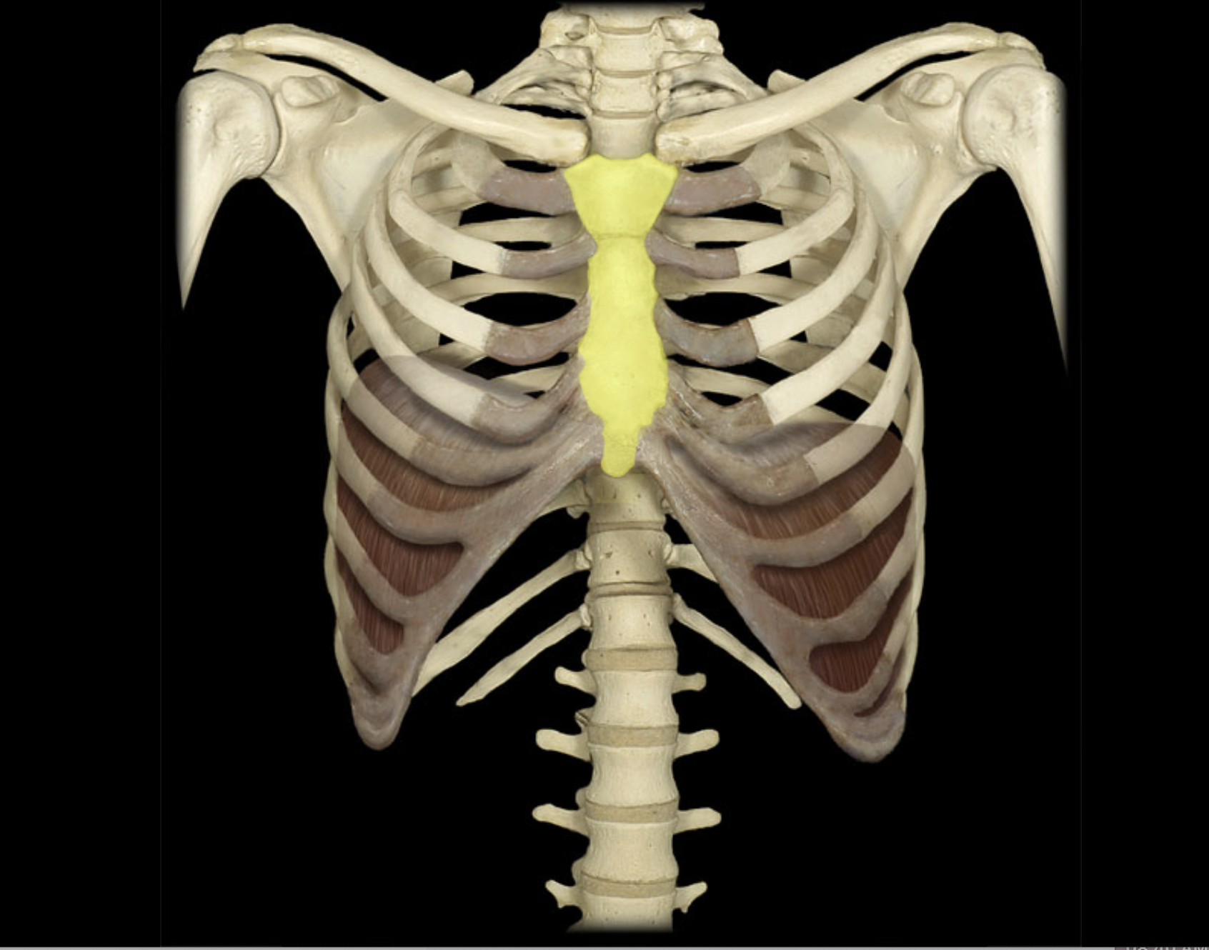
Sternum
Location:
• Thorax (anterior midline)
Description:
Flat bone with three parts: manubrium, body, and xiphoid
processCharacteristic features include jugular notch and sternal angle (angle of Louis)
Articulates with costal cartilages of true ribs (1-7), combined costal cartilages of false ribs (8-10), and clavicles
Also known as:
• "Breastbone"
Comment:
Floating ribs (ribs 11-12): not attached to sternum
Provides for attachment of sternocleidomastoid, pectoralis major, sternohyoid, and sternothyroid muscles
Thoracic vertebra
Location:
Trunk
Between C7 and L1 vertebrae
Description:
12 individual vertebrae
Characteristic features include costal demifacets (or facets) for articulation with head of rib, spinous process slopes inferiorly, and heart-shaped vertebral body

ACTIVITY 4: DISSECTION
MODULE: SKELETAL
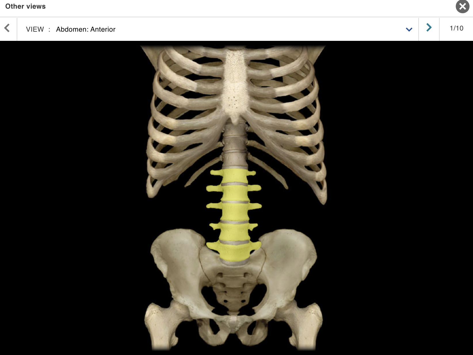
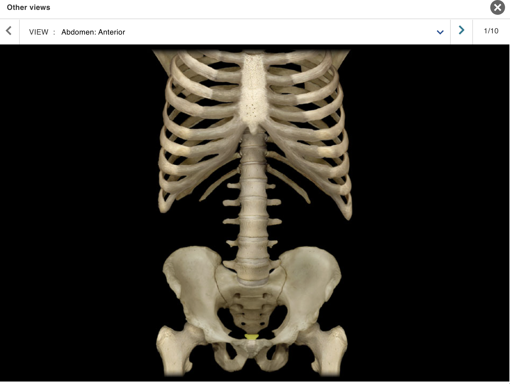
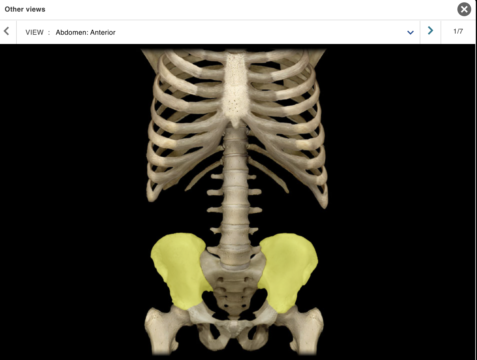
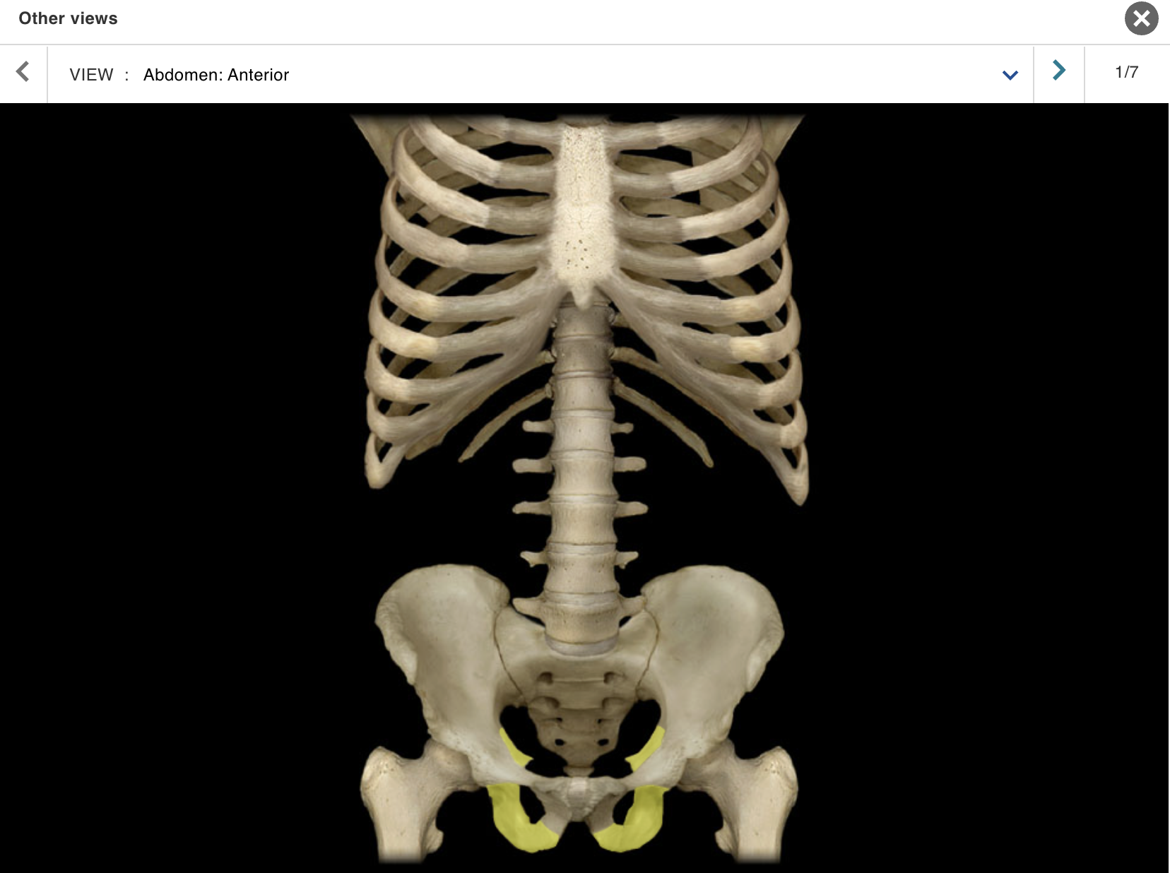
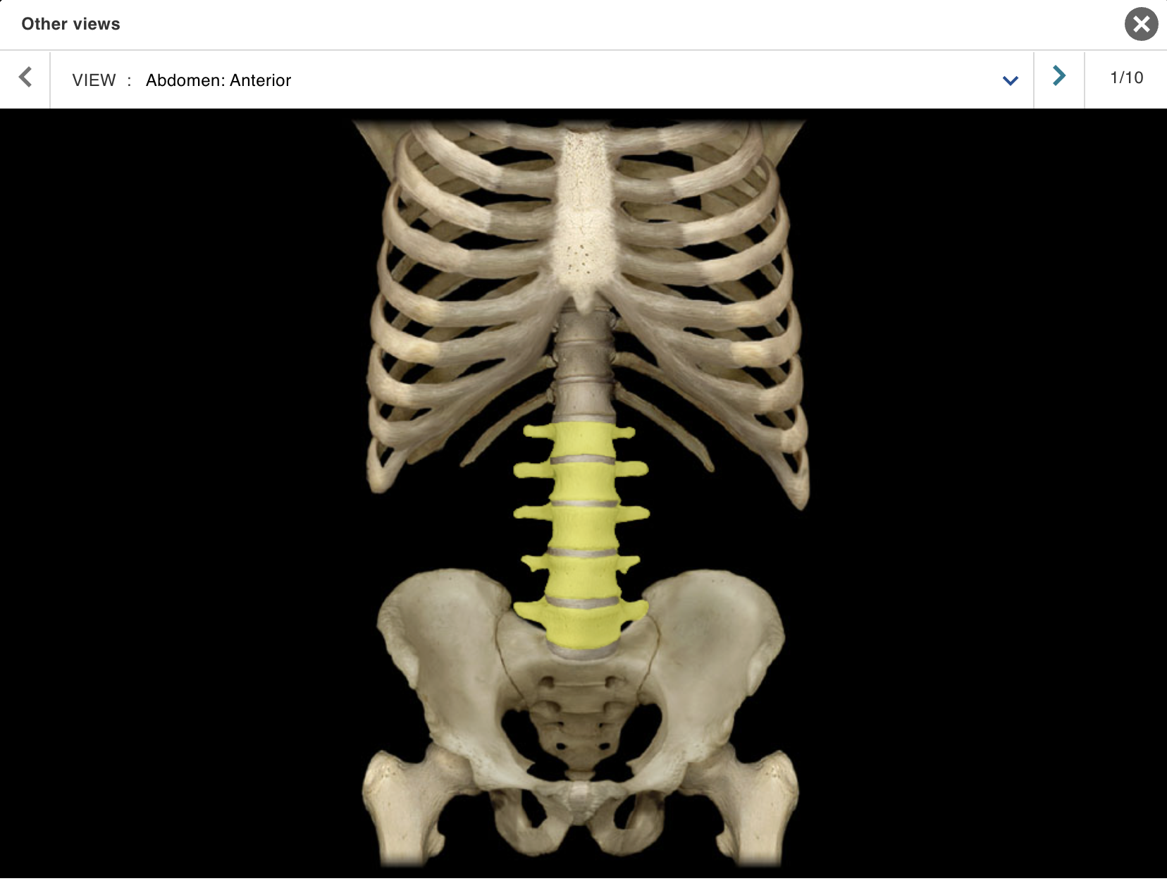
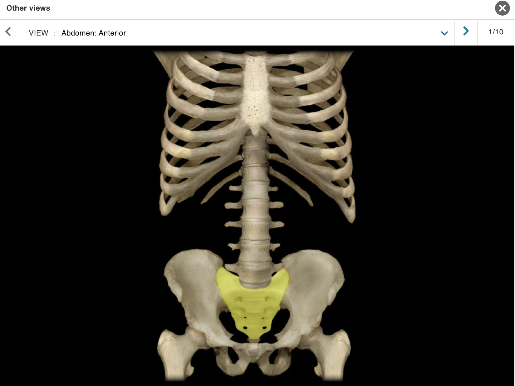
TOPIC: ABDOMEN / VIEW : ANTERIOR
Body of sternum
Соссух
Femur
lium
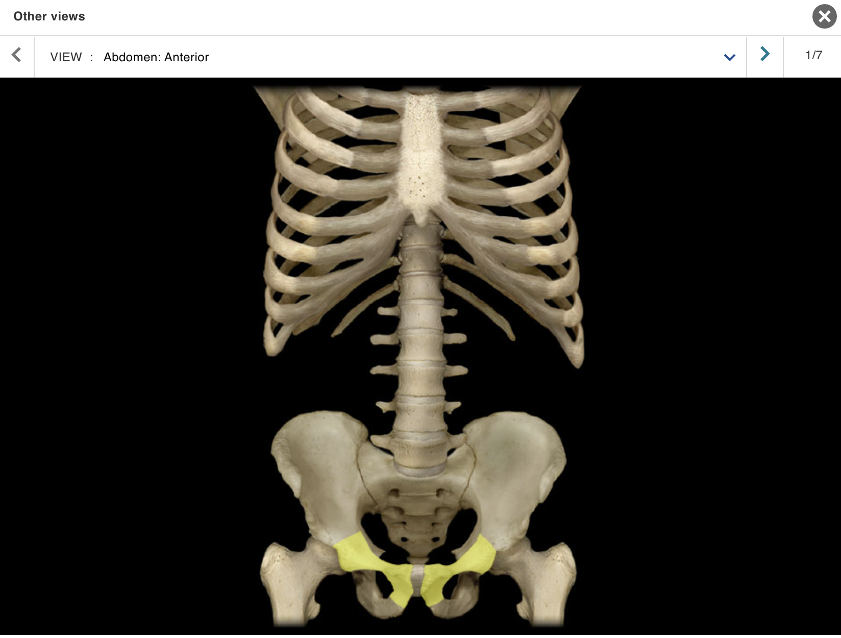
Ischium
Lumbar vertebra
Pelvic girdle
Pubic symphysis
Pubis
Ribs 2 - 12
Sacrum
Sternum
Xiphoid process
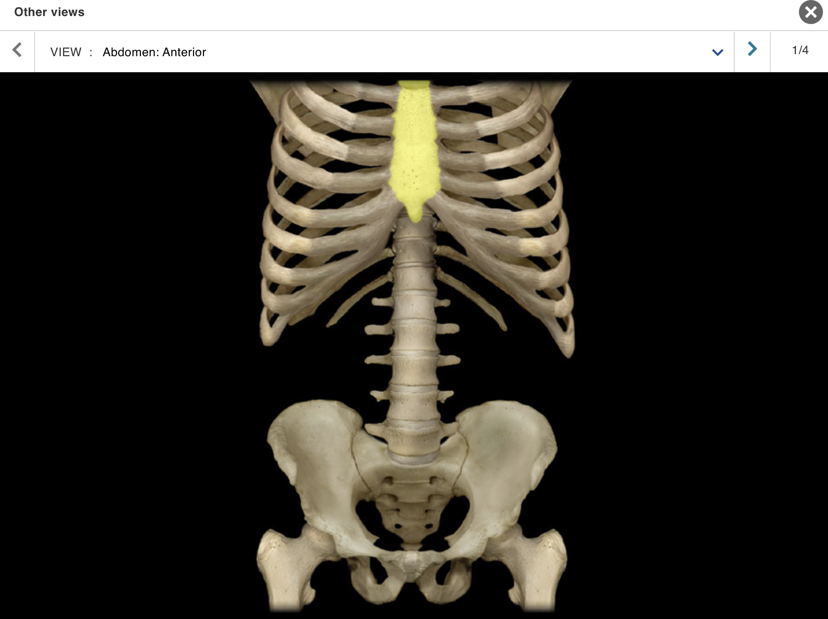
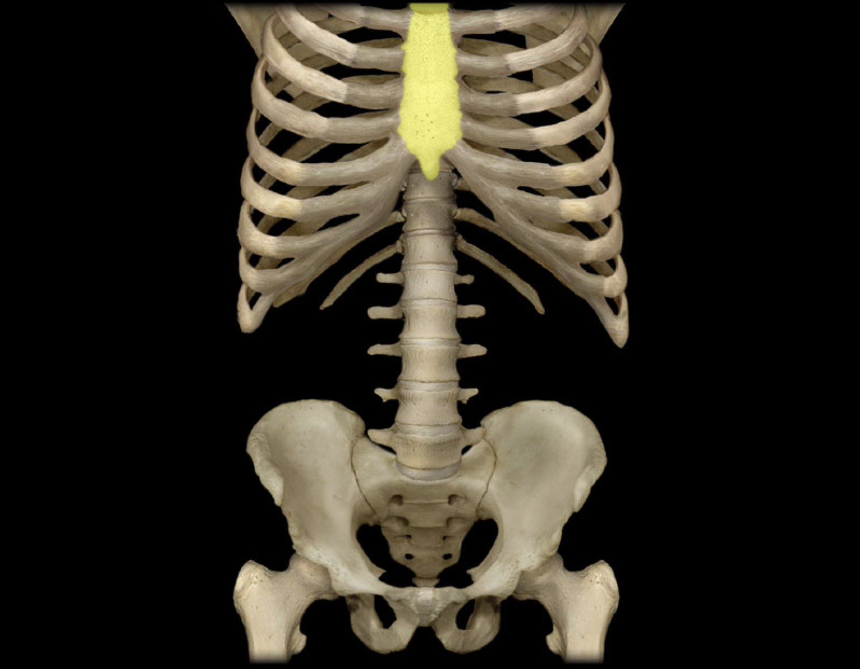
Body of sternum
Location:
• Sternum
Description:
Long flat part of sternum between manubrium and xiphoid process
Contributes to sternal angle (angle of Louis) at its superior border
Articulates with costal cartilages of ribs 2-7 and combined costal cartilages of false ribs (8-10)
Comment:
Floating ribs (ribs 11-12): not attached to sternum
Provides attachment for pectoralis major muscle
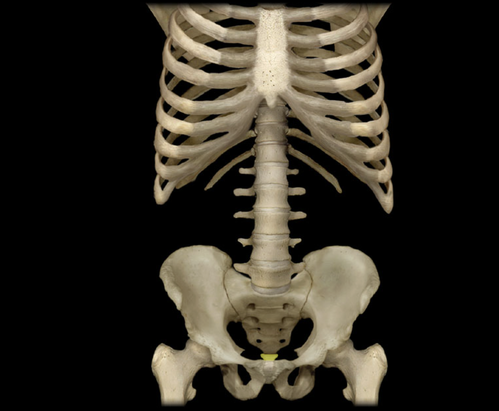
Соссух
Location:
Posterior pelvic wall
Lower back, inferior to S5 vertebra
Description:
Small, triangular bone
Consists of three to five, variably fused, poorly developed vertebrae
Also known as:
• "Tailbone"
Comment:
• Rudiment of the tail in other vertebrates

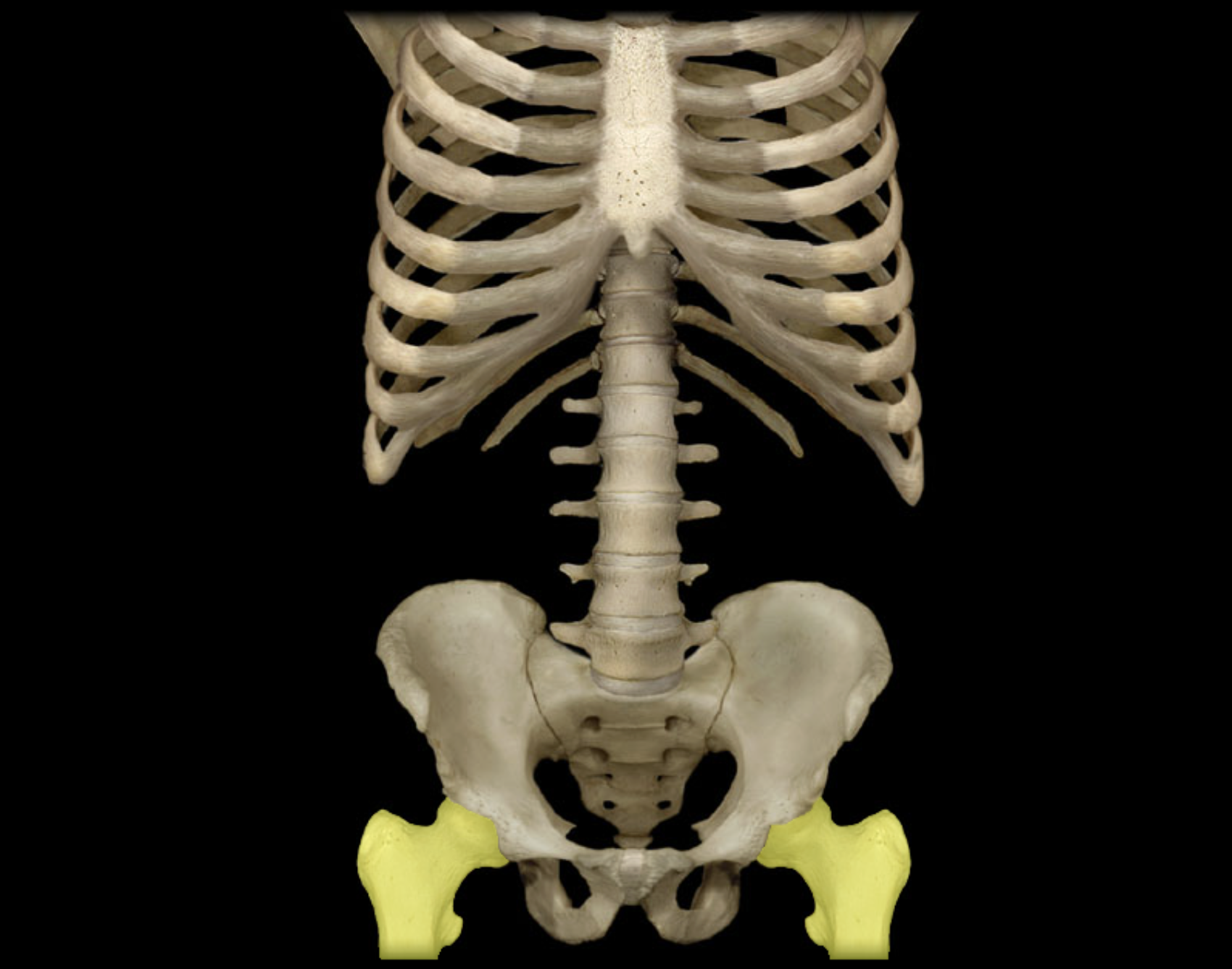
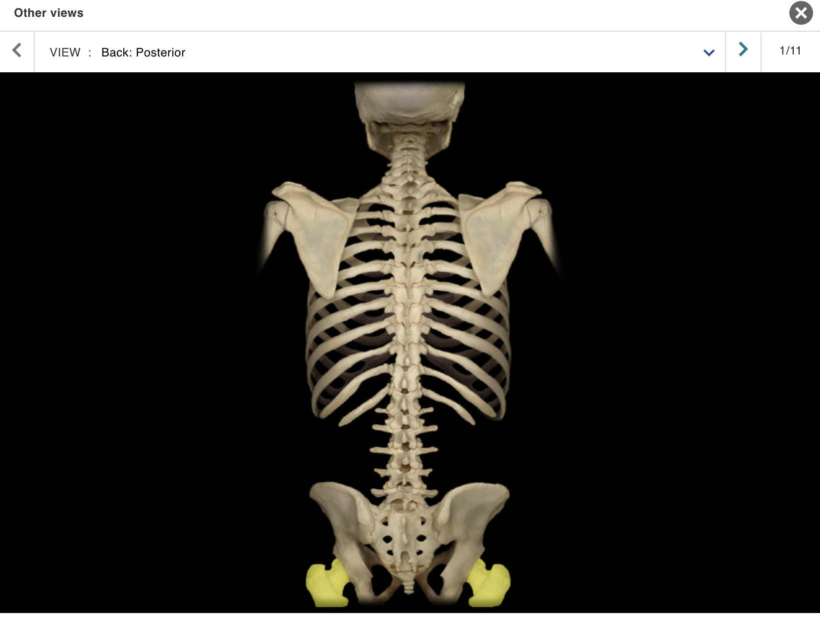
Femur
Location:
• Thigh
Description:
Long bone
Characteristic features include head, neck, shaft, greater and lesser trochanters, linea aspera, and medial and lateral condyles
Head forms part of hip joint
Distal end forms part of knee joint
Comment:
Only bone of thigh
Longest bone in body; length accurately predicts height of individual
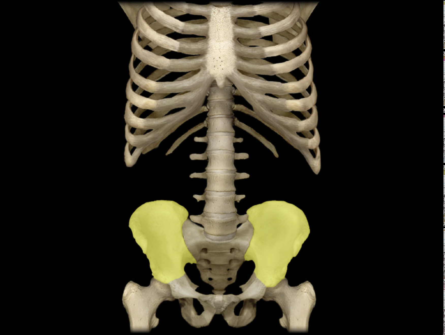
lium
Location:
• Pelvis
Description:
Largest of three coxal (hip) bones
Has large, wing-like superior extension (ala); the alae form bony walls of greater (false) pelvis
Contributes to acetabulum (hip joint socket) and wall of lesser (true) pelvis
Articulates with sacrum at sacroiliac joint
Comment:
Fused with ischium and pubis in adult to form coxal (hip) bone
Bony pelvis formed by paired hip bones and sacrum

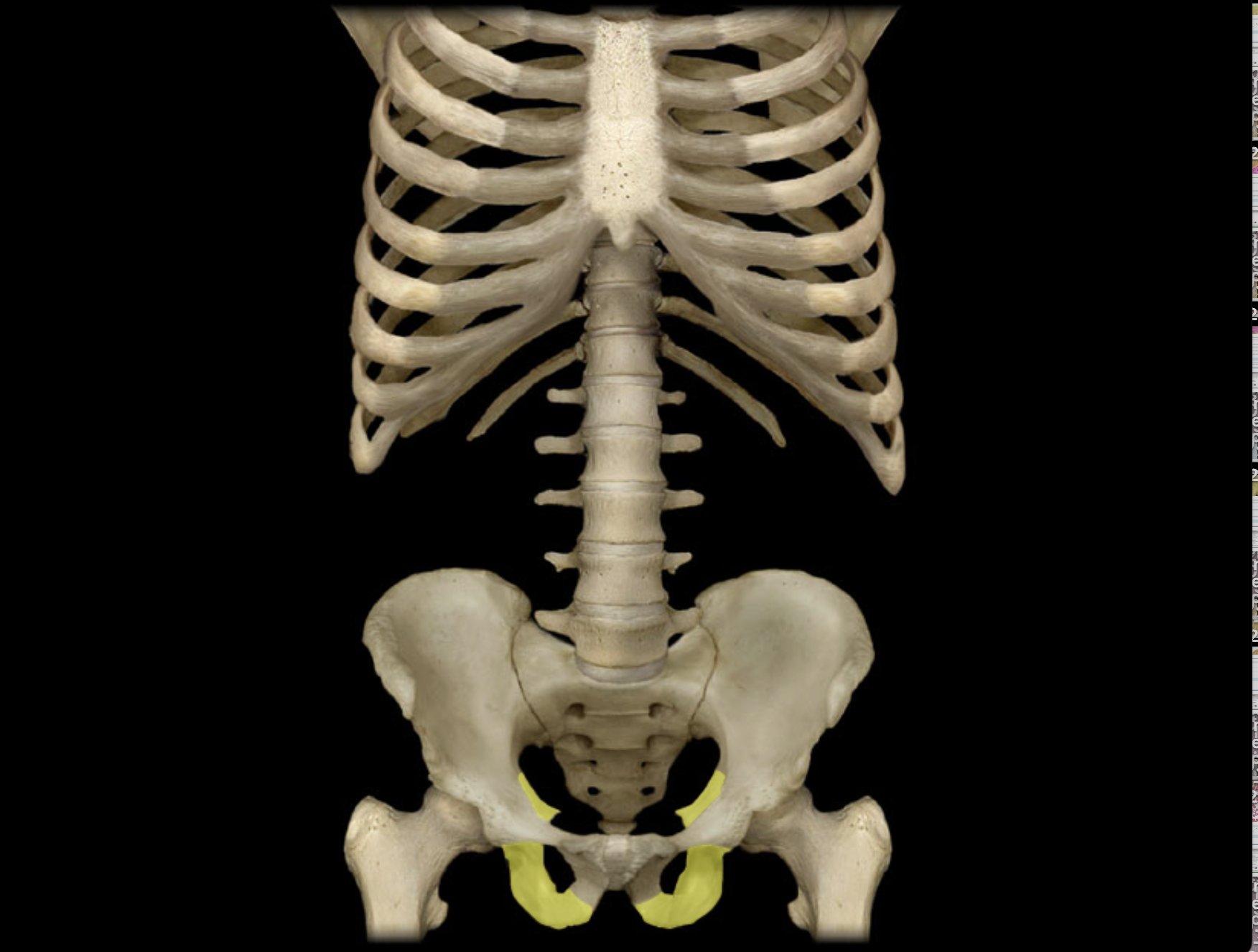
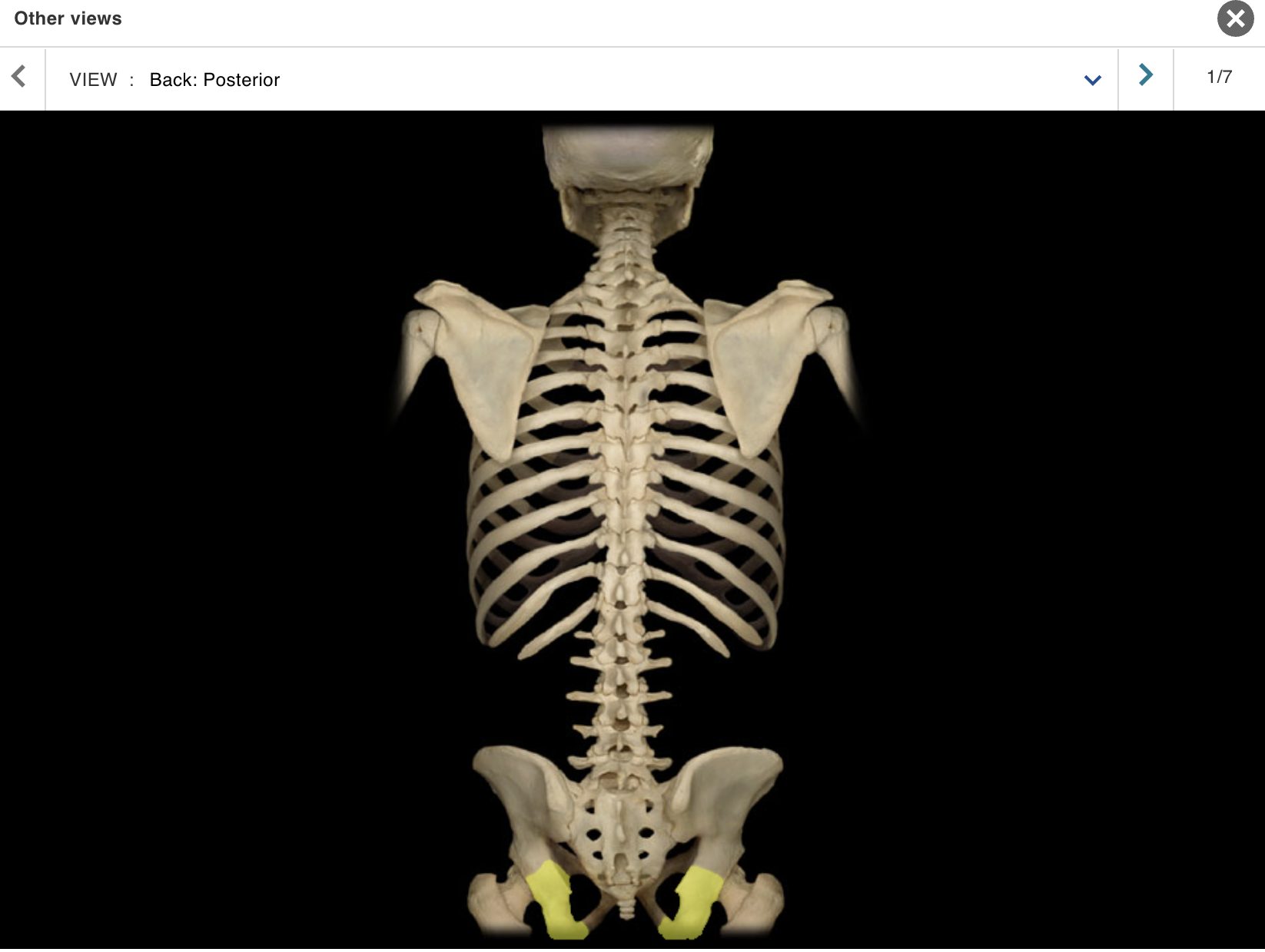
Ischium
Location:
• Pelvis
Description:
One of three coxal (hip) bones
Characteristic features include tuberosity and spine
Contributes to acetabulum (hip joint socket), obturator foramen, and wall of lesser (true) pelvis
Comment:
Fused with ilium and pubis in adult to form coxal (hip) bone
Bony pelvis formed by paired hip bones and sacrum
Obturator foramen formed by rami of pubis and ischium
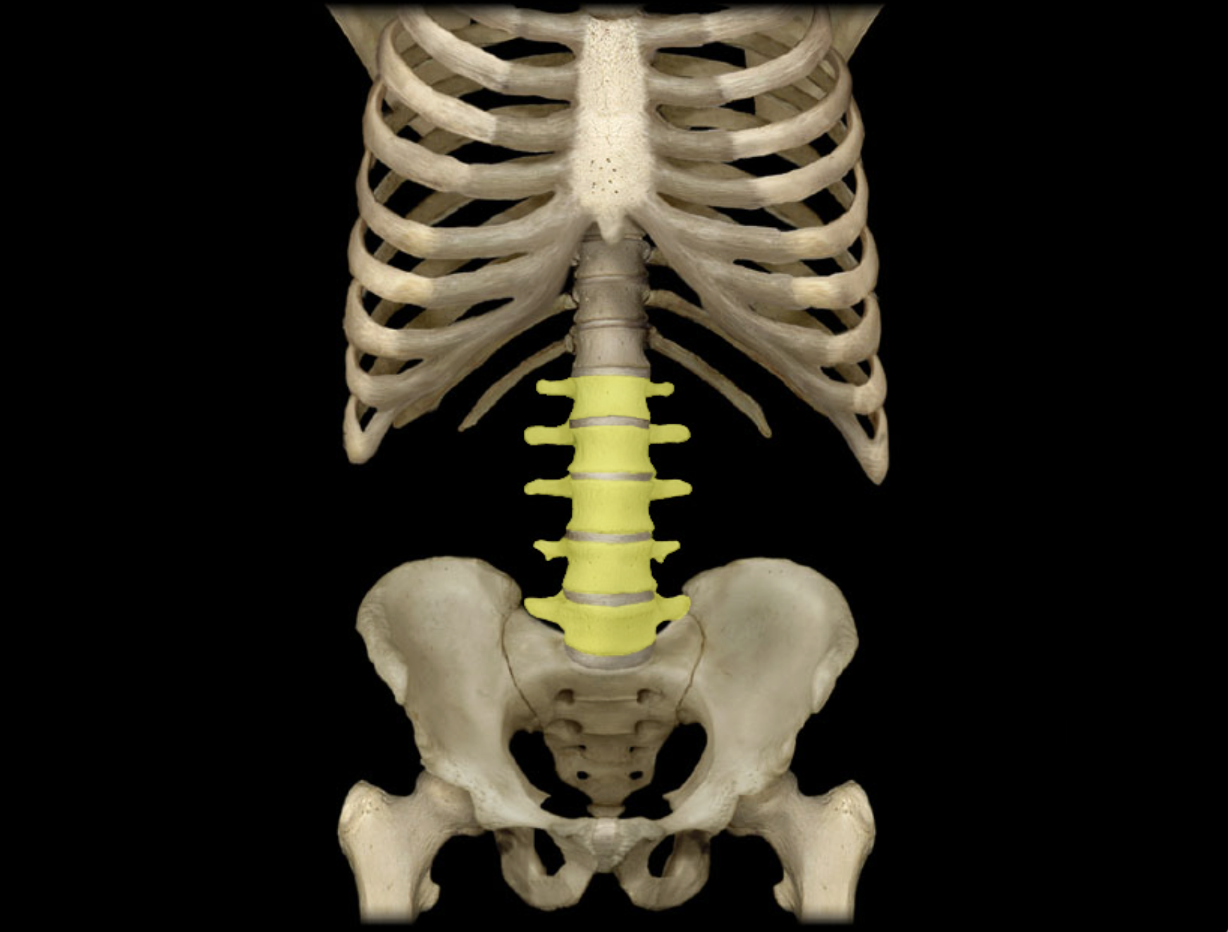
Lumbar vertebra
Location:
Lower back
Between T12 and S1 vertebrae
Description:
Five individual vertebrae
Characteristic features include large size, kidney bean-shaped body, and a thick, blunt spinous process
Comment:
Bodies arranged to form prominent anterior convexity (lumbar curvature; also known as lumbar lordosis, which can be accentuated pathologically)
Intervertebral discs between lumbar vertebrae most commonly herniate ("slipped-disk")
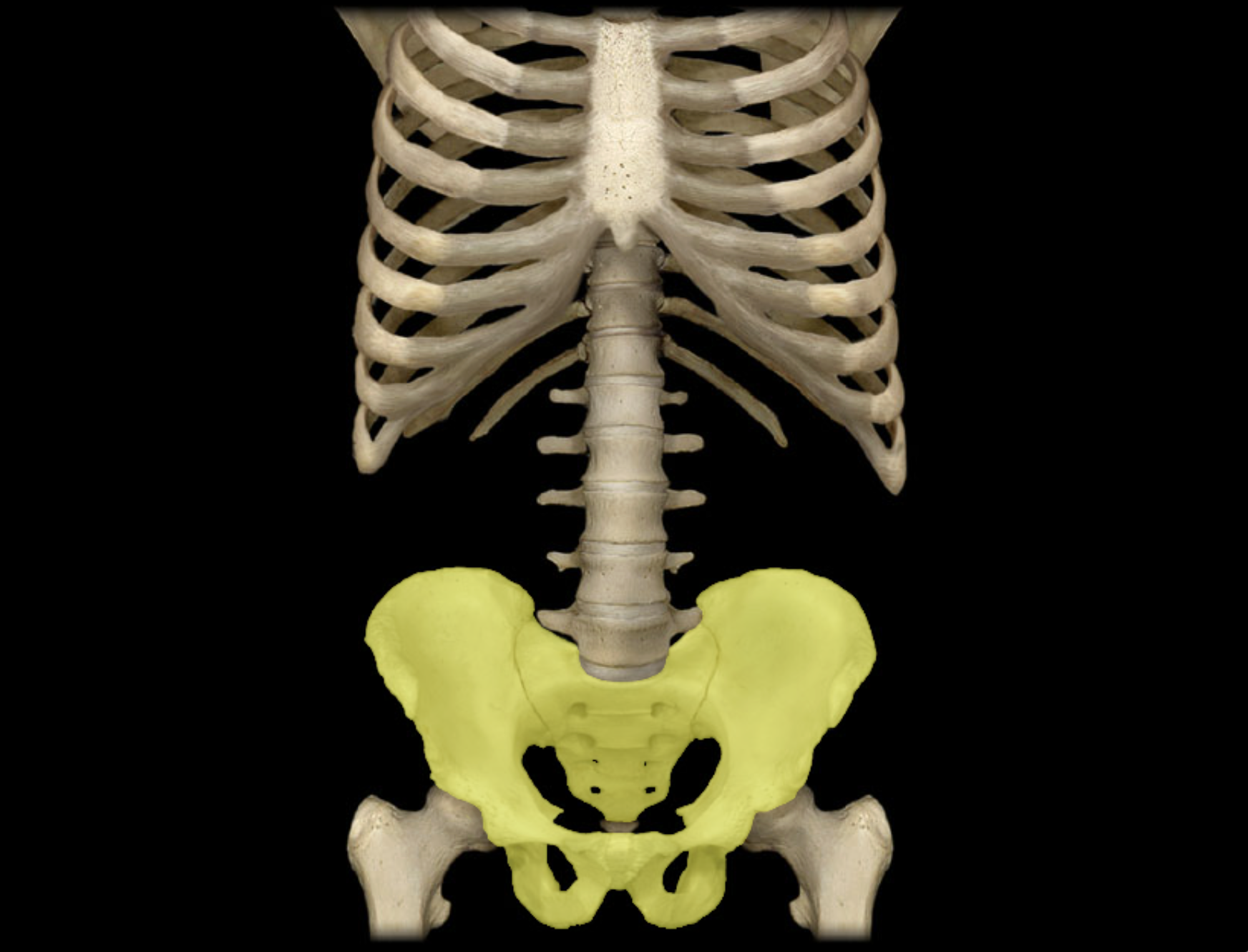
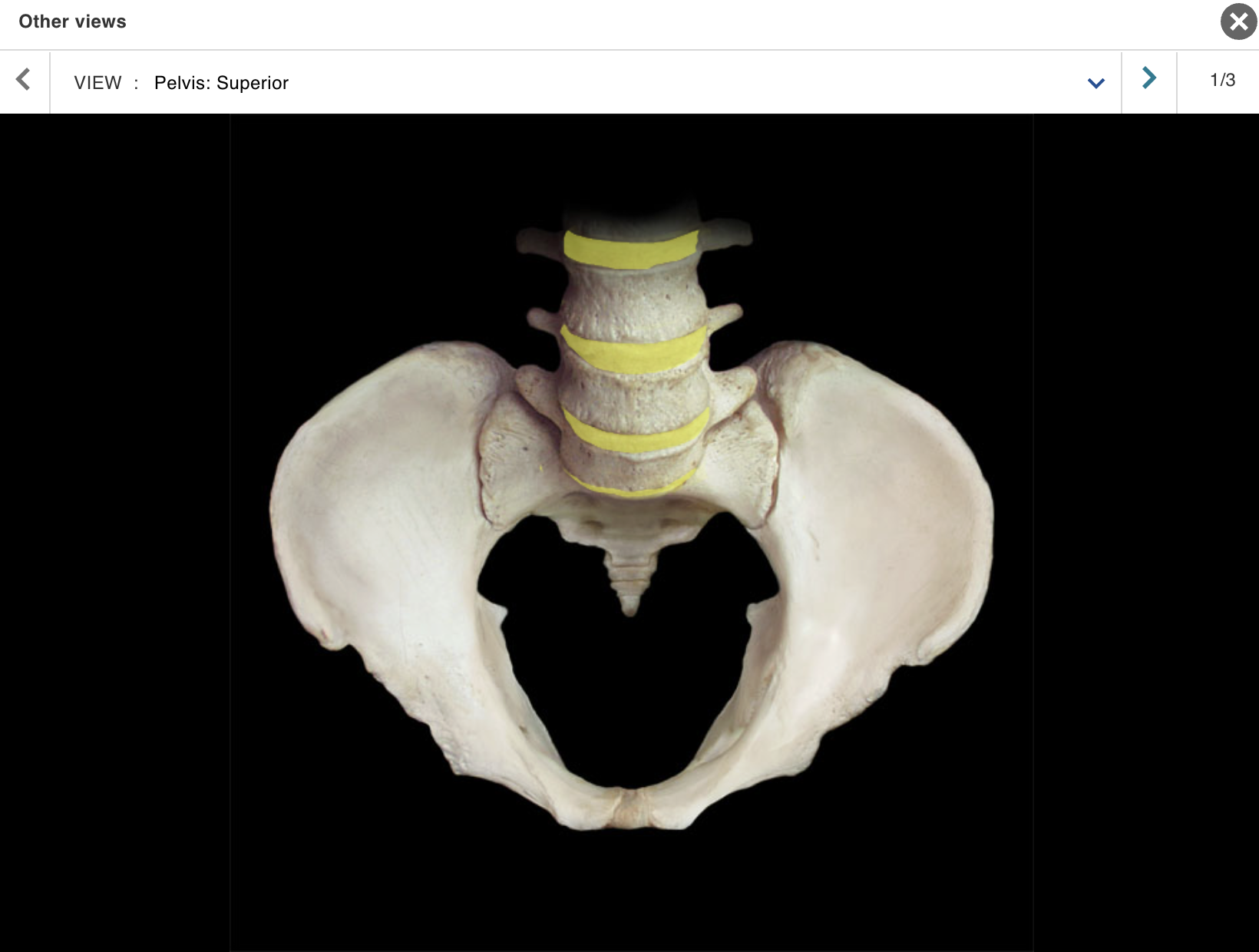
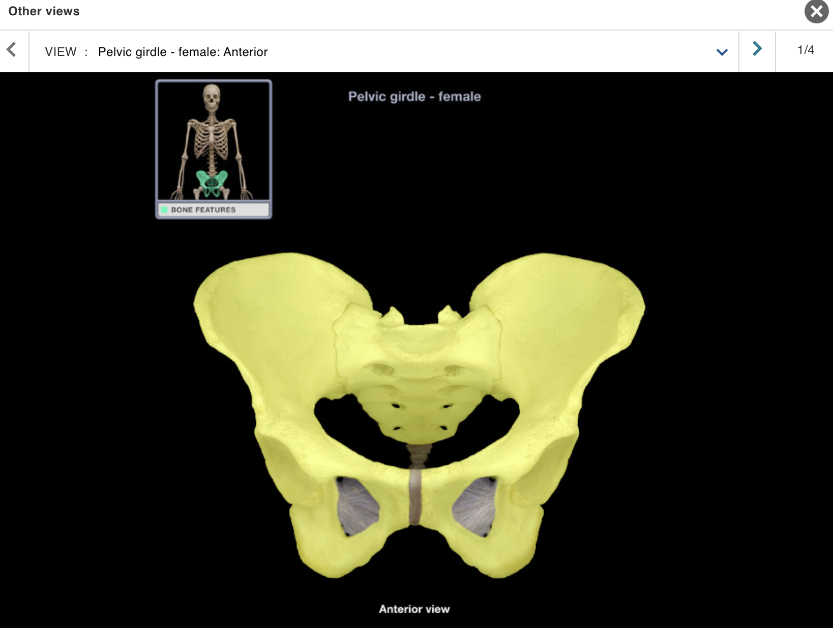
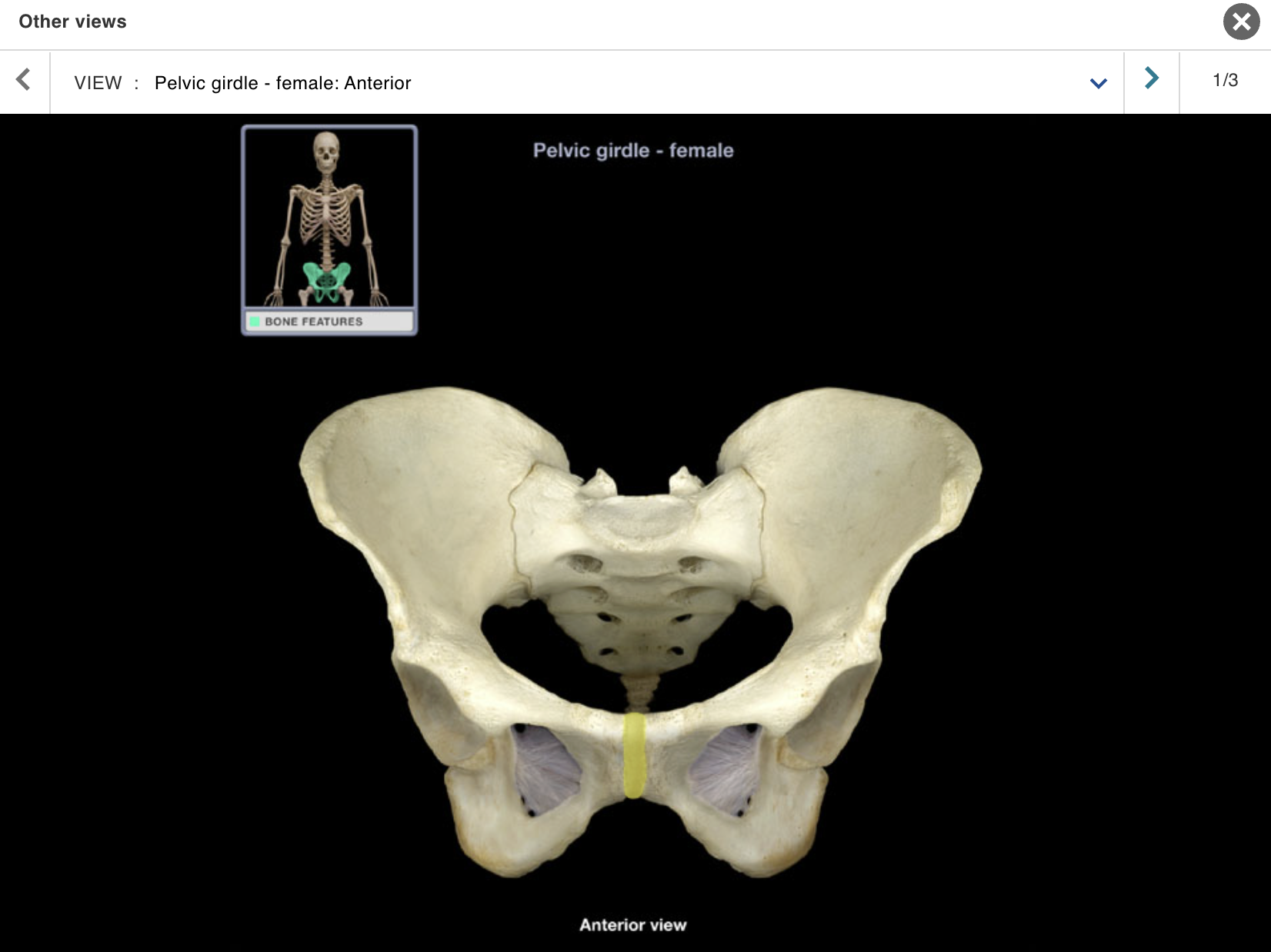
Pelvic girdle
Location:
• Pelvis
Description:
Hip bones (ilium, ischium, and pubis)
Sacrum
Comment:
joint
• Pelvic girdle articulates with axial skeleton only at sacroiliac
• Pelvic girdle sometimes considered to include hip bones only (i.e., sacrum not included)
• Hip bones also known as os coxae
Pubic symphysis
Location:
• Pelvis (anterior midline)
Description:
• Joint formed by two pubic bones and intervening fibrocartilage disc
Comment:
• In female, fibrocartilage softens in late pregnancy to allow slight separation of pubic bones
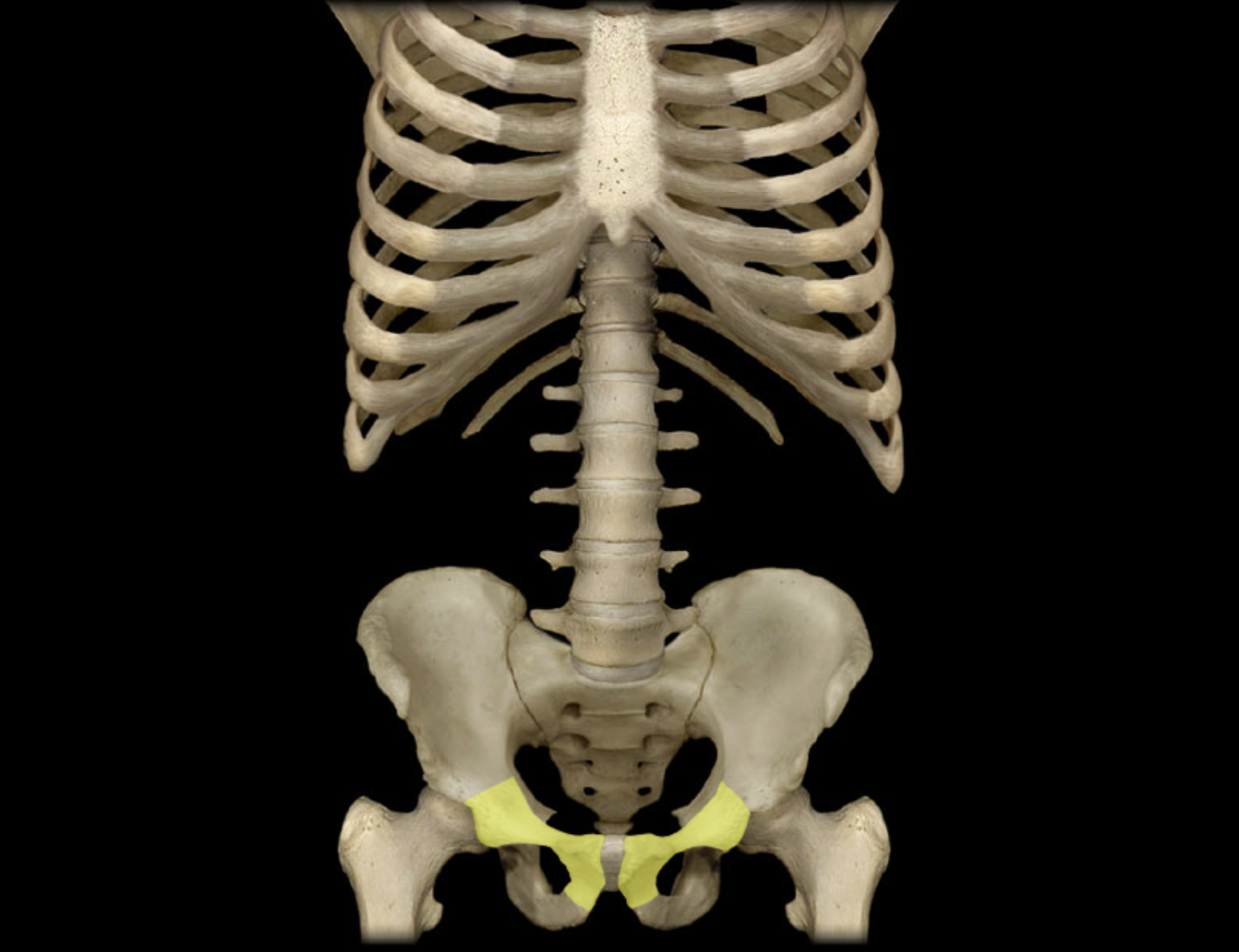
Pubis
Location:
• Pelvis
Description:
One of three coxal (hip) bones
Characteristic features include body and rami (superior and inferior)
Midline junction of pubic bones forms pubic symphysis
Contributes to acetabulum (hip joint socket), obturator foramen, and wall of lesser (true) pelvis
Comment:
Fused with ilium and ischium in adult to form coxal (hip) bone
Bony pelvis formed by paired hip bones and sacrum
Obturator foramen formed by rami of pubis and ischium
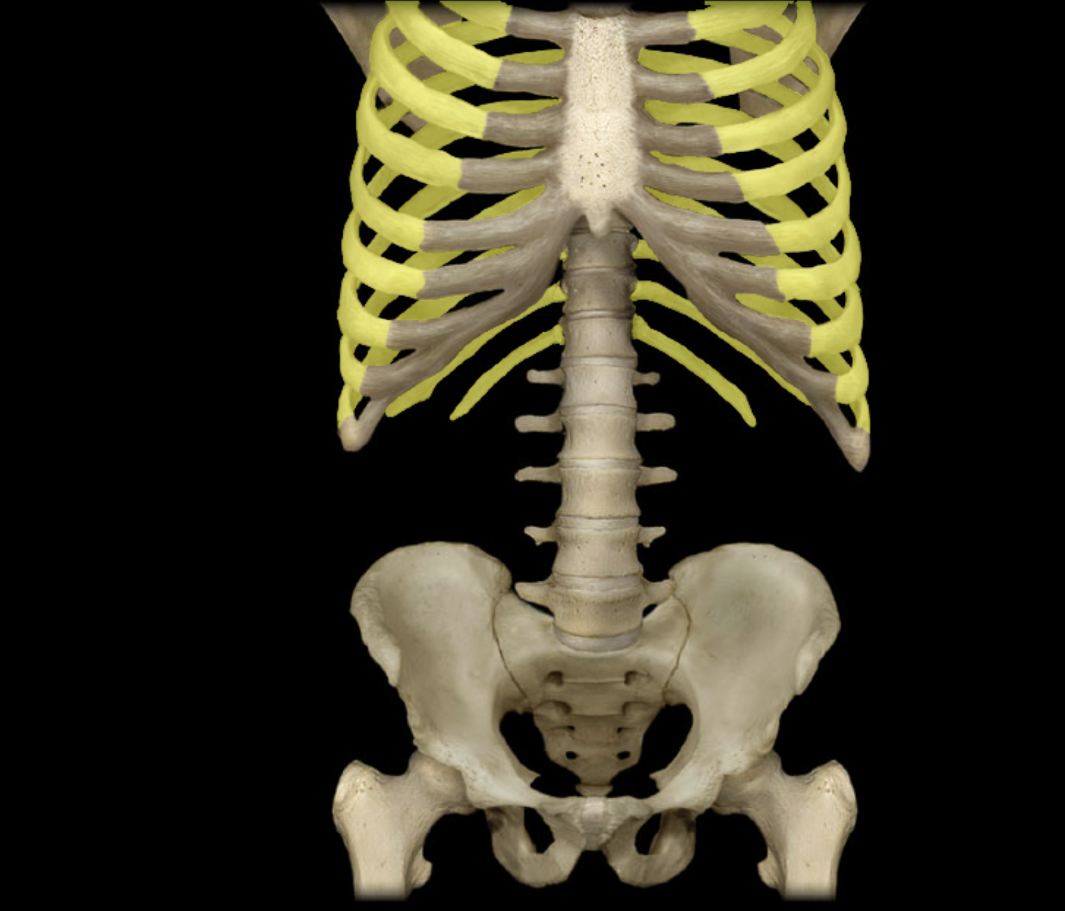
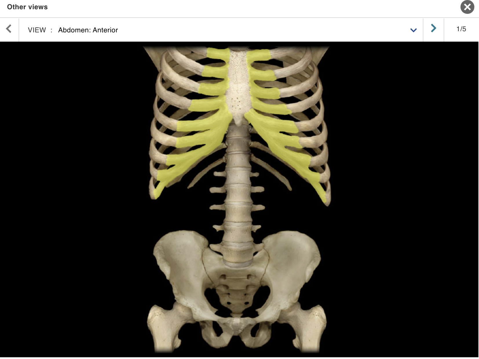
Ribs 2 - 12
Location:
• Thorax
Description:
Pairs of curved, flat bones
All ribs: articulate with thoracic vertebrae
True ribs (ribs 1-7): attached directly to sternum by costal cartilages
False ribs (ribs 8-10): attach indirectly to sternum via shared costal cartilages
Floating ribs (ribs 11-12): not attached to sternum
Comment:
• Alternate definition: some include the floating ribs (11-12) as a subcategory of false ribs
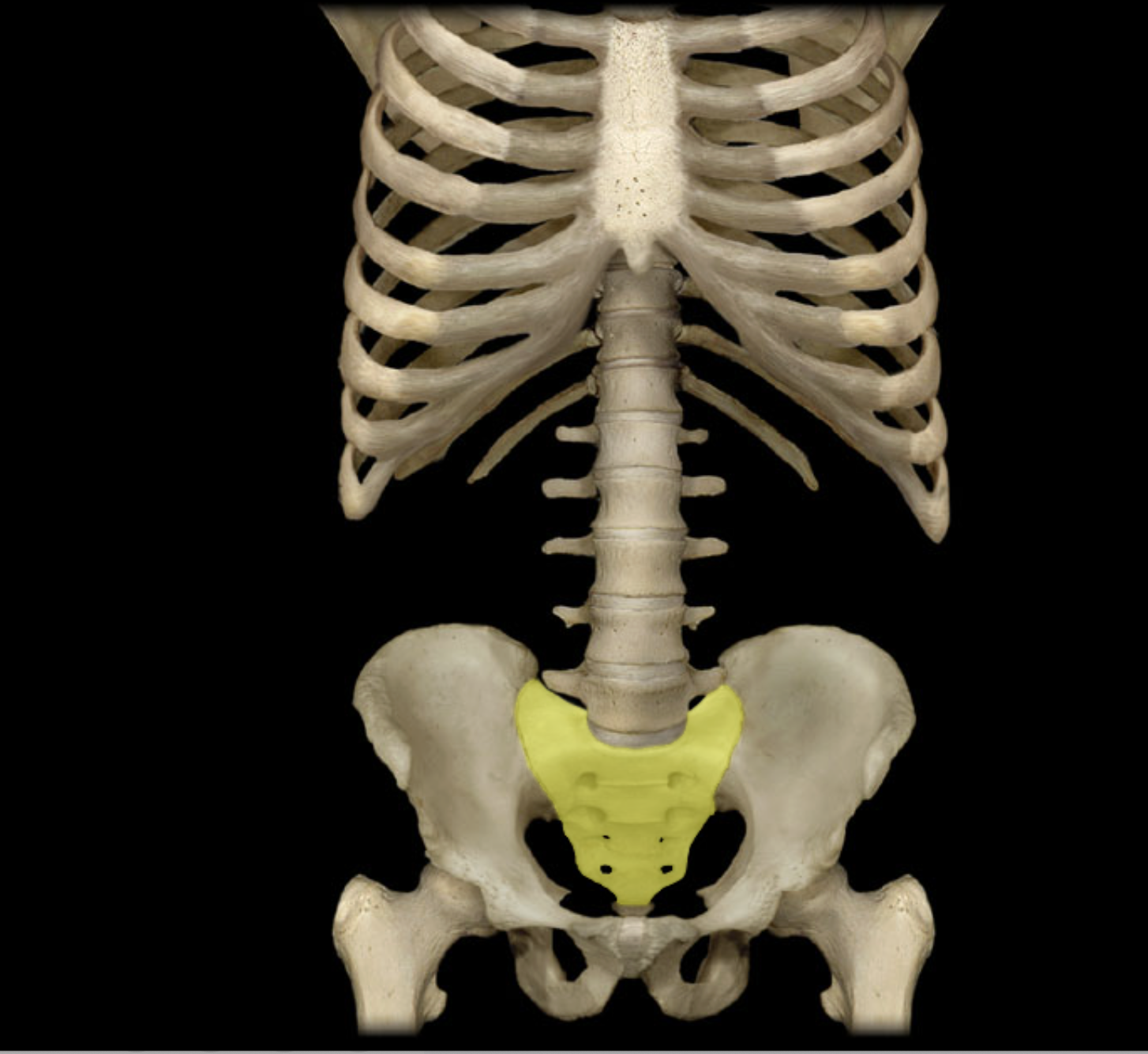
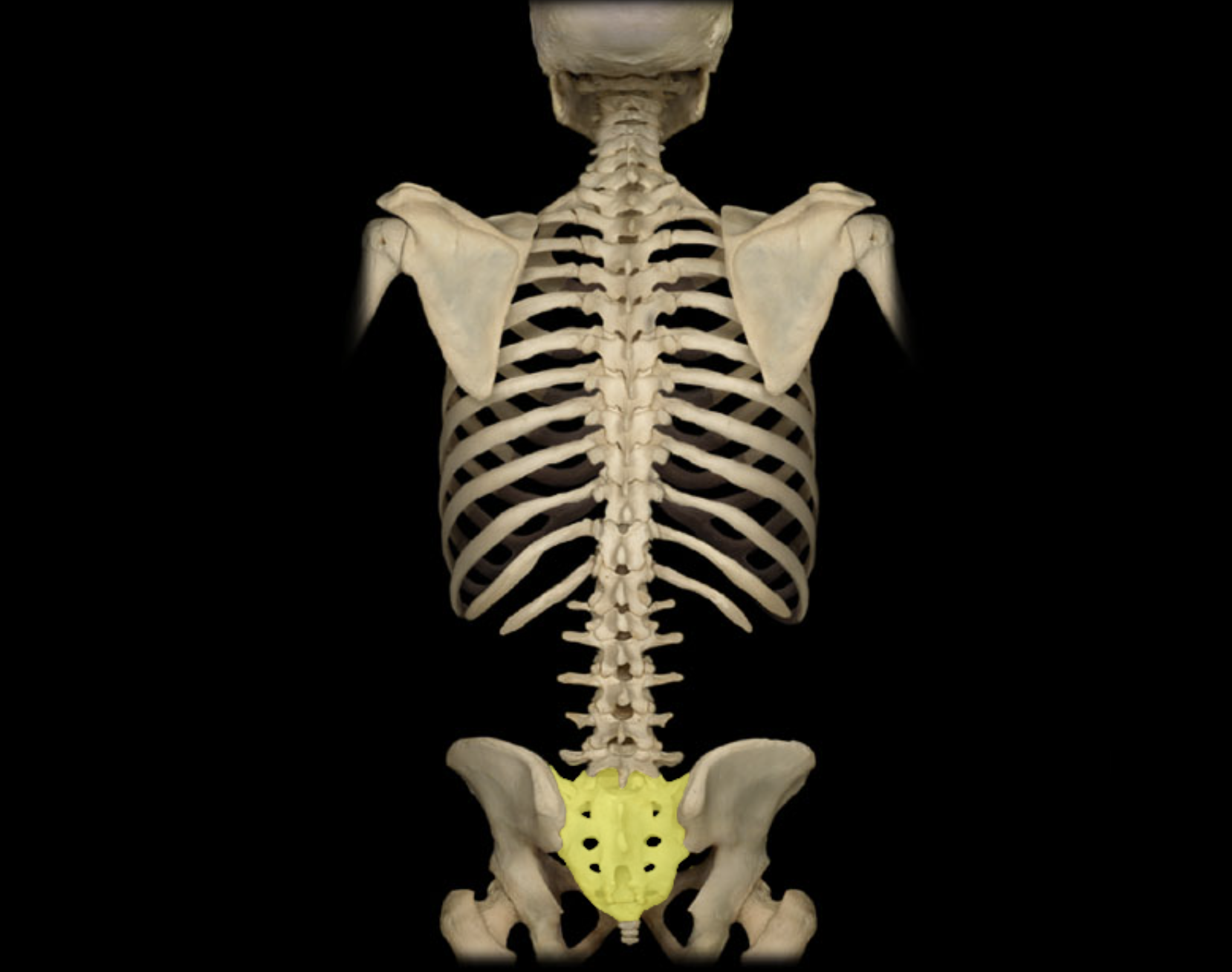
Sacrum
Location:
Lower back
Between L5 and Co1 vertebrae
Posterior wall of pelvis
Description:
Five fused vertebrae
Triangular bone wedged between hip bones
Comment:
• Sacral promontory (the prominent, projecting edge of base of sacrum formed by superior border of S1 vertebral body) is landmark for establishing female pelvic dimensions

Sternum
Location:
• Thorax (anterior midline)
Description:
Flat bone with three parts: manubrium, body, and xiphoid process
Characteristic features include jugular notch and sternal angle (angle of Louis)
Articulates with costal cartilages of true ribs (1-7), combined costal cartilages of false ribs (8-10), and clavicles
Also known as:
• "Breastbone"
Comment:
Floating ribs (ribs 11-12): not attached to sternum
Provides for attachment of sternocleidomastoid, pectoralis major, sternohyoid, and sternothyroid muscles
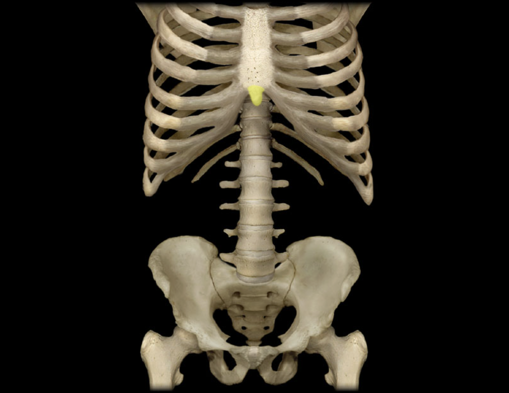
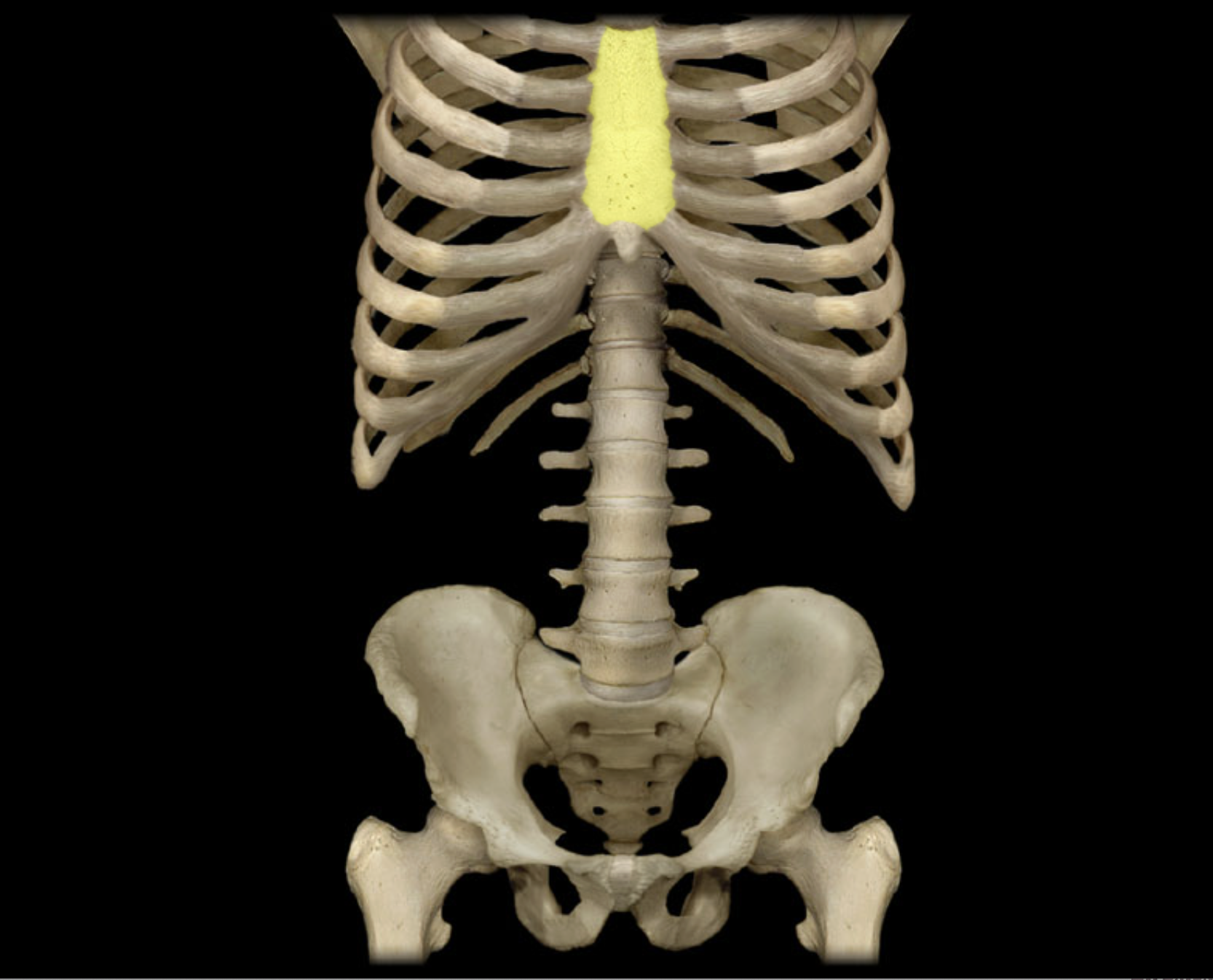
Xiphoid process
Location:
• Sternum (inferior part)
Description:
Midline shallow depression
Cartilage until approximately 5th decade of life
Comment:
Provides attachment for anterior abdominal wall muscles
Landmark for cardiopulmonary resuscitation (CPR)
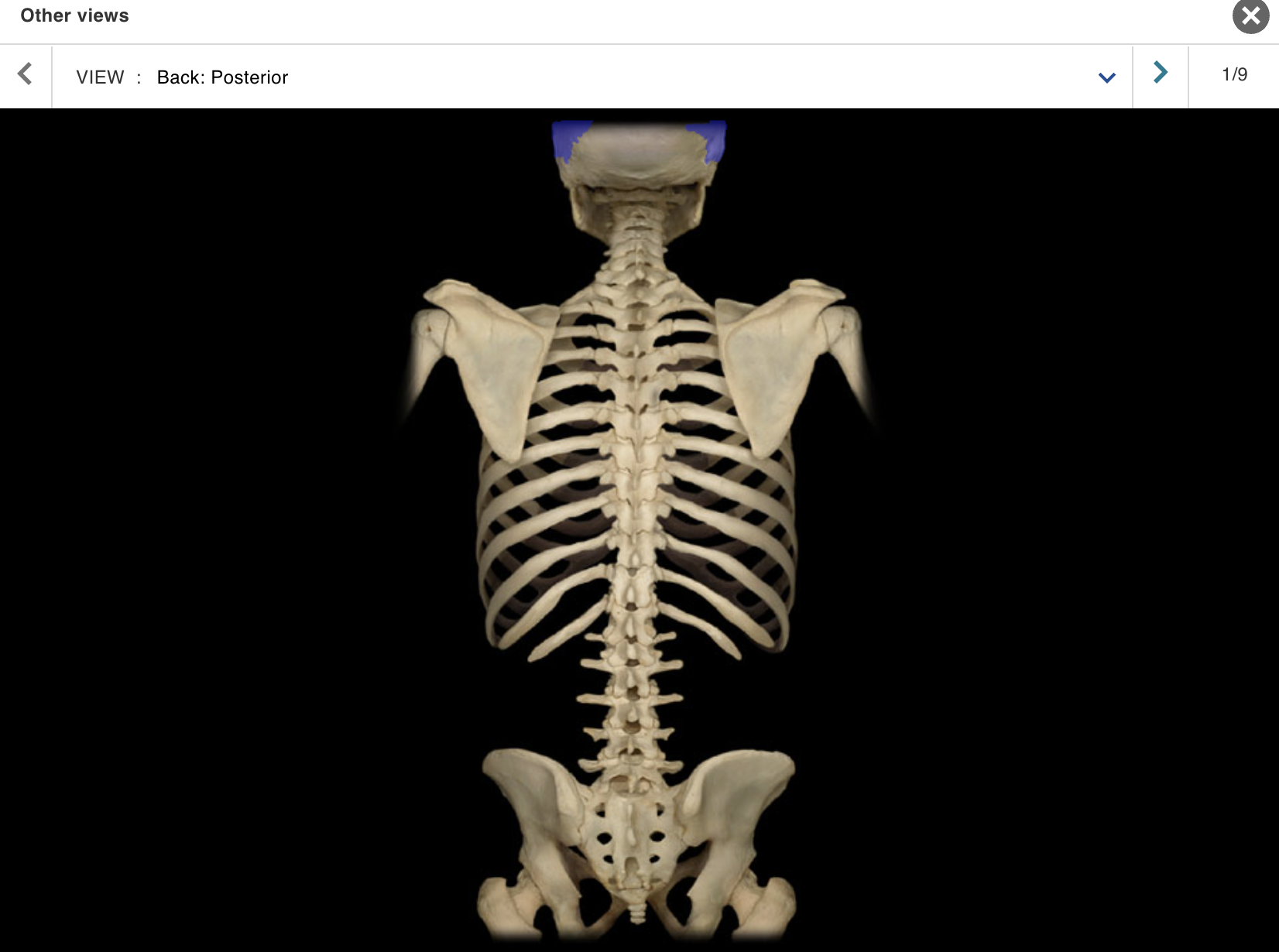
ACTIVITY 4: DISSECTION
MODULE: SKELETAL
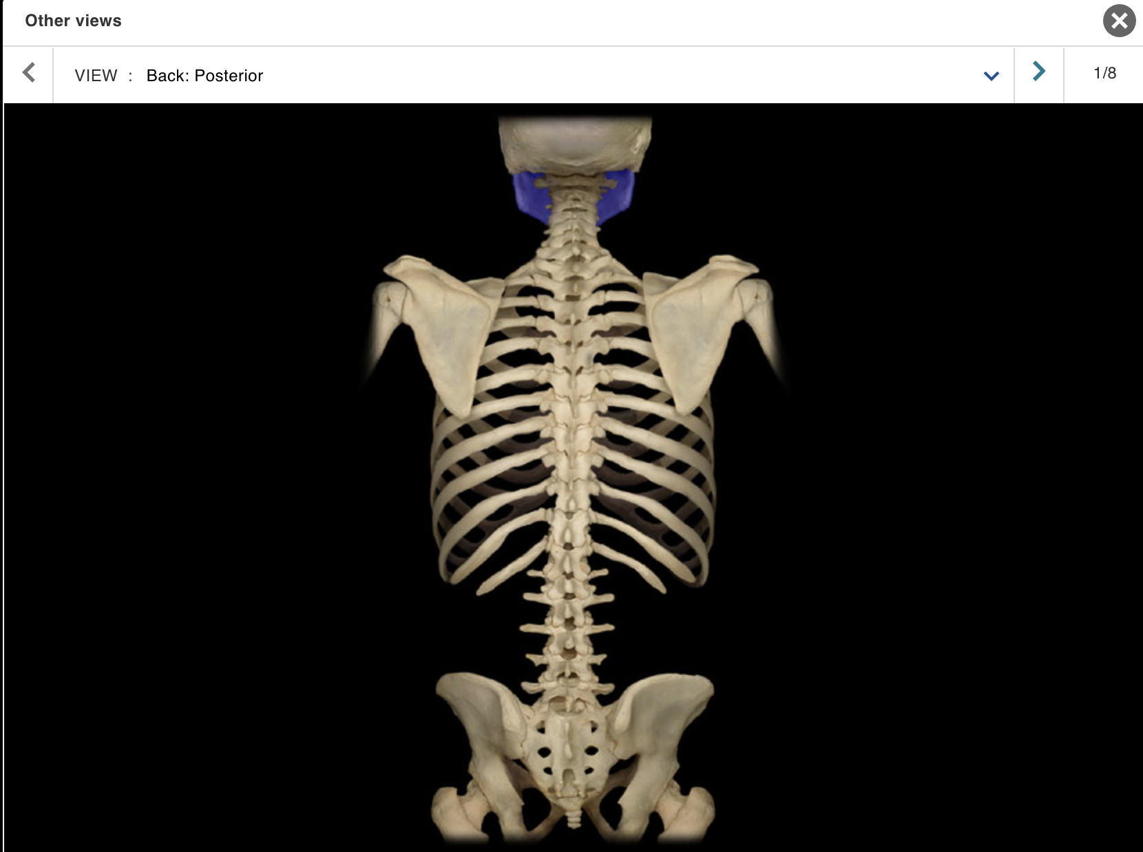
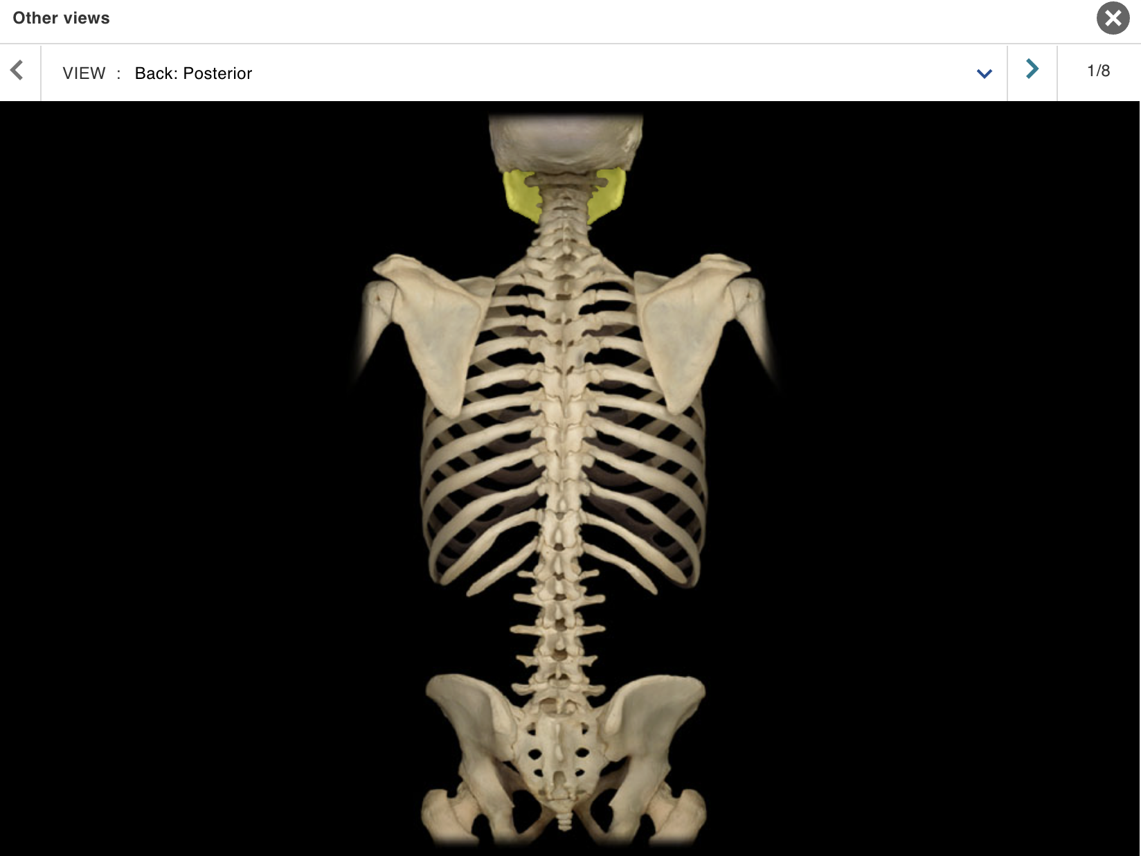
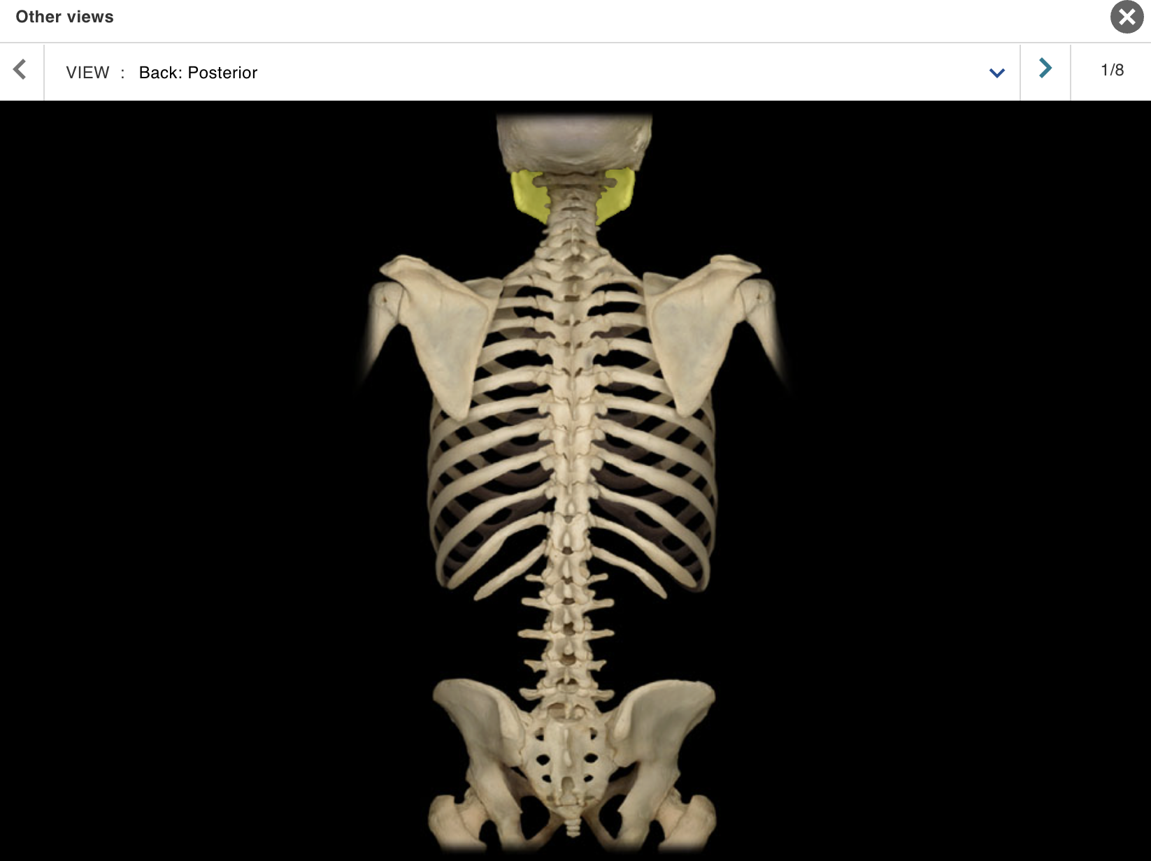
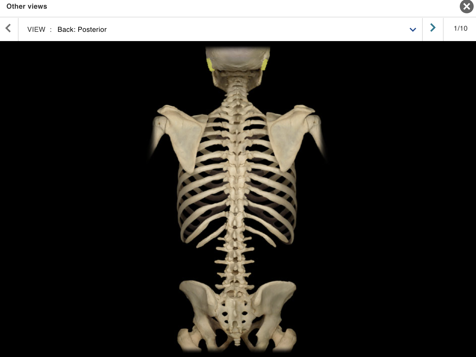
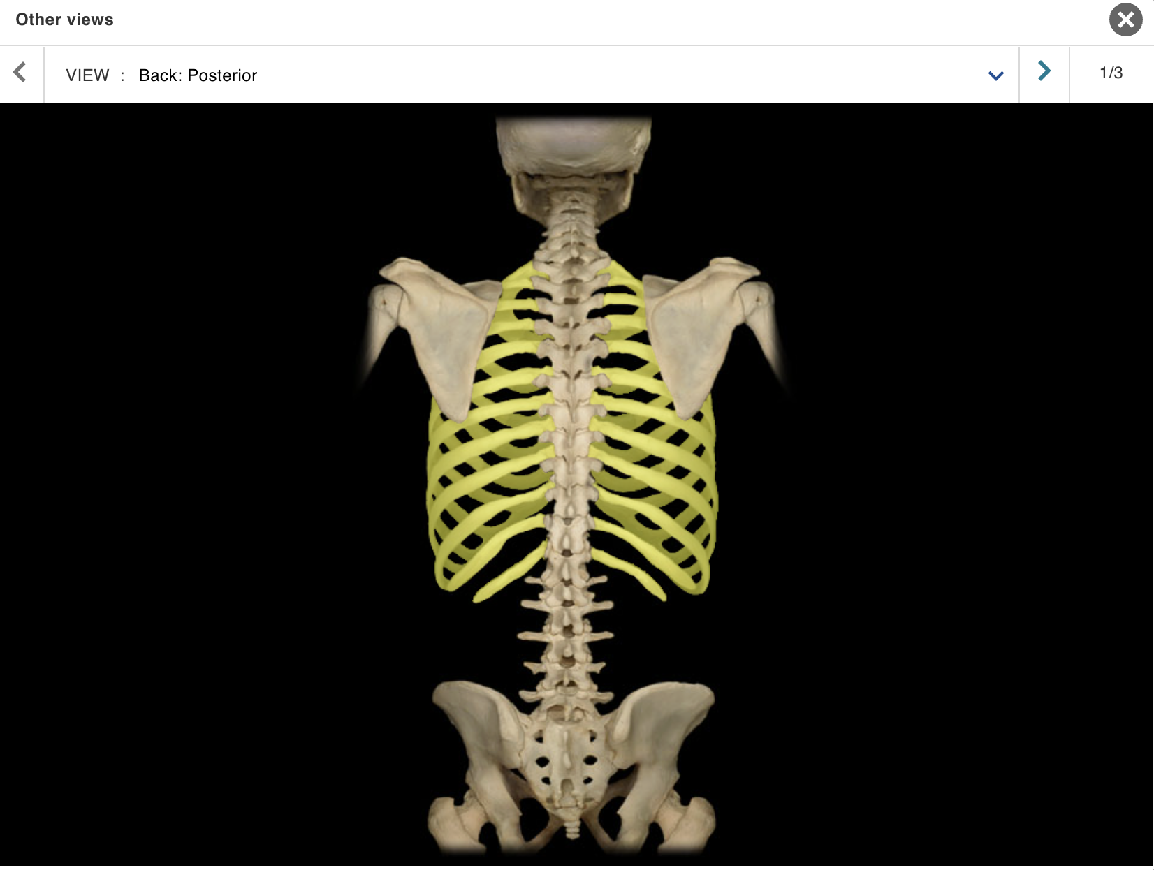
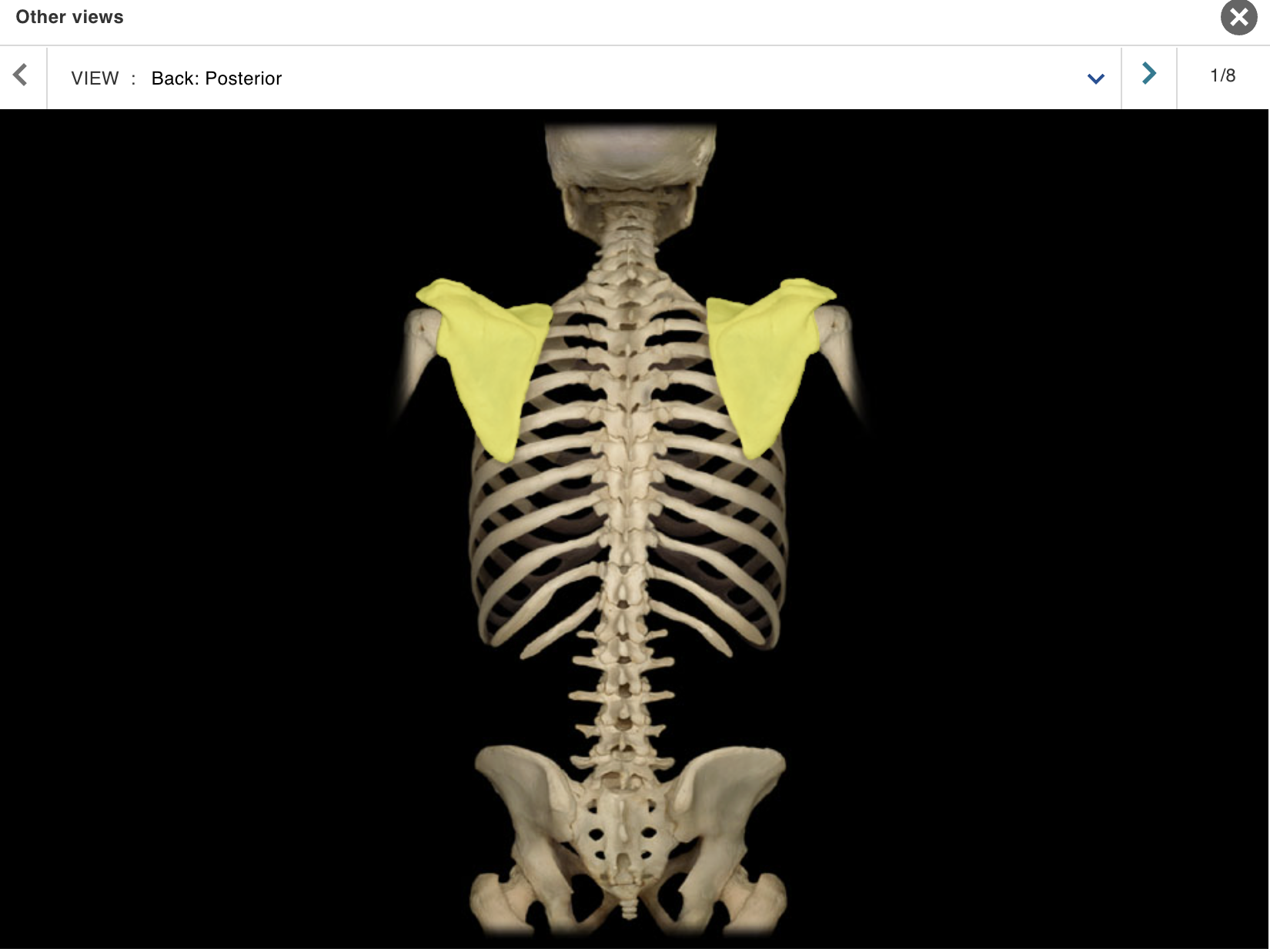
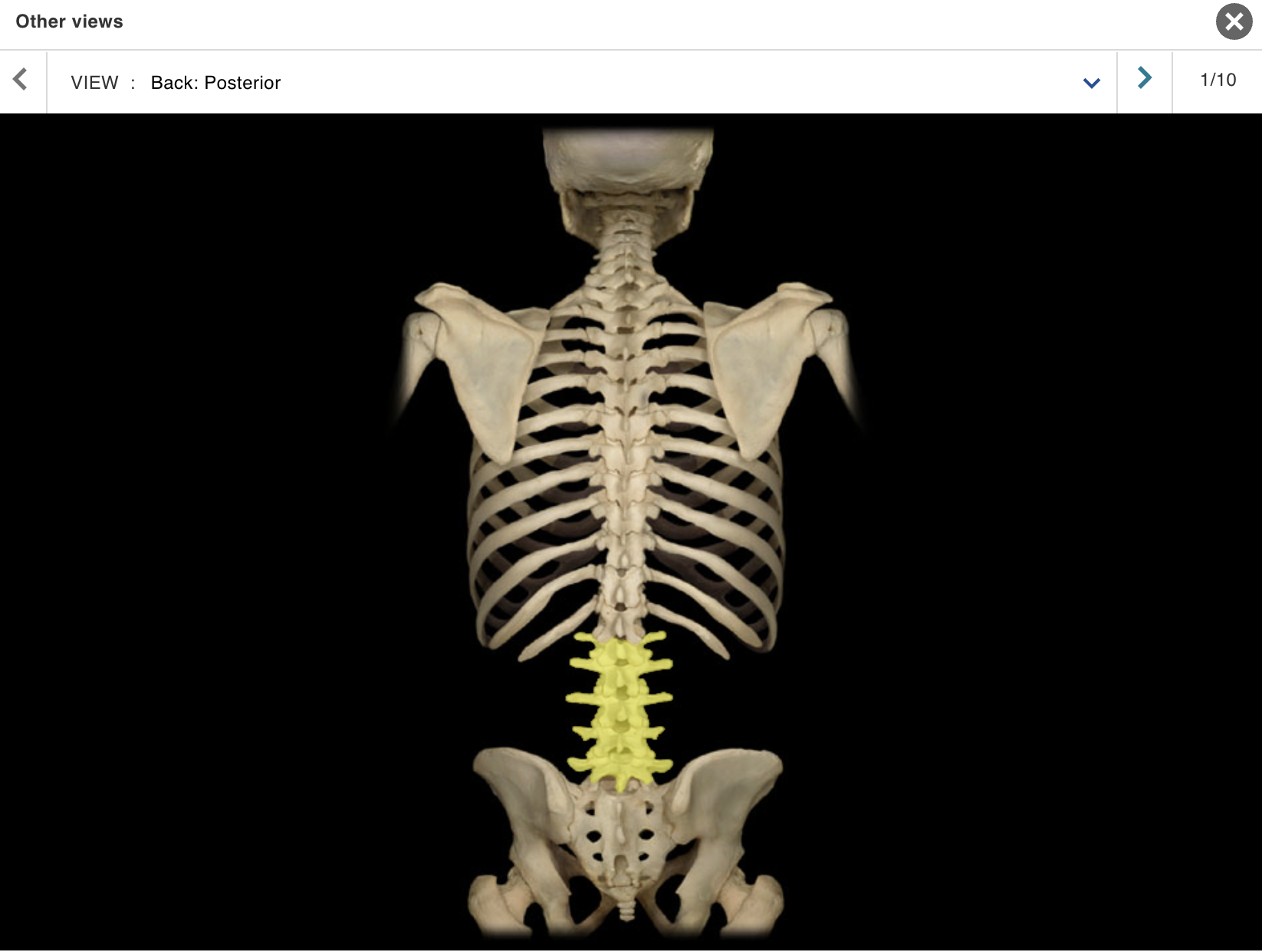
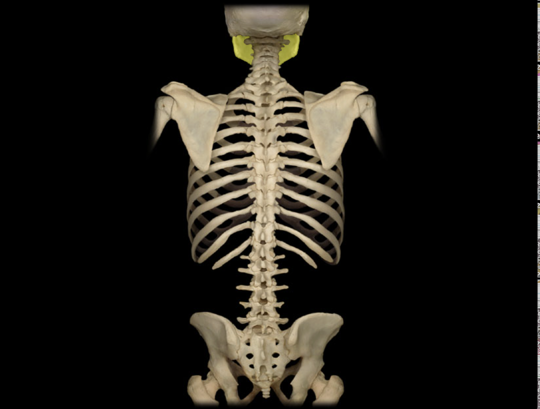
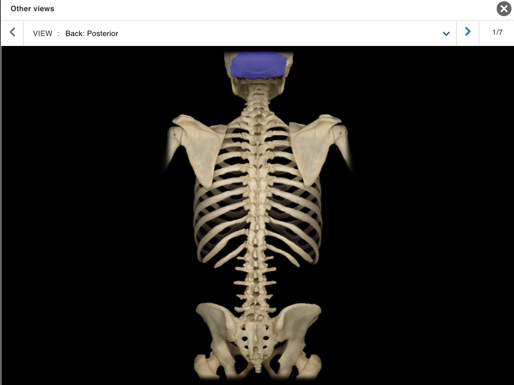
TOPIC: BACK / VIEW : POSTERIOR
Cervical vertebra
Соссух
lium
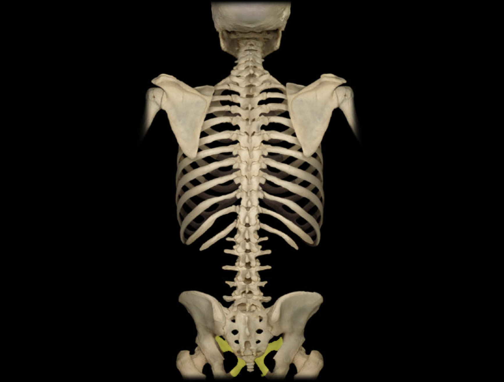
Ischium
Lumbar vertebra
Mandible
Occipital bone
Parietal bone
Pubis
Ribs 1 - 12
Sacrum
Scapula
Spinous process of lumbar vertebra
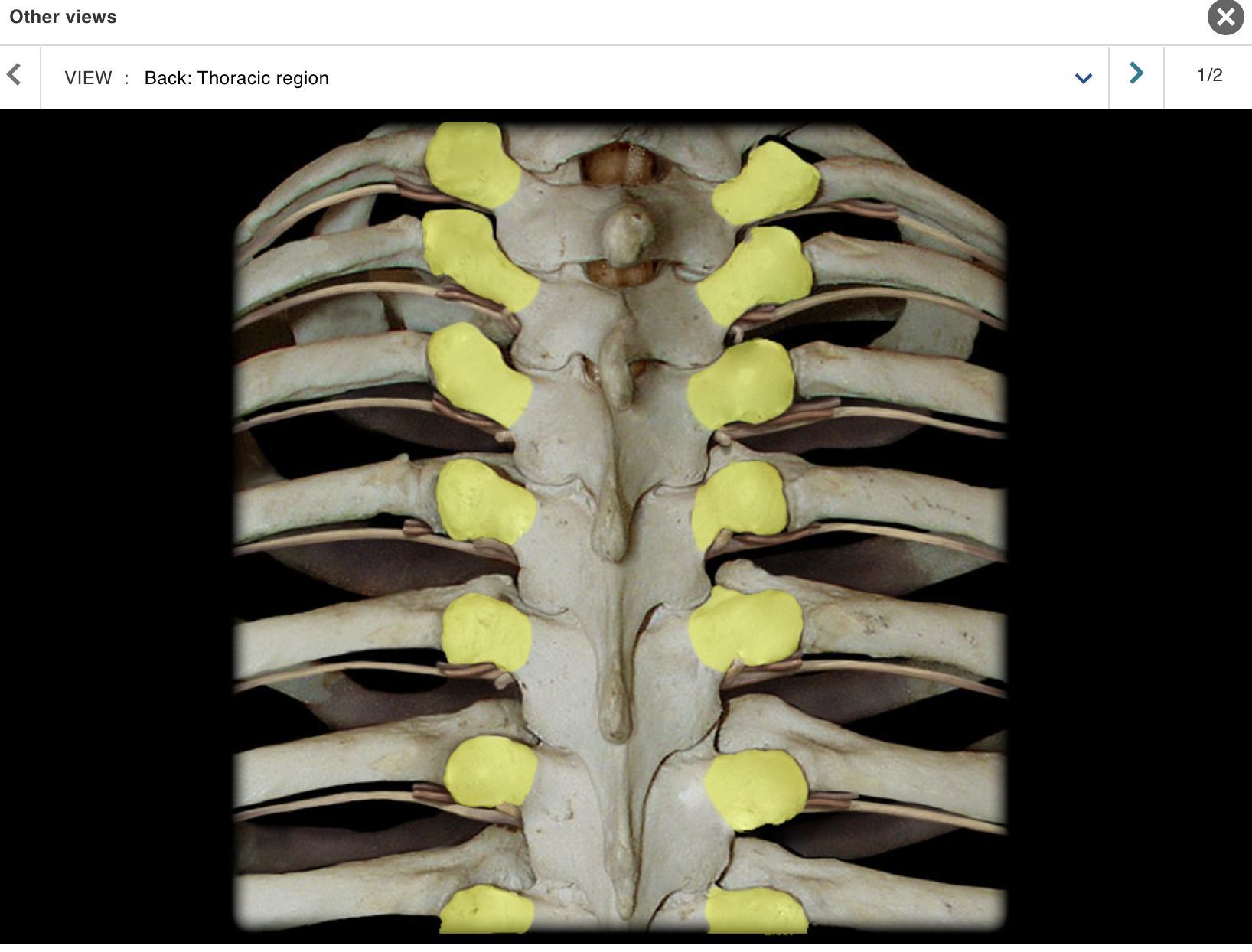
Spinous process of thoracic vertebra
Temporal bone
Thoracic vertebra
Transverse process of lumbar vertebra
Transverse process of thoracic vertebra
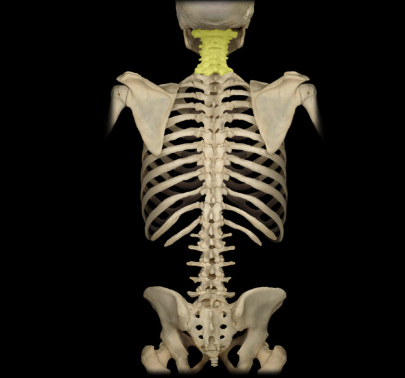
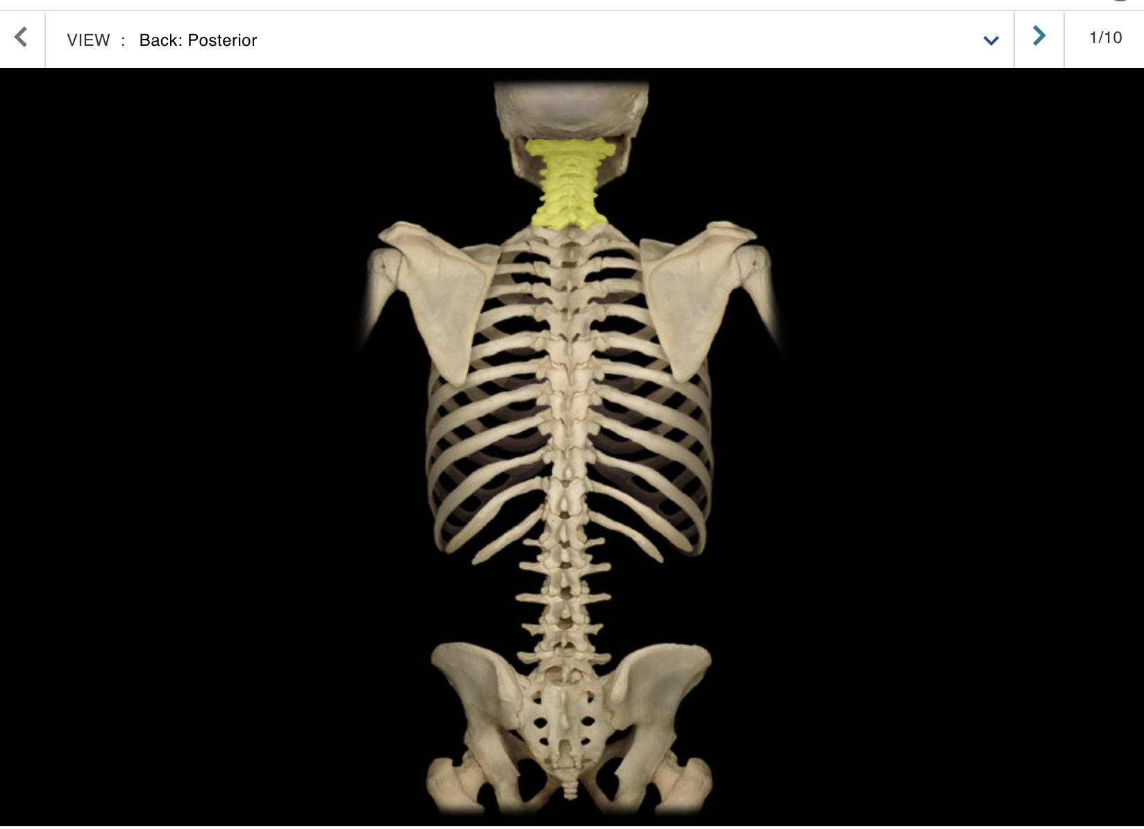
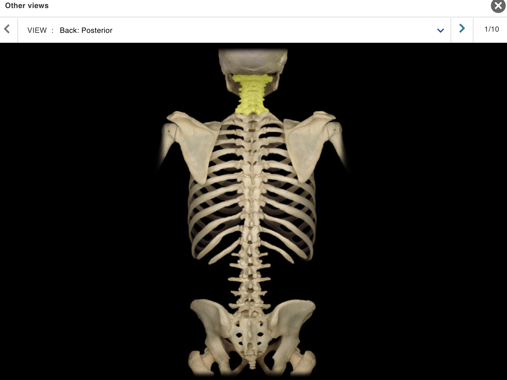
Cervical vertebra
Location:
Neck
Between occipital bone and T1 vertebra
Description:
Seven individual vertebrae
Characteristic features include transverse foramen and bifid
(split) spinous process on C3-C6
Comment:
• Atlas (C1 vertebra) articulates with skull
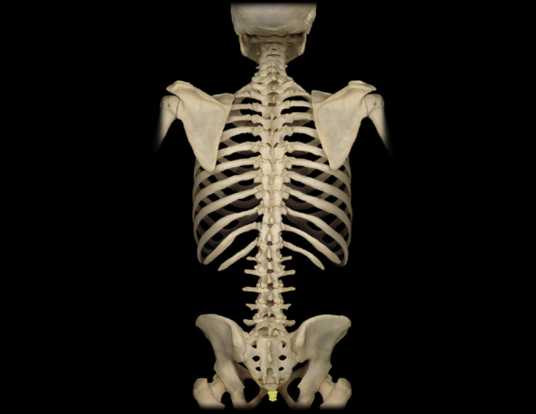
Соссух
Location:
Posterior pelvic wall
Lower back, inferior to S5 vertebra
Description:
Small, triangular bone
Consists of three to five, variably fused, poorly developed vertebrae
Also known as:
• "Tailbone"
Comment:
• Rudiment of the tail in other vertebrates
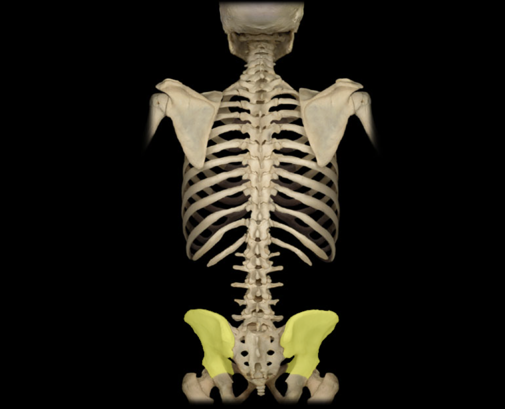
lium
Location:
• Pelvis
Description:
Largest of three coxal (hip) bones
Has large, wing-like superior extension (ala); the alae form bony walls of greater (false) pelvis
Contributes to acetabulum (hip joint socket) and wall of lesser (true) pelvis
Articulates with sacrum at sacroiliac joint
Comment:
Fused with ischium and pubis in adult to form coxal (hip) bone
Bony pelvis formed by paired hip bones and sacrum
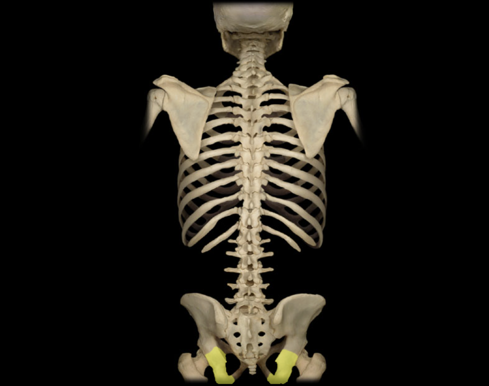
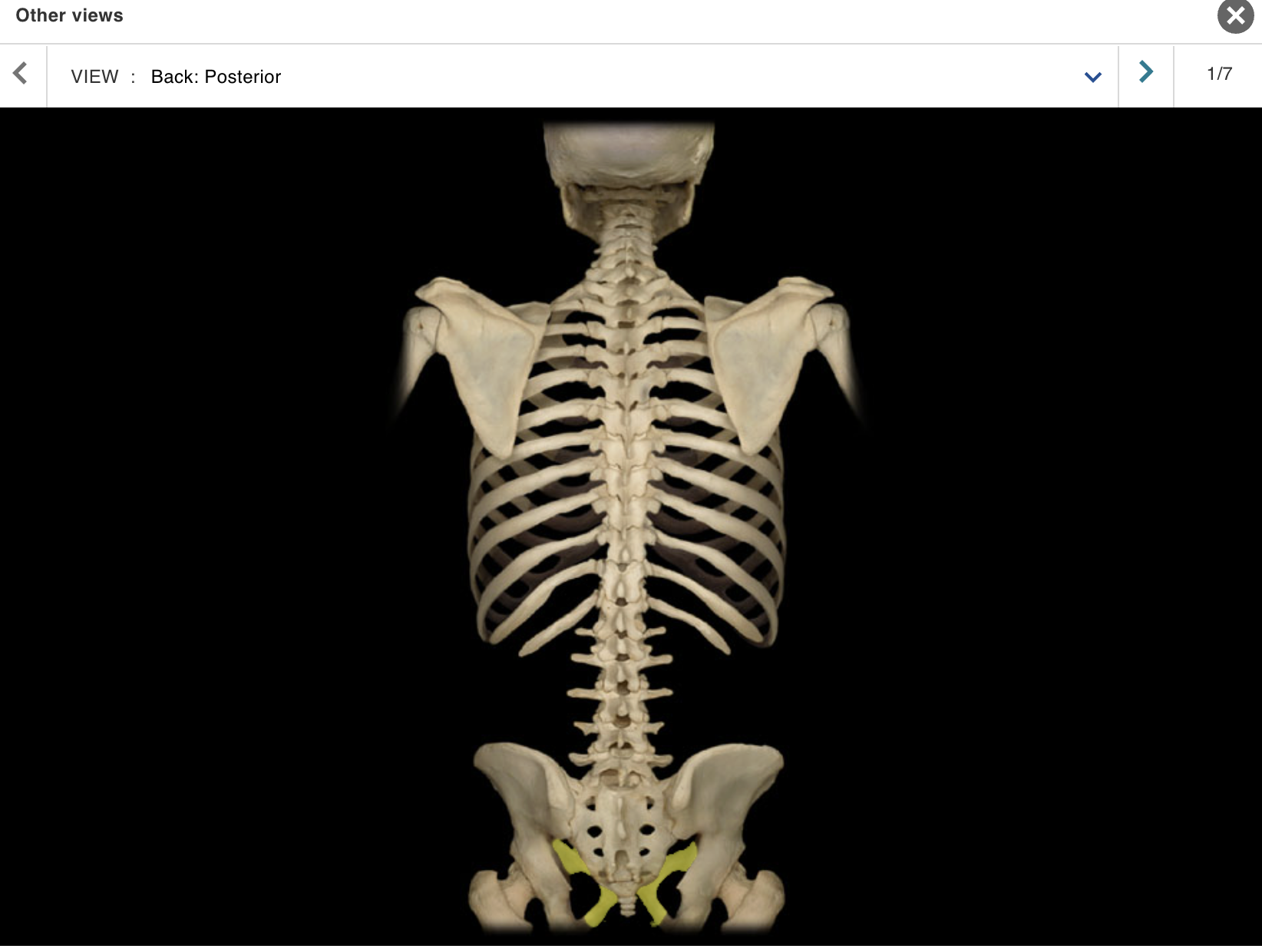
Ischium
Location:
• Pelvis
Description:
One of three coxal (hip) bones
Characteristic features include tuberosity and spine
Contributes to acetabulum (hip joint socket), obturator foramen, and wall of lesser (true) pelvis
Comment:
Fused with ilium and pubis in adult to form coxal (hip) bone
Bony pelvis formed by paired hip bones and sacrum
Obturator foramen formed by rami of pubis and ischium
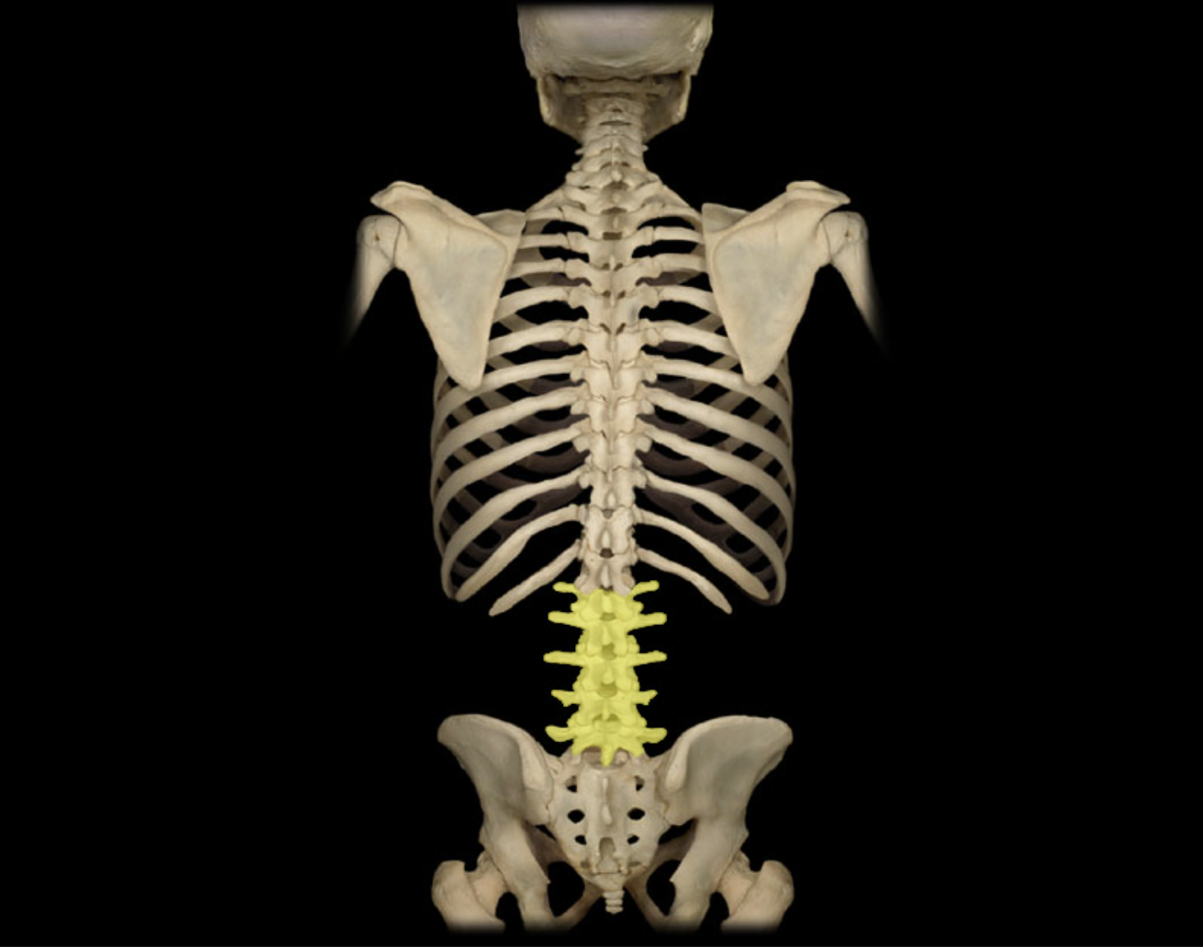
Lumbar vertebra
Location:
Lower back
Between T12 and S1 vertebrae
Description:
Five individual vertebrae
Characteristic features include large size, kidney bean-shaped body, and a thick, blunt spinous process
Comment:
Bodies arranged to form prominent anterior convexity (lumbar curvature; also known as lumbar lordosis, which can be accentuated pathologically)
Intervertebral discs between lumbar vertebrae most commonly herniate ("slipped-disk")
Mandible
Location:
• Skull (anterior)
Description:
U-shaped bone
Each side consists of body (horizontal) and ramus (vertical) with coronoid and condylar processes
Mental protuberance forms point of chin
Contains alveoli ("sockets") for teeth
Also known as:
• "Lower jaw"
Comment:
• Contributes to temporomandibular joint (TMJ)
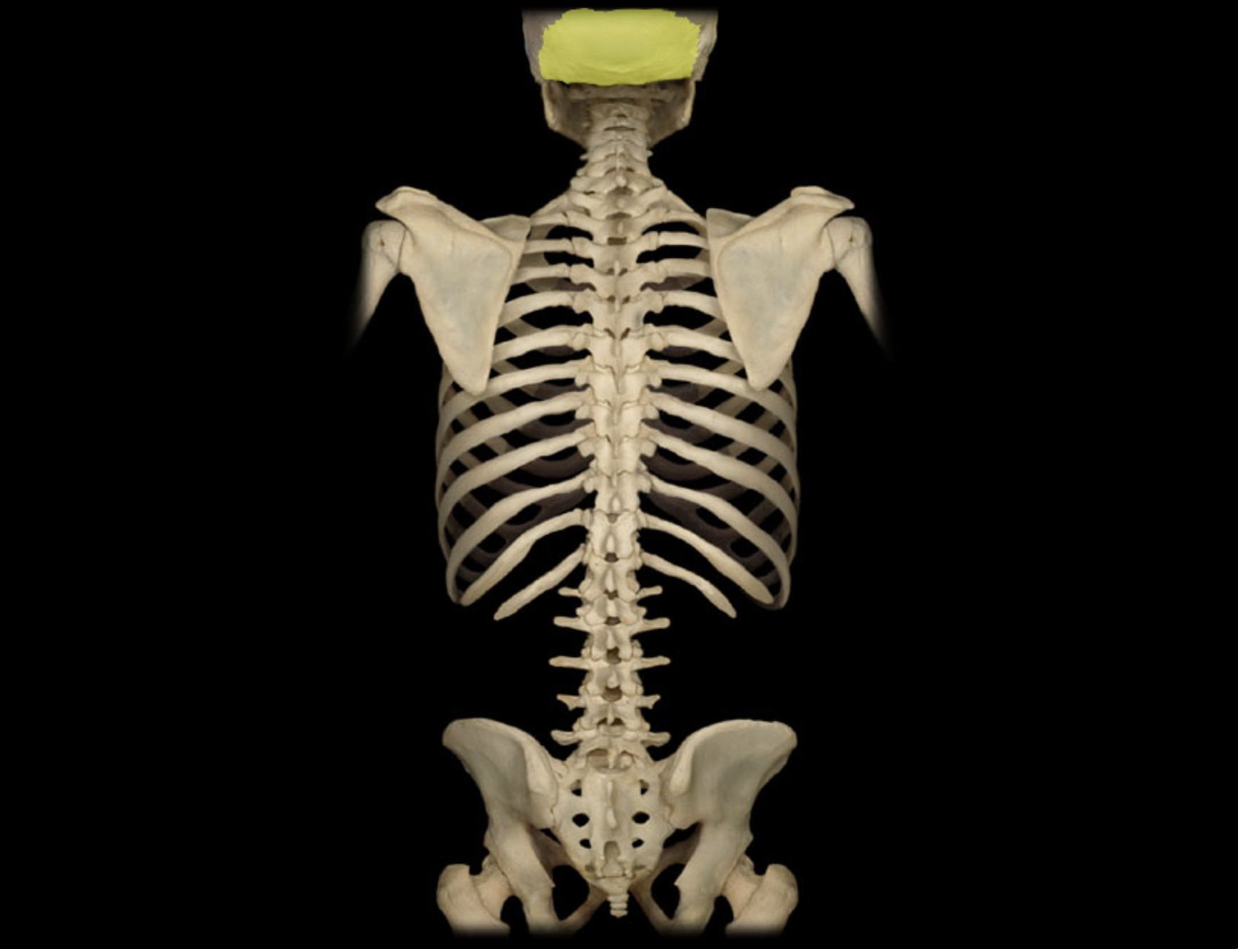
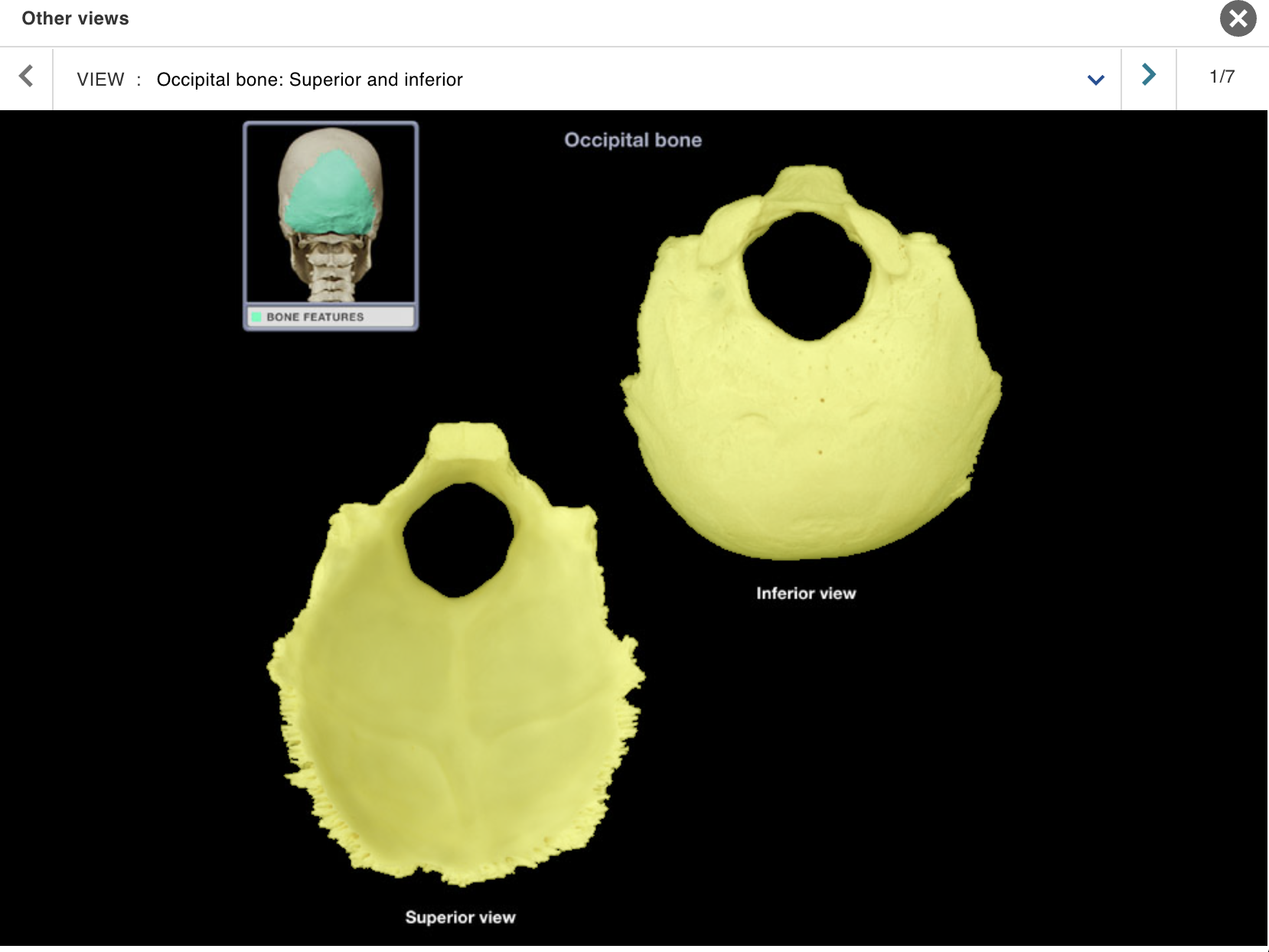
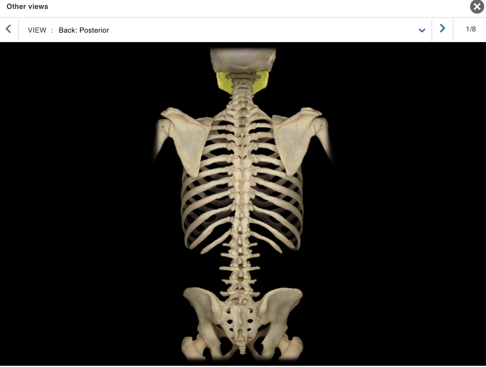
Occipital bone
Location:
• Skull (posterior and inferior)
Description:
Irregular-shaped bone
Contains foramen magnum in central portion
Comment:
Forms most of posterior skull
Forms most of posterior cranial fossa
Articulates with parietal bone at lambdoid suture, temporal bone at occipitomastoid and other sutures, and sphenoid bone at spheno-occipital synchondrosis
Articulates with atlas (C1 vertebra)
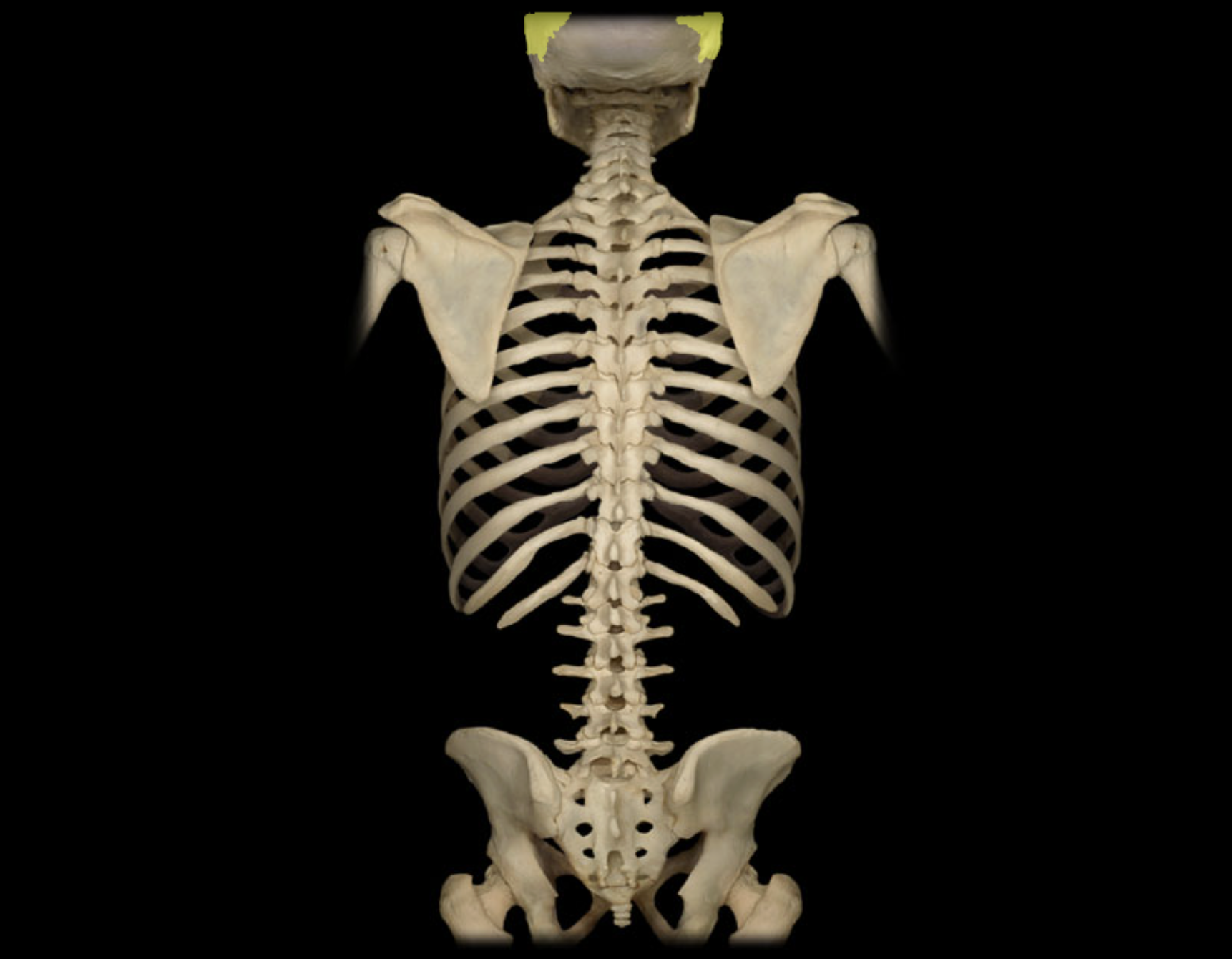
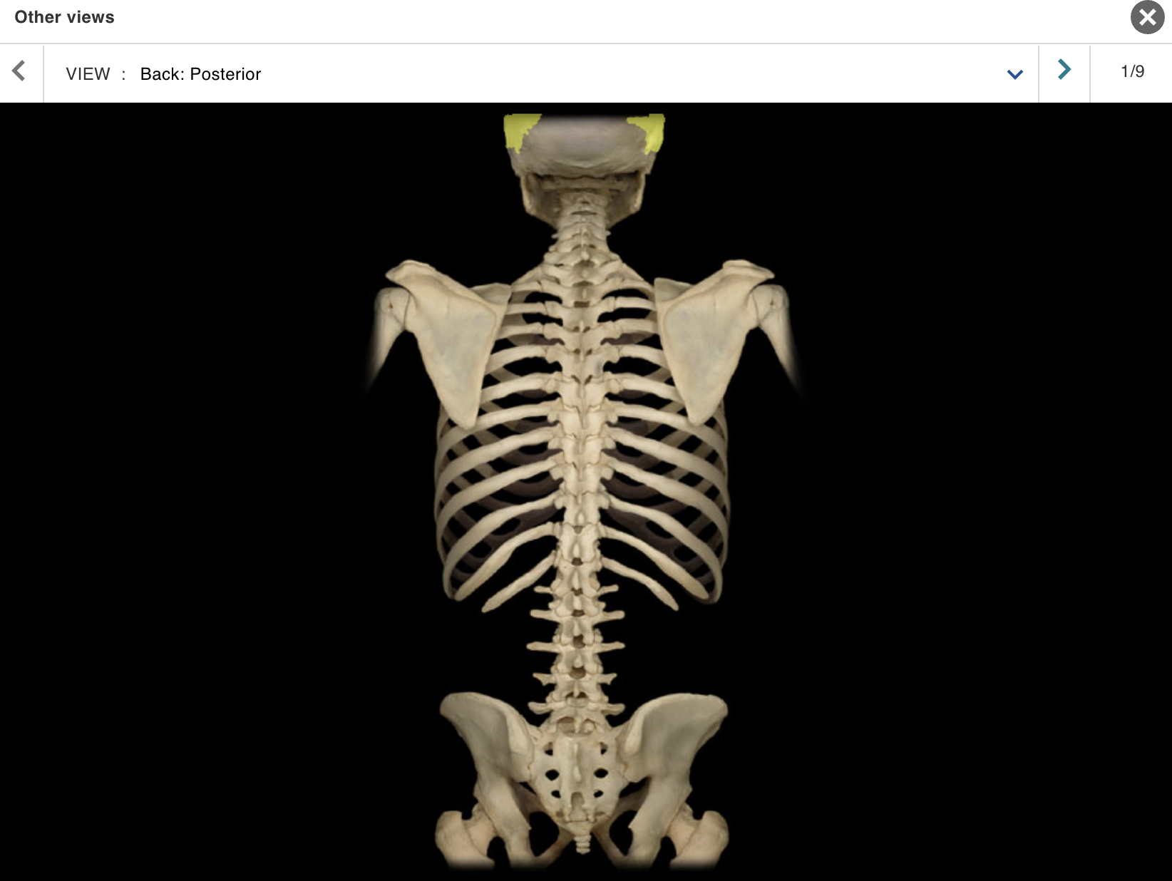
Parietal bone
Location:
• Skull (lateral)
Description:
Paired, flat bone
Forms most of lateral skull
Comment:
Parietal bones articulate at sagittal suture (midline)
Articulates with frontal bone at coronal suture, occipital bone at lambdoid suture, and temporal bone at squamosal suture
Pubis
Location:
• Pelvis
Description:
One of three coxal (hip) bones
Characteristic features include body and rami (superior and inferior)
Midline junction of pubic bones forms pubic symphysis
Contributes to acetabulum (hip joint socket), obturator foramen, and wall of lesser (true) pelvis
Comment:
Fused with ilium and ischium in adult to form coxal (hip) bone
Bony pelvis formed by paired hip bones and sacrum
Obturator foramen formed by rami of pubis and ischium
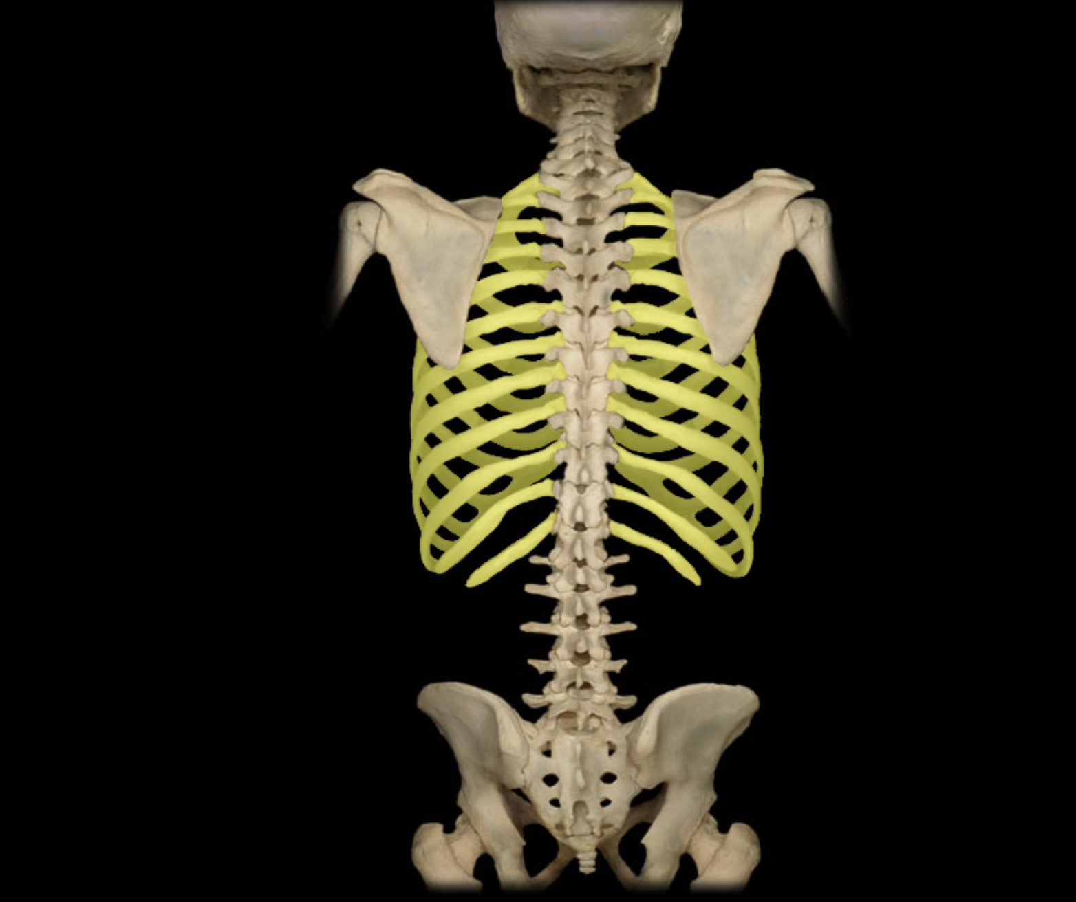
Ribs 1 - 12
Location:
• Thorax
Description:
Twelve pairs of curved, flat bones
All ribs: articulate with thoracic vertebrae
True ribs (ribs 1-7): attached directly to sternum by costal cartilages
False ribs (ribs 8-10): attach indirectly to sternum via shared costal cartilages
Floating ribs (ribs 11-12): not attached to sternum
Comment:
• Alternate definition: some include the floating ribs (11-12) as a subcategory of false ribs
Sacrum
Location:
Lower back
Between L5 and Co1 vertebrae
Posterior wall of pelvis
Description:
Five fused vertebrae
Triangular bone wedged between hip bones
Comment:
• Sacral promontory (the prominent, projecting edge of base of sacrum formed by superior border of S1 vertebral body) is landmark for establishing female pelvic dimensions
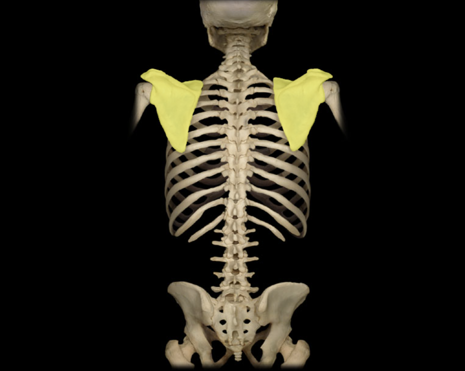
Scapula
Location:
Posterior thorax
Overlies ribs 2-7
Description:
Large, triangular, flat bone
Characteristic features include spine, acromion, coracoid process, and glenoid cavity
Spinous process of lumbar vertebra
Location:
• Vertebrae (posterior aspect)
Description:
Unpaired, posterior projection from midline of vertebral arch
Has characteristic thick, blunt form
Comment:
Spinous process present on all vertebrae except the atlas (C1 vertebra) and coccygeal vertebrae
Provides attachment for muscles and ligaments
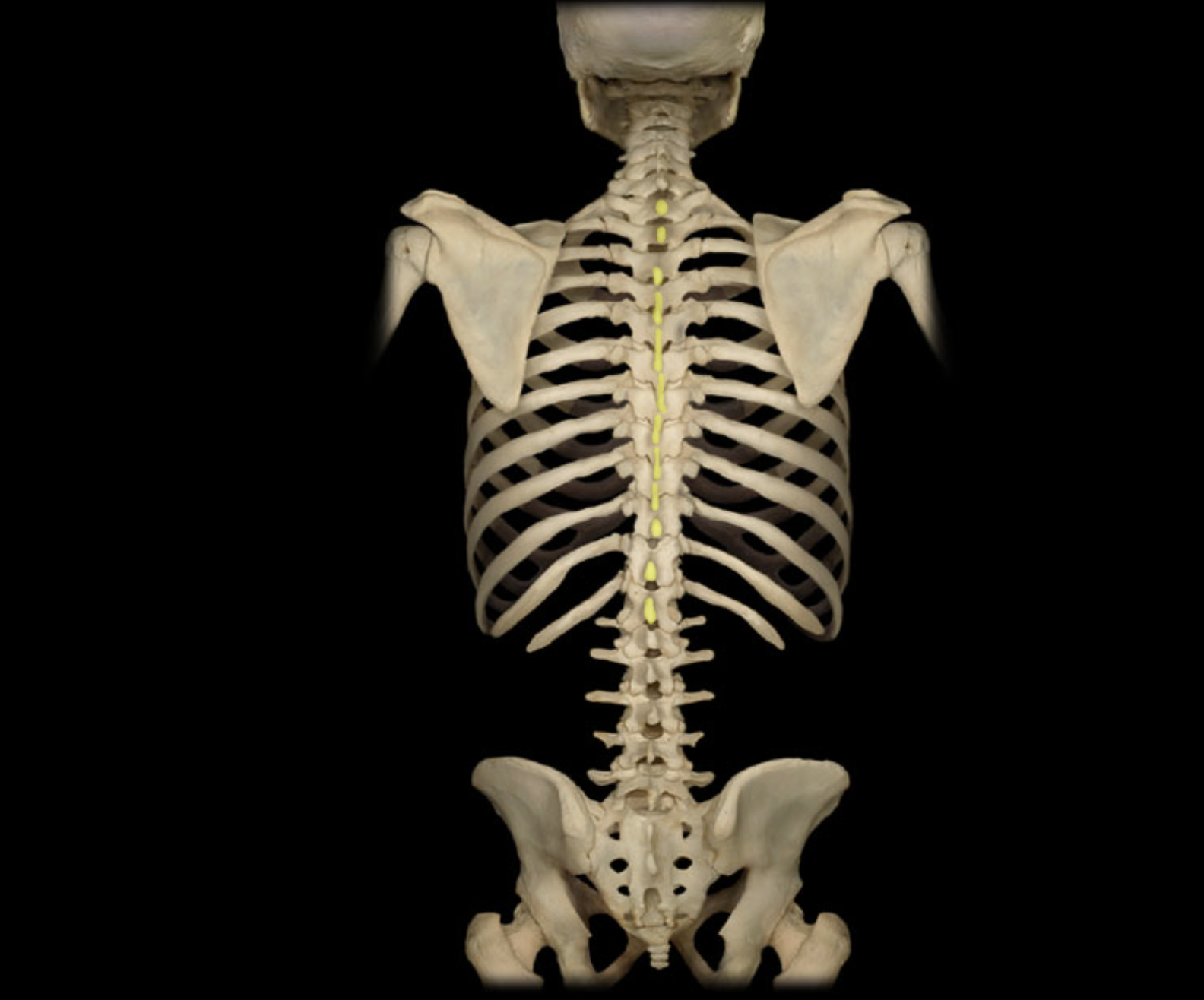
Spinous process of thoracic vertebra
Location:
• Thoracic vertebra
Description:
Unpaired posterior midline projection from vertebral arch
Has characteristic long, slender form
Processes of inferior thoracic vertebrae directed inferiorly
Comment:
• Spinous process present on all vertebrae except atlas (C1 vertebra) and coccygeal vertebrae
• Provides attachment for muscles and ligaments
Temporal bone
Location:
• Skull (lateral and inferior)
Description:
Paired, irregular-shaped, flat bone
Consists of squamous, petrous, mastoid, and tympanic parts
Comment:
Forms part of lateral skull (temple)
Forms inferior and lateral walls of middle cranial fossa
Articulates with sphenoid bone at sphenosquamosal suture, occipital bone at lambdoid suture, and parietal bone at squamosal suture
Has mandibular fossa that forms temporomandibular joint with
head of mandibleForms zygomatic process that forms zygomatic arch with temporal process of zygomatic bone
Thoracic vertebra
Location:
Trunk
Between C7 and L1 vertebrae
Description:
12 individual vertebrae
Characteristic features include costal demifacets (or facets) for articulation with head of rib, spinous process slopes inferiorly, and heart-shaped vertebral body
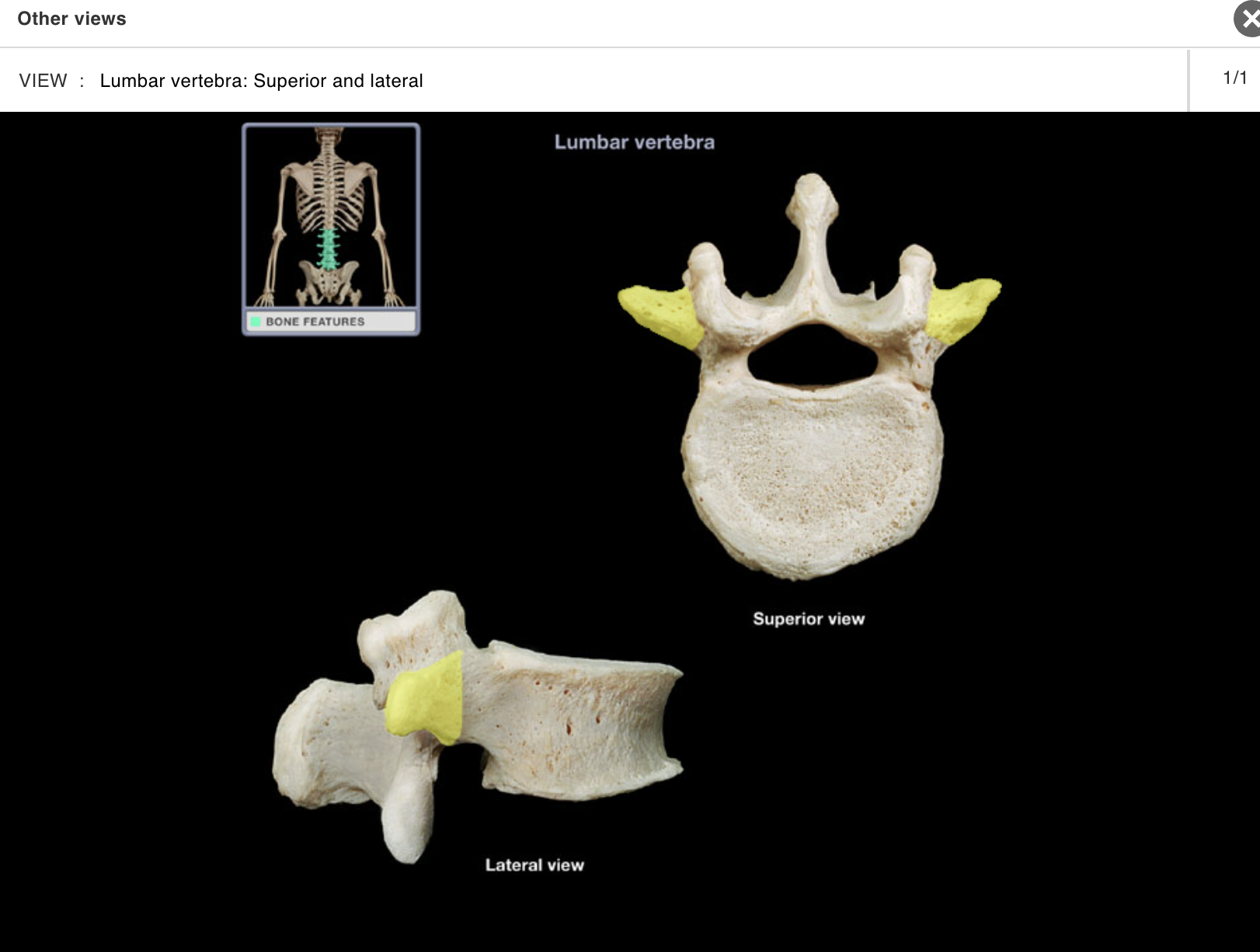
Transverse process of lumbar vertebra
Location:
• Lateral aspect of lumbar vertebra
Description:
• Prominent, paired, laterally-directed process
Comment:
• Provides attachment for intrinsic back muscles and ligaments
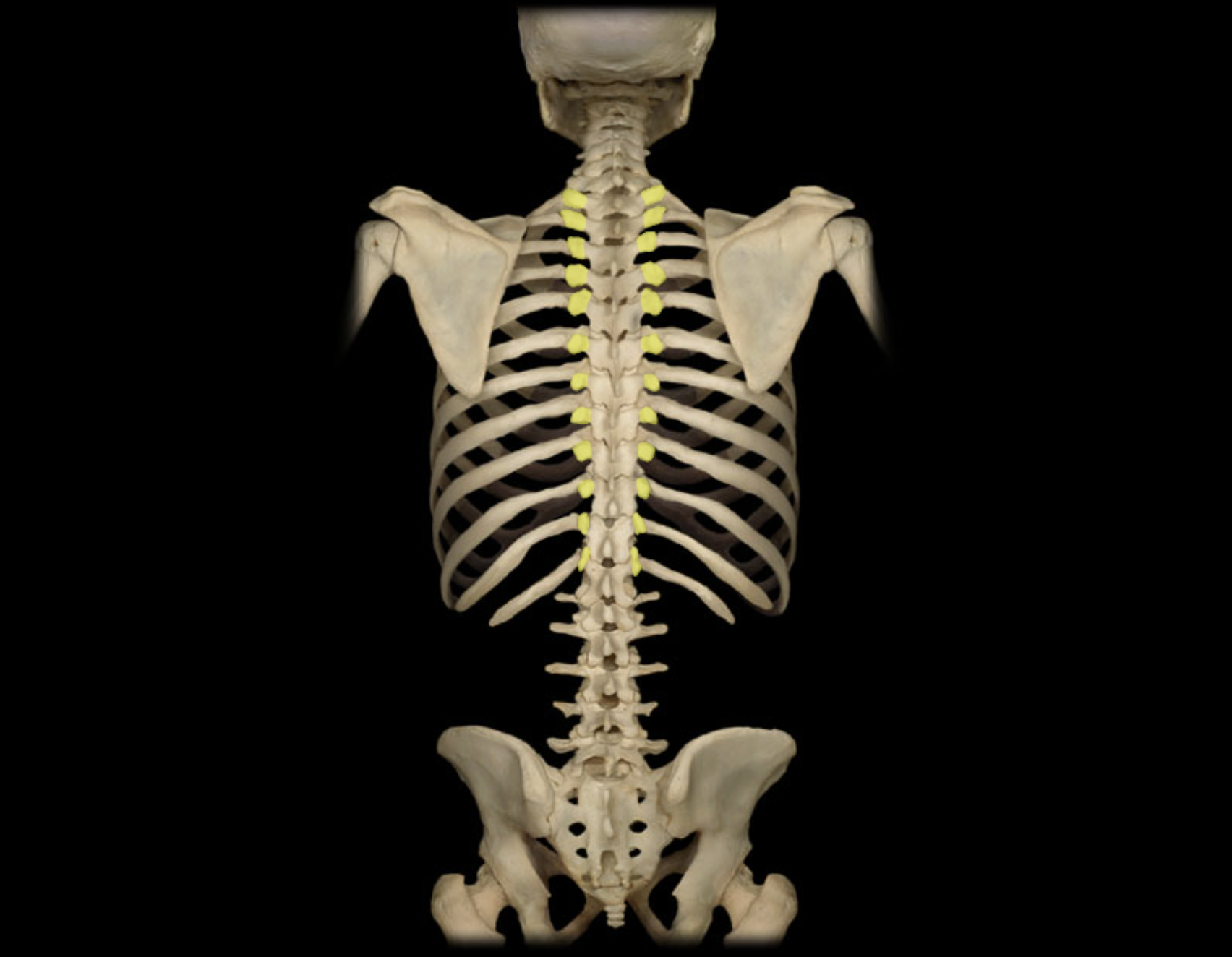
Transverse process of thoracic vertebra
Location:
• Lateral aspect of thoracic vertebra
Description:
• Prominent, paired, laterally-directed process
Comment:
• Provides attachment for intrinsic back muscles and ligaments
ACTIVITY 6 : DISSECTION
MODULE: SKELETAL
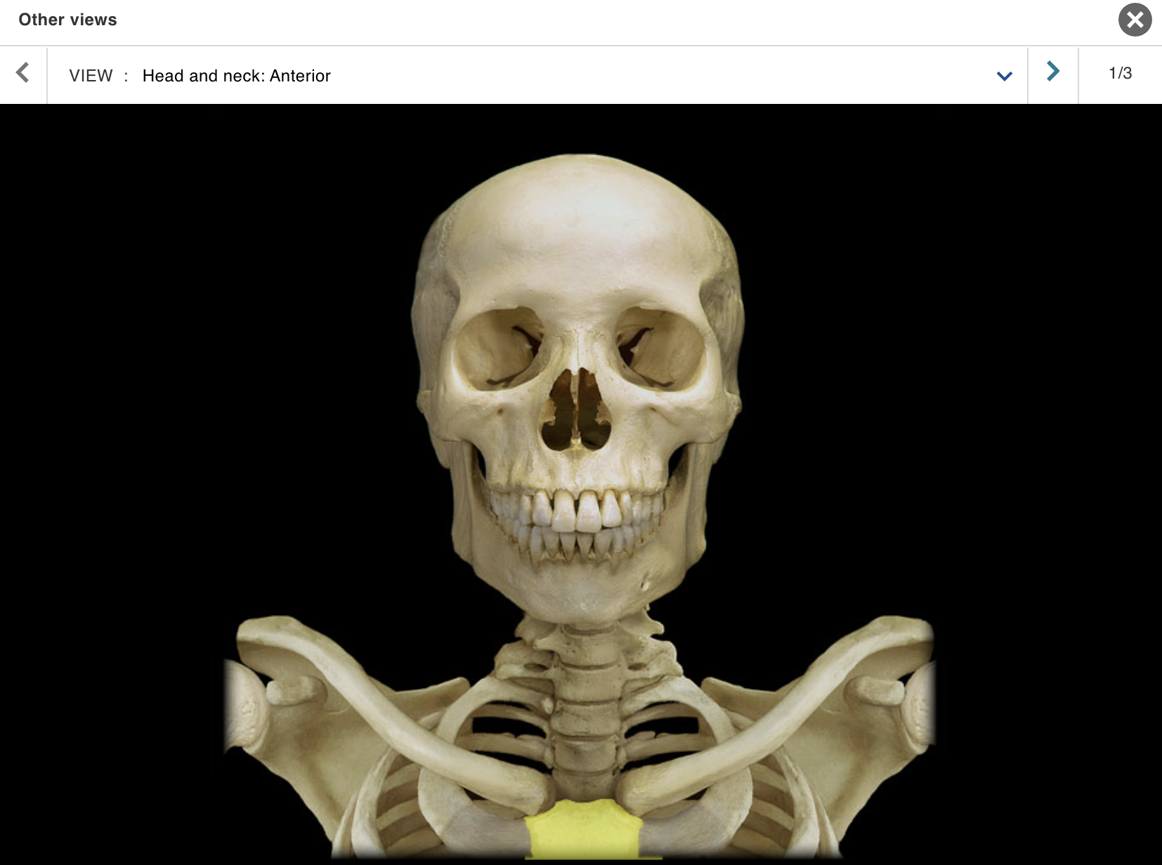
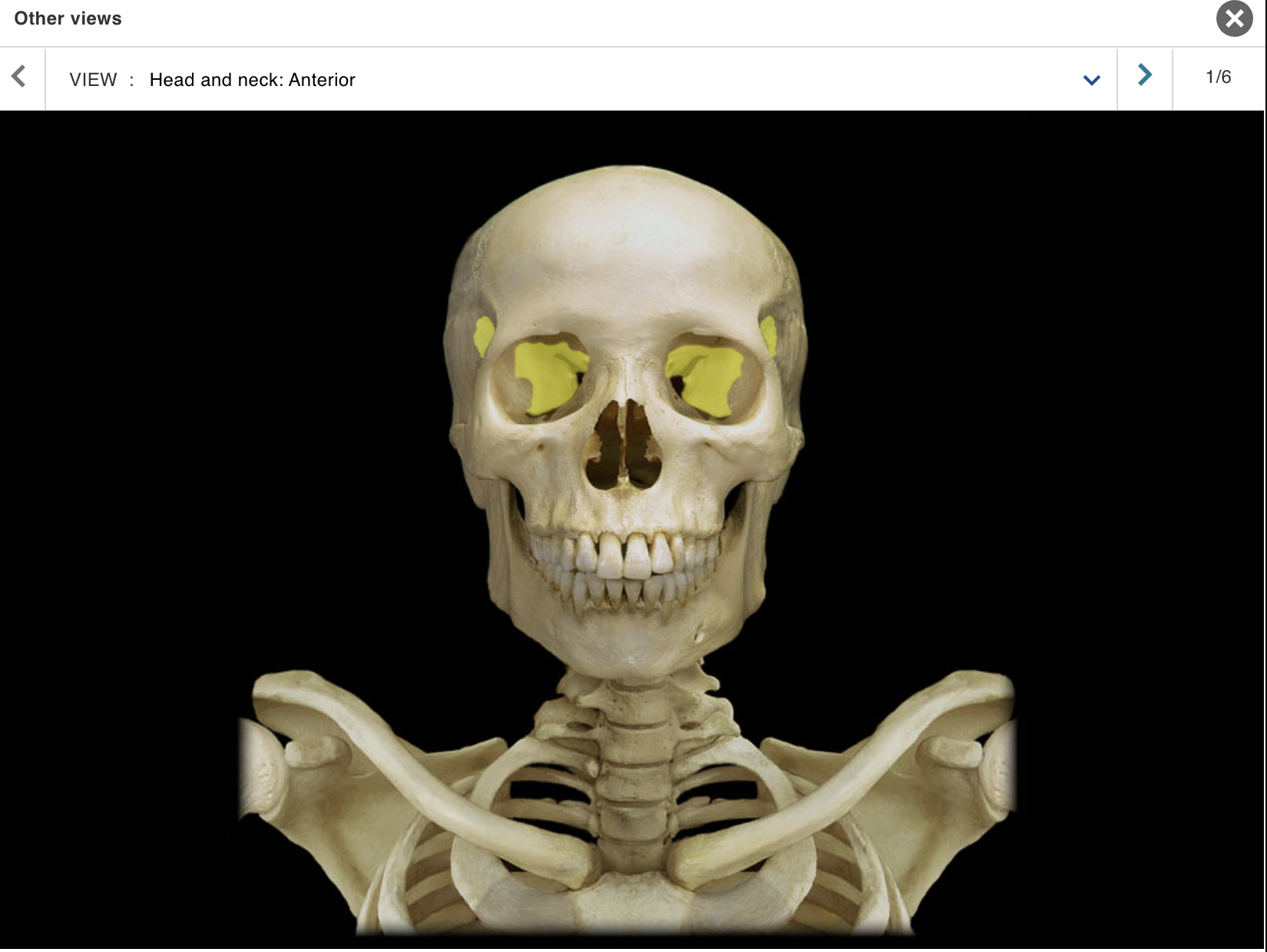
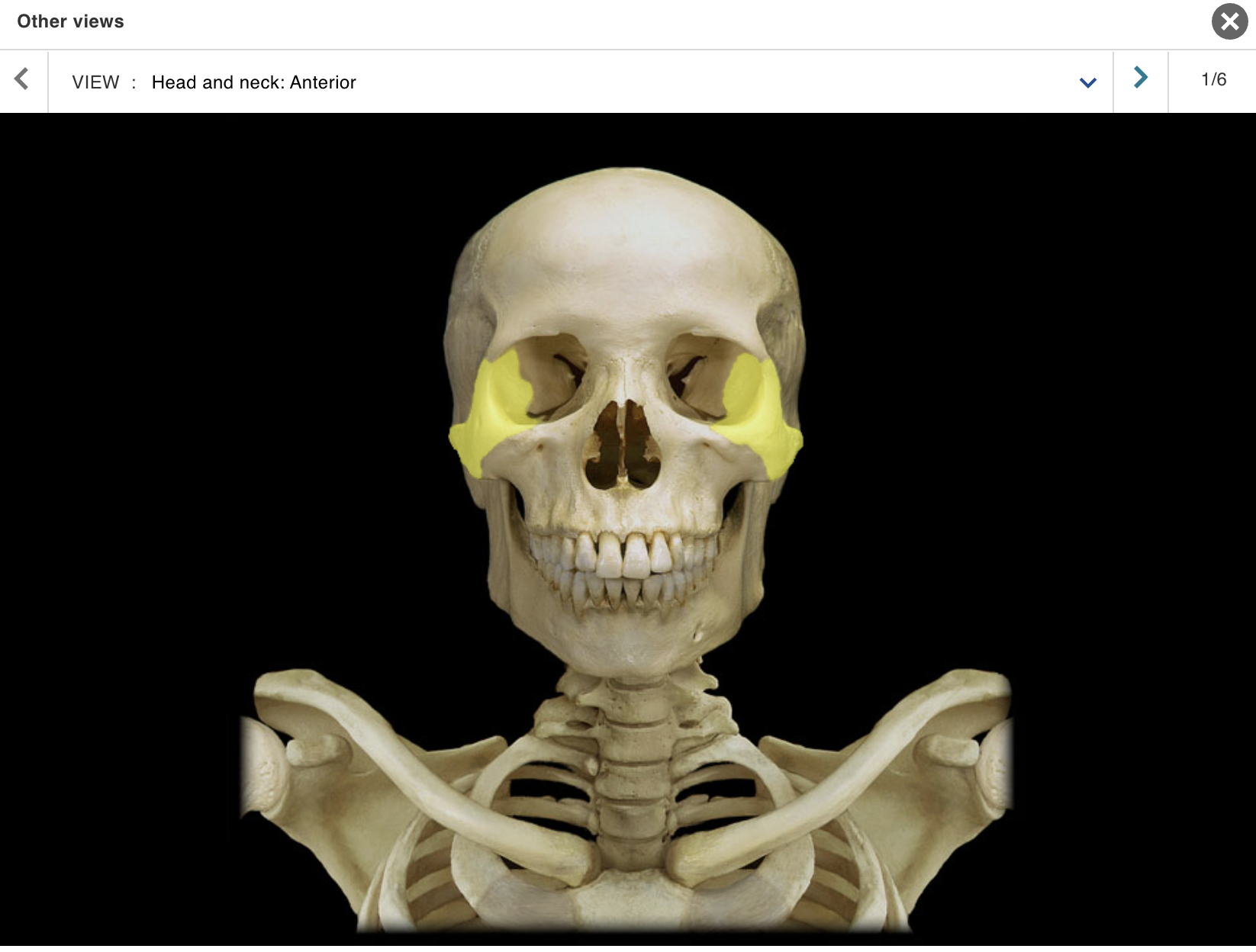
TOPIC: SKULL / VIEW : ANTERIOR
Ethmoid bone
Frontal bone
Lacrimal bone
Mandible
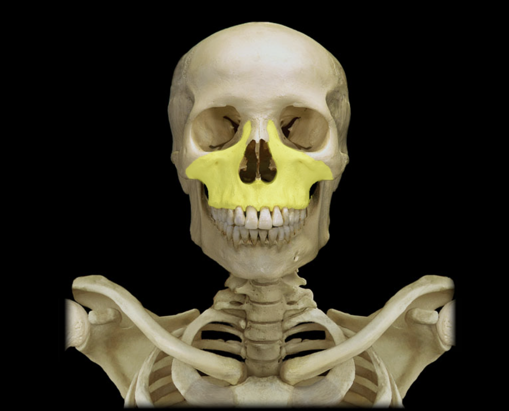
Maxilla
Nasal bone
Orbit
Parietal bone
Sphenoid bone
Temporal bone
Zygomatic bone
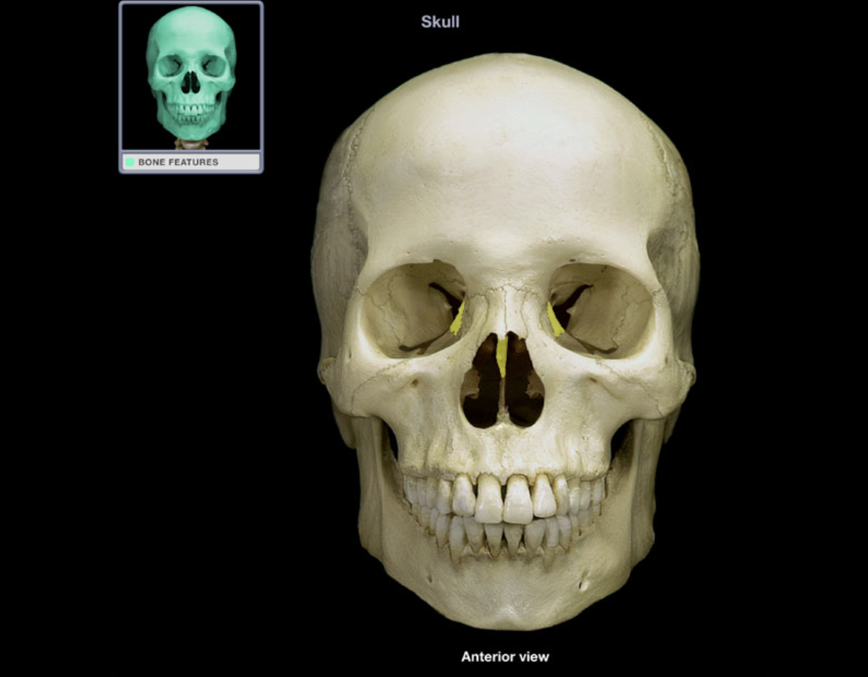
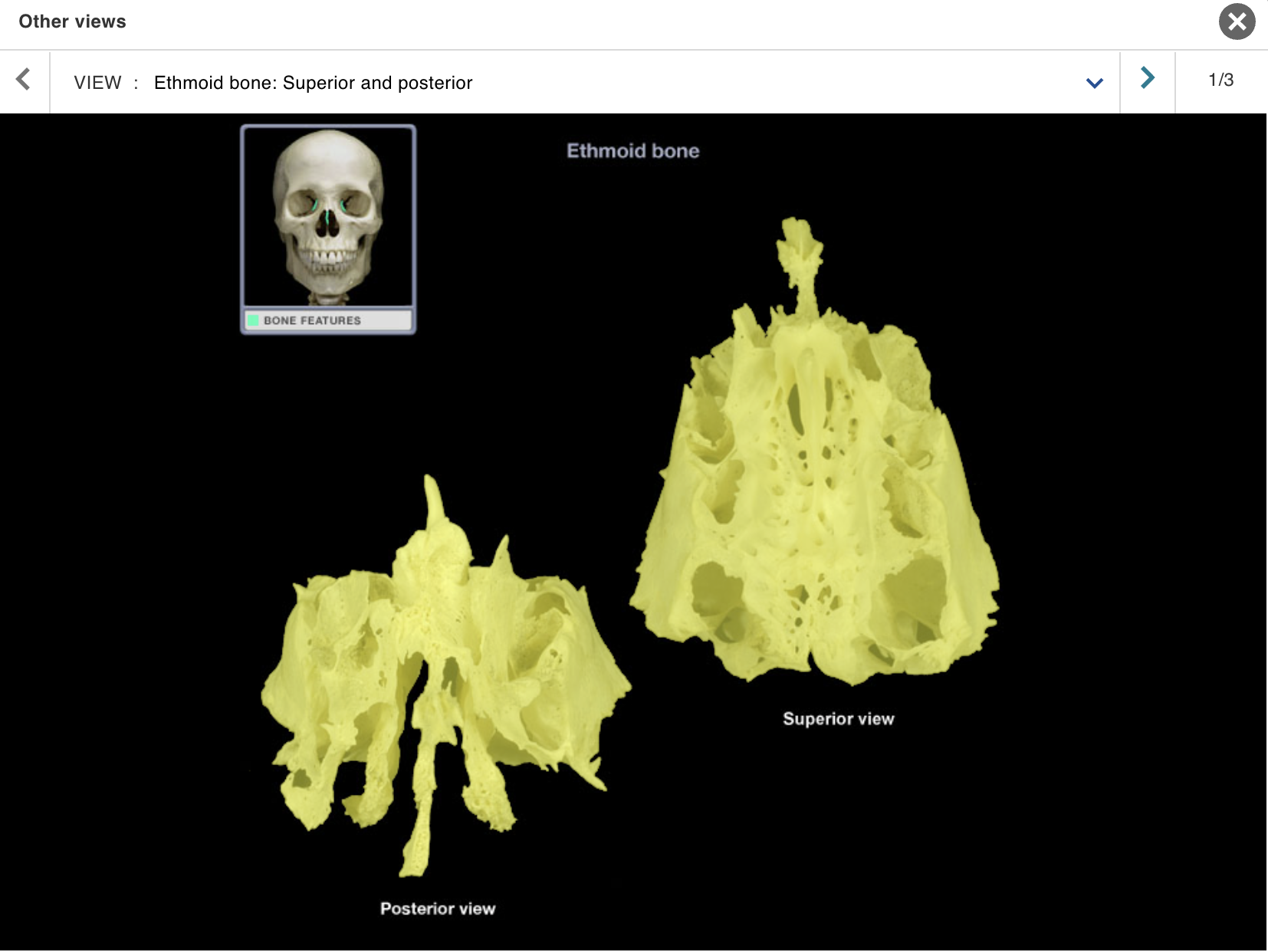
Ethmoid bone
Location:
• Nasal cavity
Description:
Irregular-shaped bone
Characteristic features include cribriform plate, crista galli, and perpendicular plate
Comment:
Contributes to anterior cranial fossa, nasal cavity, and orbit
Contains numerous sinuses (air cells)
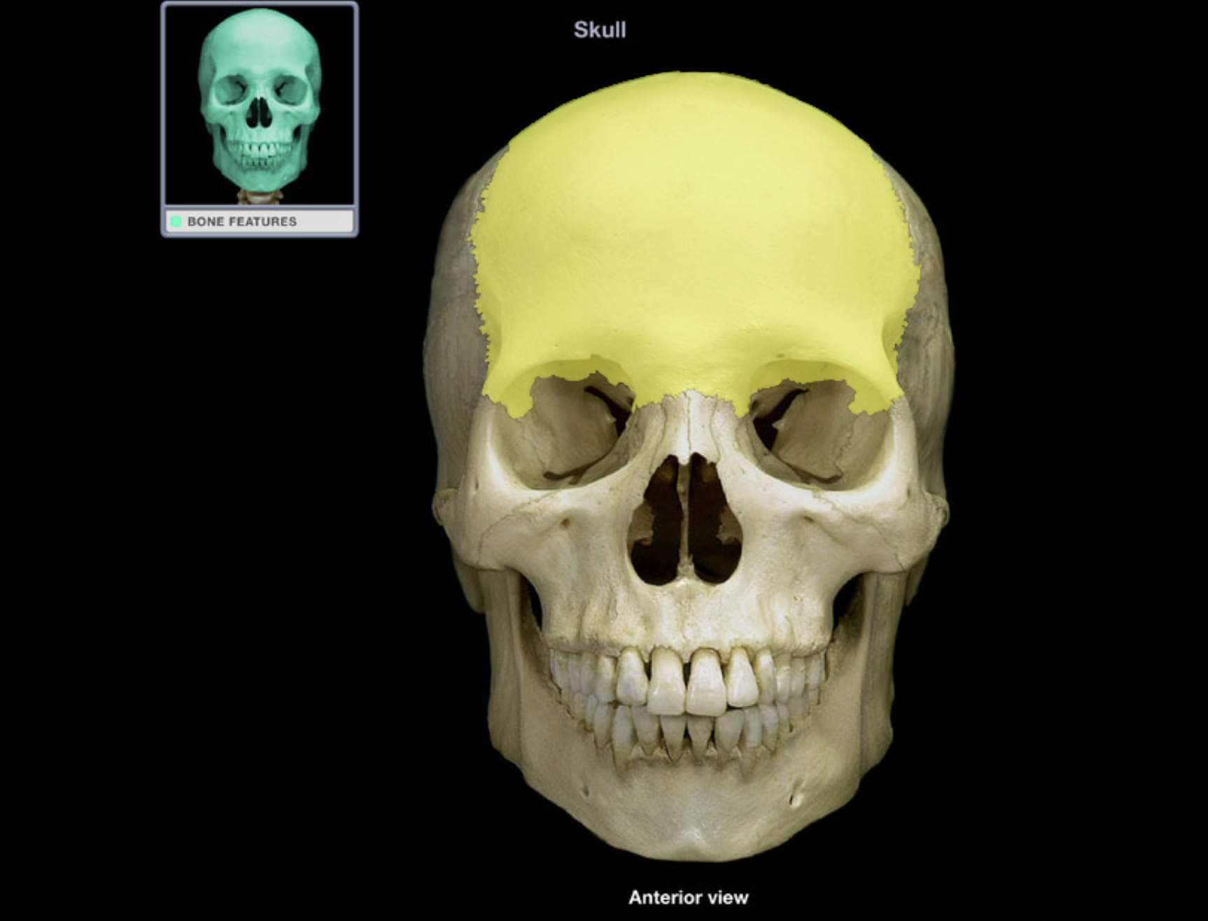
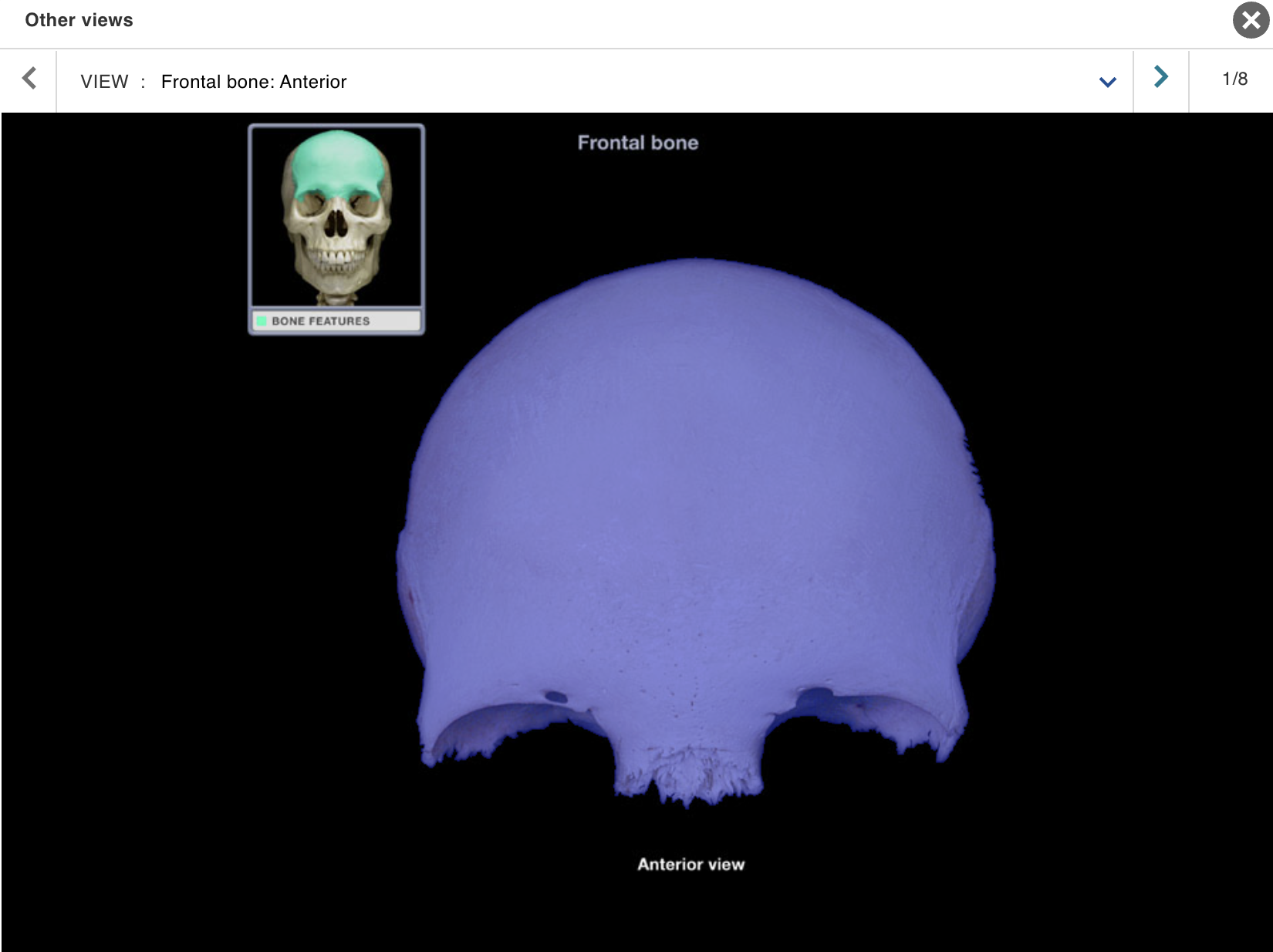
Frontal bone
Location:
• Skull (anterior superior part)
Description:
Unpaired, irregular-shaped, flat bone
Forms forehead, roof of orbits, and most of anterior cranial fossa
Contains frontal air sinuses
Comment:
• Articulates with parietal bone at coronal suture and sphenoid bone at sphenosquamosal suture

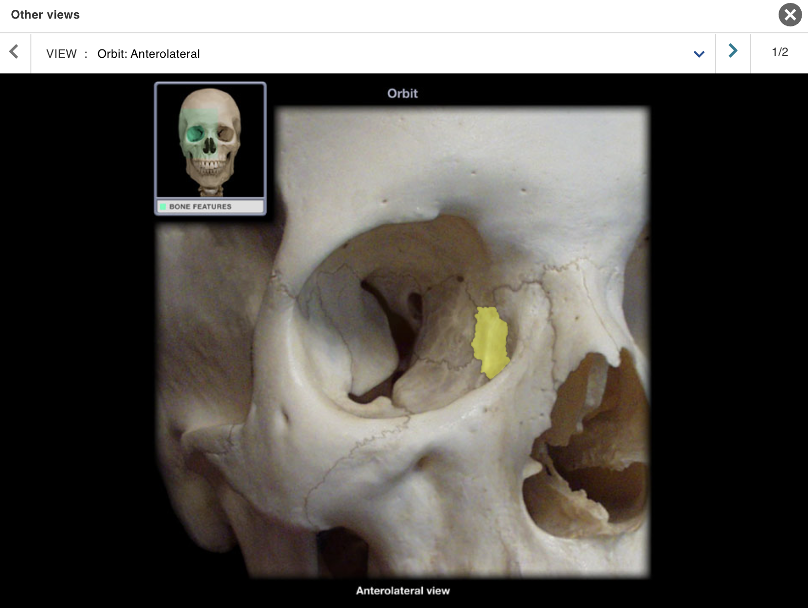
Lacrimal bone
Location:
Orbit (anterior and medial)
Nasal cavity (lateral wall)
Description:
Smallest and most delicate bone of skull
Orbital surface contributes to fossa for lacrimal sac and
nasolacrimal grooveNasal surface contributes to middle nasal concha
Comment:
• Some fibers of orbicularis oculi attach to lacrimal bone
Mandible
Location:
• Skull (anterior)
Description:
U-shaped bone
Each side consists of body (horizontal) and ramus (vertical) with coronoid and condylar processes
Mental protuberance forms point of chin
Contains alveoli ("sockets") for teeth
Also known as:
• "Lower jaw"
Comment:
• Contributes to temporomandibular joint (TMJ)
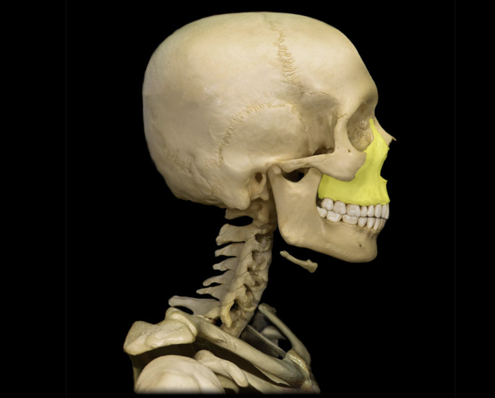
Maxilla
Location:
• Skull (anterior)
Description:
Paired, irregular-shaped bone
Left and right maxillae unite to form "upper jaw"
Contains alveoli ("sockets") for teeth
Contains maxillary air sinus on each side
Also known as:
• "Upper jaw"
Comment:
Forms part of floor of orbit and anterior part of hard palate
Contributes to upper face
Nasal bone
Location:
• Skull (anterior)
Description:
Small, paired bone
Articulates at midline with nasal bone from opposite side
Also known as:
• "Bridge of nose"
Comment:
• Forms bony part of nose
Orbit
Location:
Skull (anterior)
Lateral to nasal cavity
Description:
Pyramidal space with roof, floor, and medial and lateral walls
Roof: frontal and sphenoid bones
Floor: palatine and zygomatic bones, and maxilla
Lateral wall: zygomatic, sphenoid, and frontal bones
Medial wall: ethmoid and lacrimal bones, and maxilla
• Openings include superior and inferior orbital fissures, and optic canal
Also known as:
• Eye socket
Comment:
• Contains eyeball and its muscles; cranial nerves II, III, IV, V1, and VI; vasculature; lacrimal apparatus; and fat
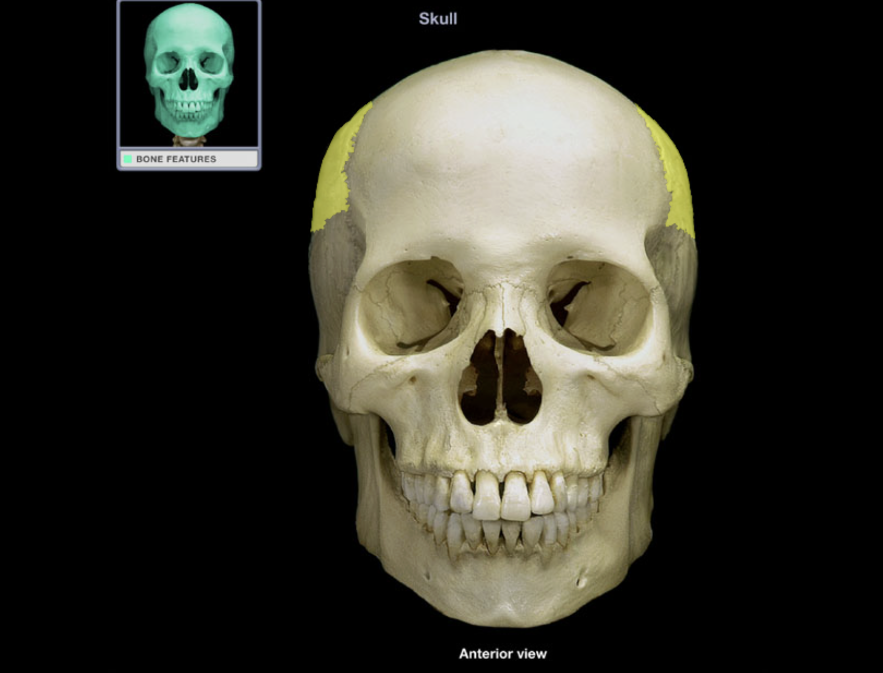
Parietal bone
Location:
• Skull (lateral)
Description:
Paired, flat bone
Forms most of lateral skull
Comment:
Parietal bones articulate at sagittal suture (midline)
Articulates with frontal bone at coronal suture, occipital bone at lambdoid suture, and temporal bone at squamosal suture
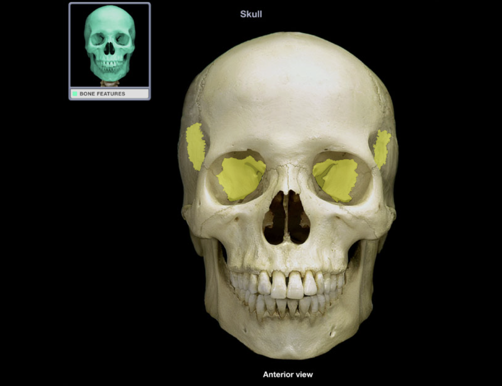
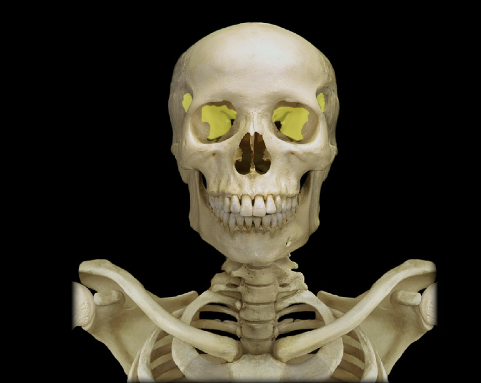
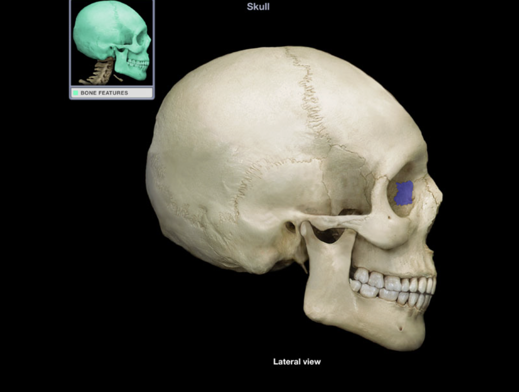
Sphenoid bone
Location:
• Skull
Description:
Unpaired, irregular-shaped bone
Shape resembles a butterfly
Consists of body, and greater and lesser wings
Comment:
• Contributes to middle cranial fossa, nasal cavities, orbits, and lateral skull (temples)
• Contains sella turcica ("Turkish saddle"), sphenoidal air sinus, and many foramina
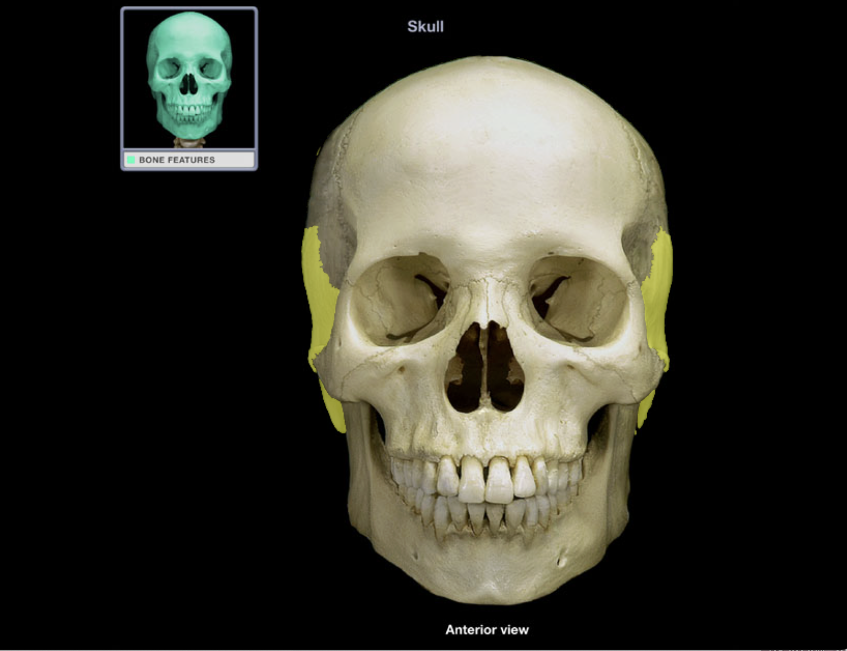
Temporal bone
Location:
• Skull (lateral and inferior)
Description:
Paired, irregular-shaped, flat bone
Consists of squamous, petrous, mastoid, and tympanic parts
Comment:
Forms part of lateral skull (temple)
Forms inferior and lateral walls of middle cranial fossa
Articulates with sphenoid bone at sphenosquamosal suture, occipital bone at lambdoid suture, and parietal bone at squamosal suture
Has mandibular fossa that forms temporomandibular joint with
head of mandibleForms zygomatic process that forms zygomatic arch with temporal process of zygomatic bone

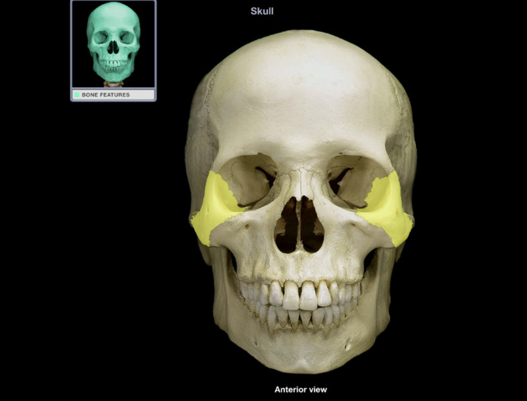
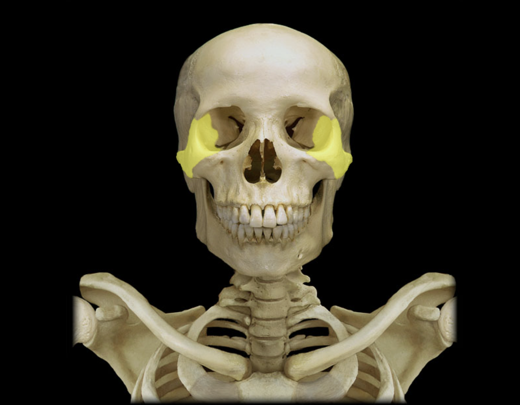
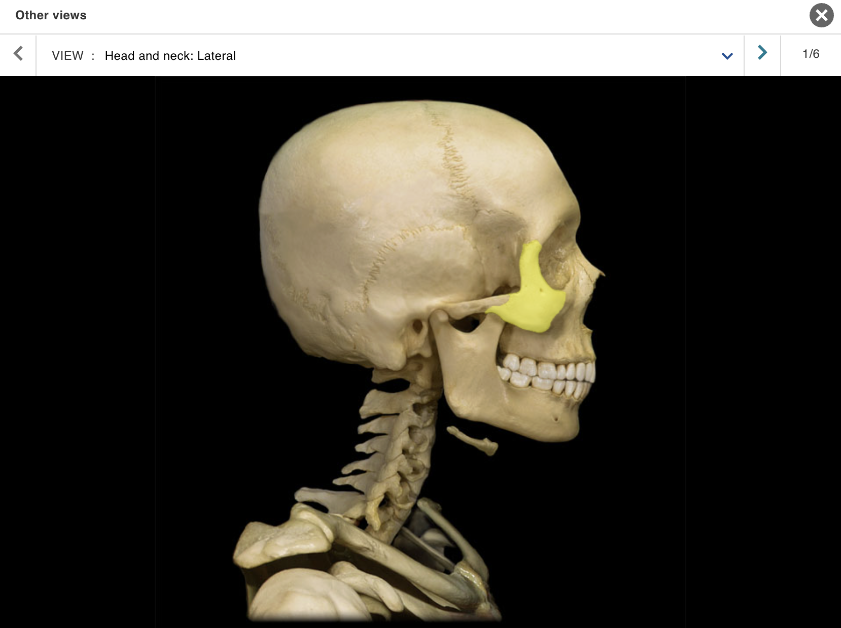
Zygomatic bone
Location:
• Skull (anterior and lateral)
Description:
Paired, irregular-shaped bone
Temporal process contributes to zygomatic arch
Also known as:
• "Cheekbone"
Comment:
• Forms part of floor and lateral wall of orbit
ACTIVITY 6 : DISSECTION
MODULE: SKELETAL
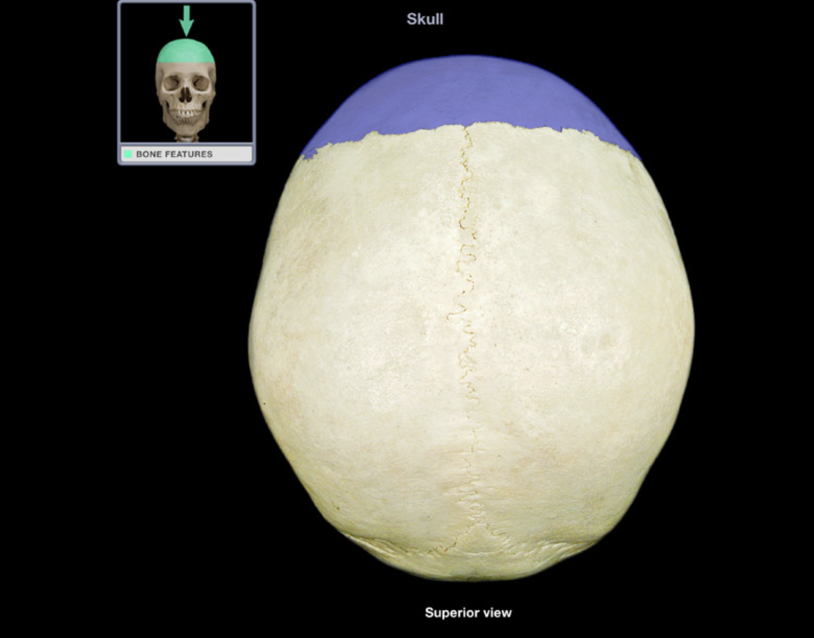
TOPIC: SKULL / VIEW : SUPERIOR
Coronal suture
Frontal bone
Lambdoid suture
Occipital bone
Parietal bone
Sagittal suture
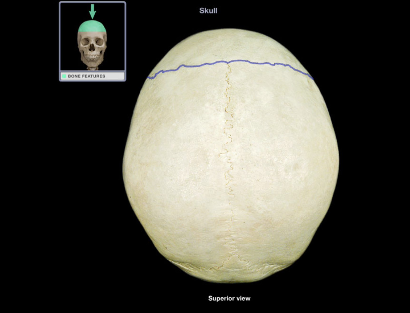
Coronal suture
Location:
• Skull (superior)
Description:
• Joint between frontal and parietal bones

Frontal bone
Location:
• Skull (anterior superior part)
Description:
Unpaired, irregular-shaped, flat bone
Forms forehead, roof of orbits, and most of anterior cranial fossa
Contains frontal air sinuses
Comment:
• Articulates with parietal bone at coronal suture and sphenoid bone at sphenosquamosal suture

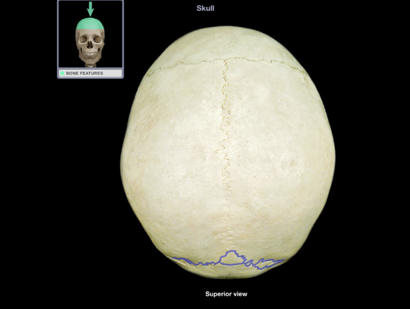
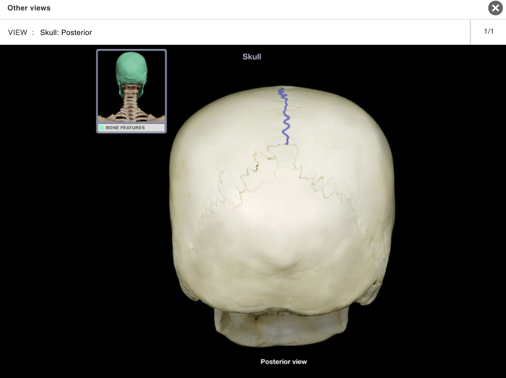
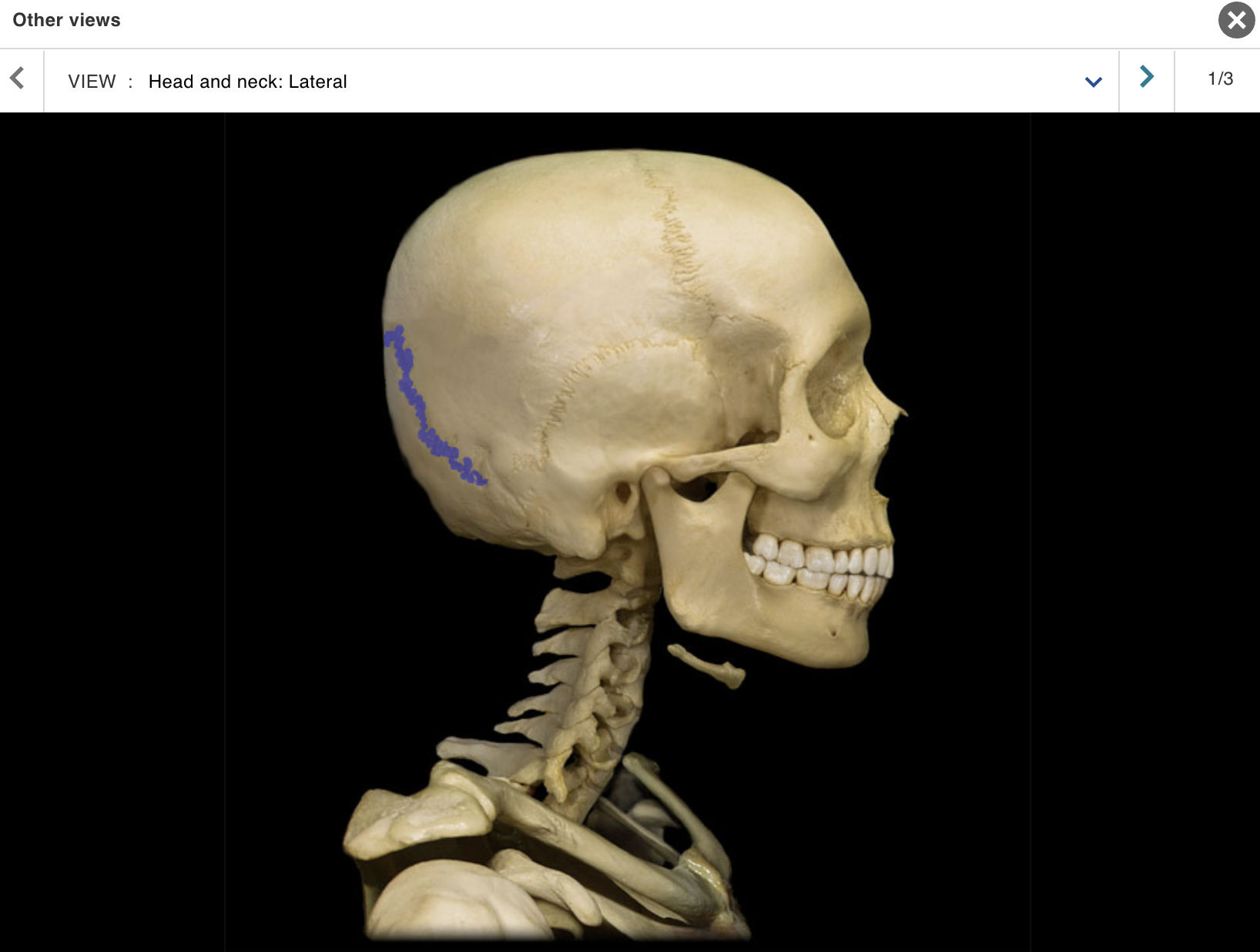
Lambdoid suture
Location:
• Skull (posterior)
Description:
• Joint between parietal and occipital bones
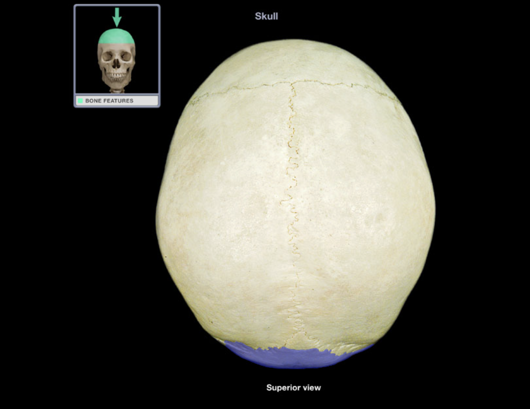
Occipital bone
Location:
• Skull (posterior and inferior)
Description:
Irregular-shaped bone
Contains foramen magnum in central portion
Comment:
Forms most of posterior skull
Forms most of posterior cranial fossa
Articulates with parietal bone at lambdoid suture, temporal bone at occipitomastoid and other sutures, and sphenoid bone at spheno-occipital synchondrosis
Articulates with atlas (C1 vertebra)
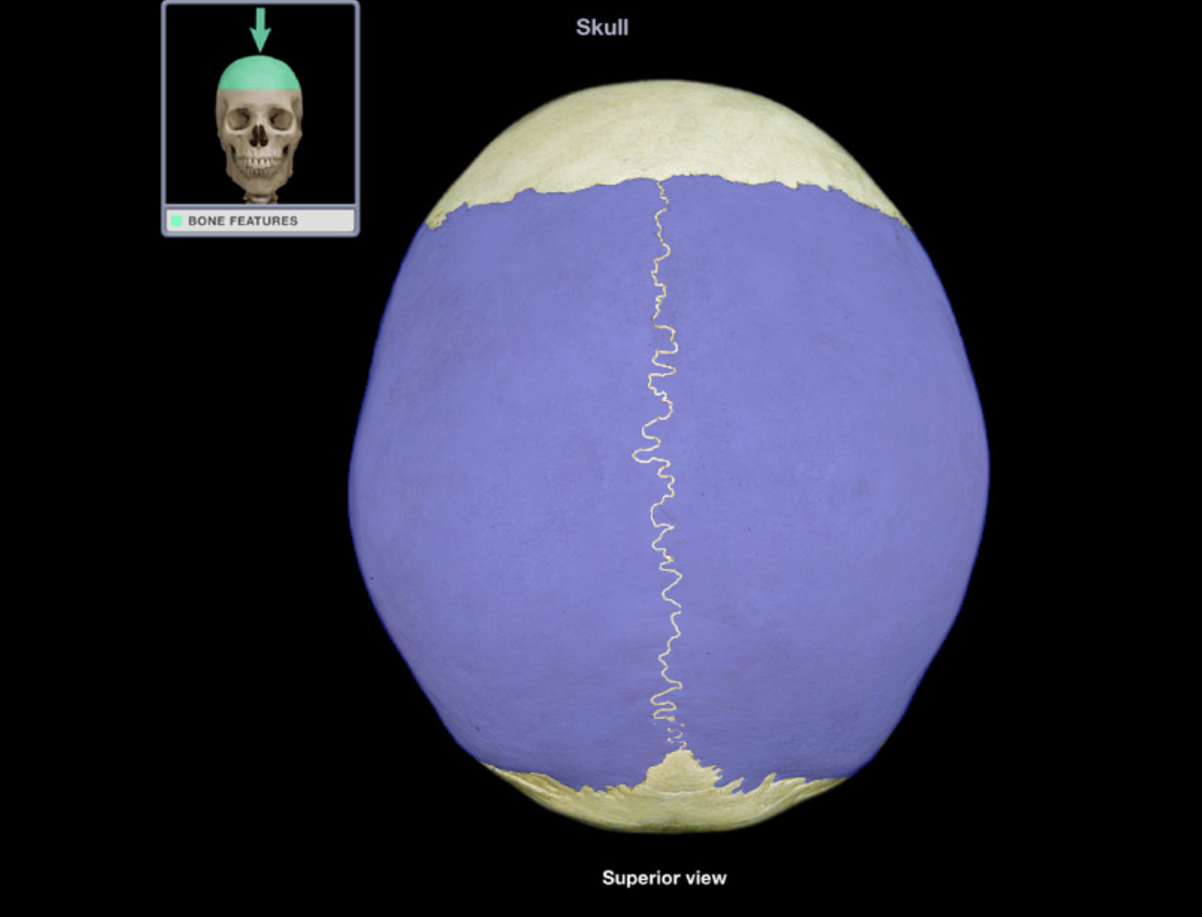
Parietal bone
Location:
• Skull (lateral)
Description:
Paired, flat bone
Forms most of lateral skull
Comment:
Parietal bones articulate at sagittal suture (midline)
Articulates with frontal bone at coronal suture, occipital bone at lambdoid suture, and temporal bone at squamosal suture
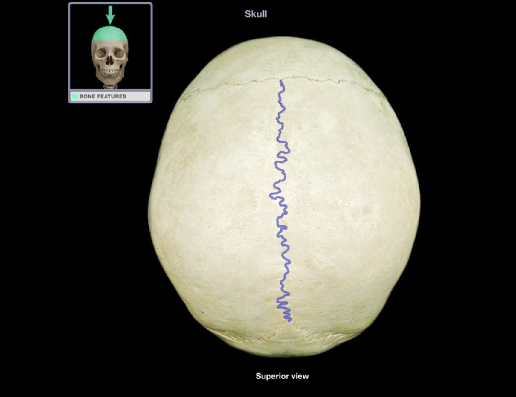
Sagittal suture
Location:
• Skull (superior)
Description:
• Joint between parietal bones
ACTIVITY 8 : DISSECTION
MODULE: SKELETAL
TOPIC: SKULL / VIEW : LATERAL
Coronal suture
Frontal bone
Lacrimal bone
Lambdoid suture
Mandible
Maxilla
Nasal bone
Occipital bone
Parietal bone
Temporal bone
Zygomatic bone
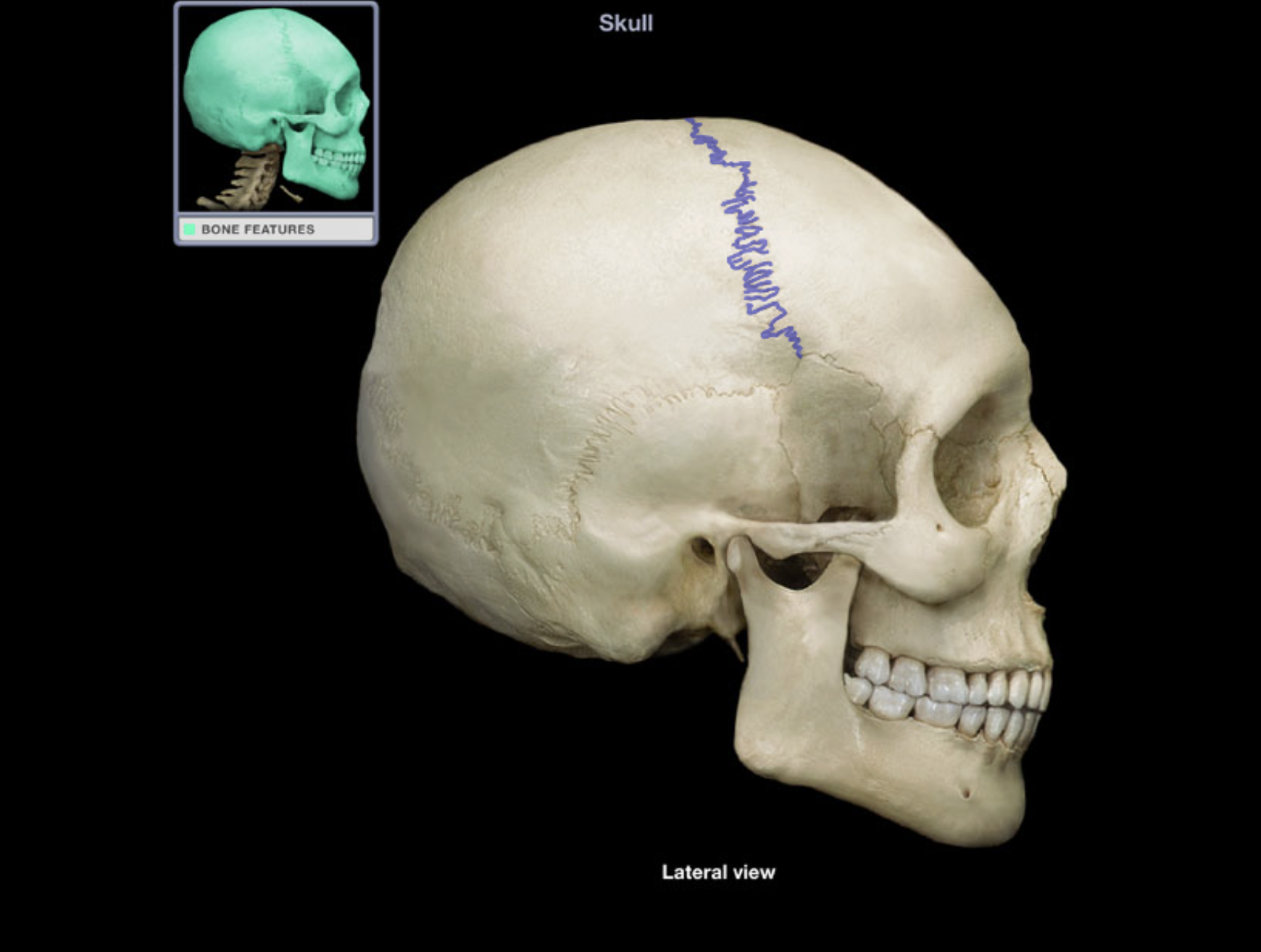
Coronal suture
Location:
• Skull (superior)
Description:
• Joint between frontal and parietal bones
Frontal bone
Location:
• Skull (anterior superior part)
Description:
Unpaired, irregular-shaped, flat bone
Forms forehead, roof of orbits, and most of anterior cranial fossa
Contains frontal air sinuses
Comment:
• Articulates with parietal bone at coronal suture and sphenoid bone at sphenosquamosal suture
Lacrimal bone
Location:
Orbit (anterior and medial)
Nasal cavity (lateral wall)
Description:
Smallest and most delicate bone of skull
Orbital surface contributes to fossa for lacrimal sac and
nasolacrimal grooveNasal surface contributes to middle nasal concha
Comment:
• Some fibers of orbicularis oculi attach to lacrimal bone
Lambdoid suture
Location:
• Skull (posterior)
Description:
• Joint between parietal and occipital bones
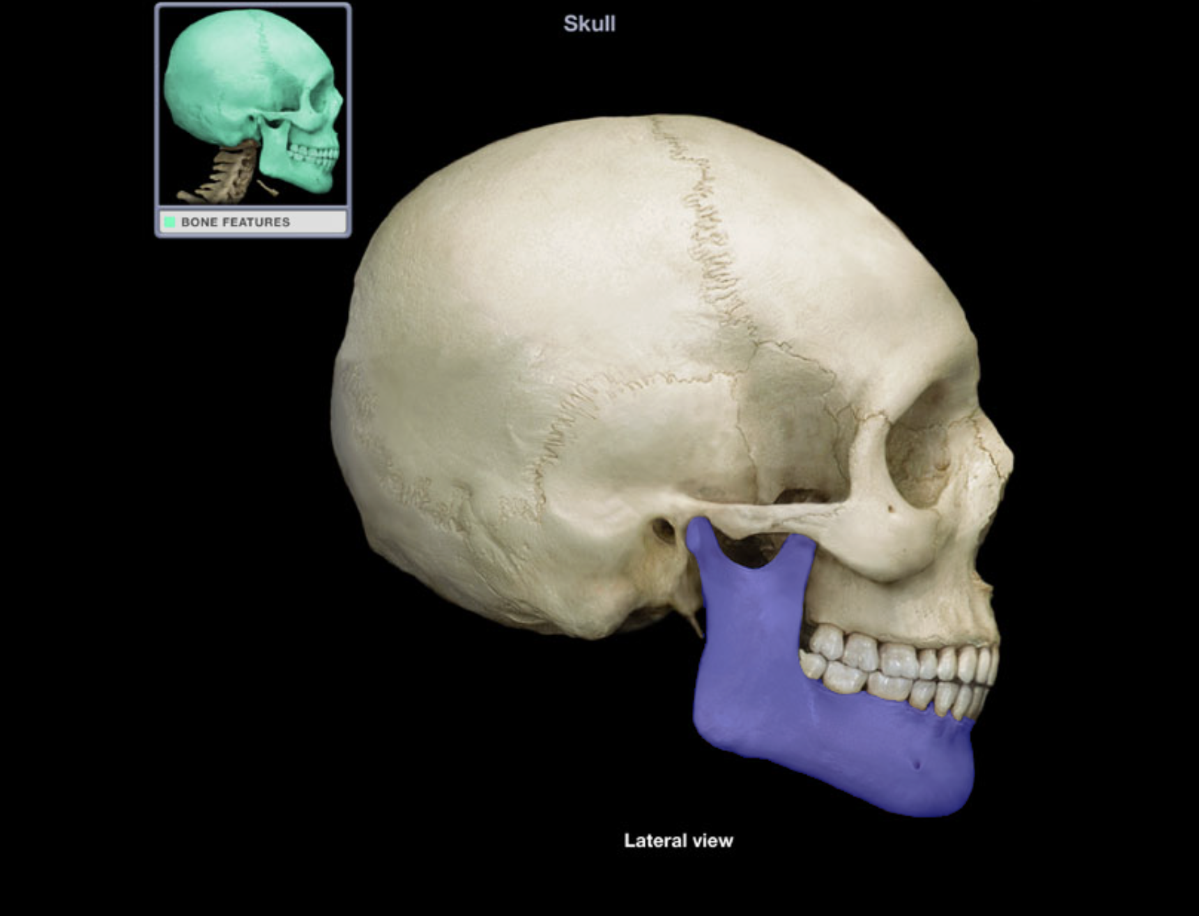
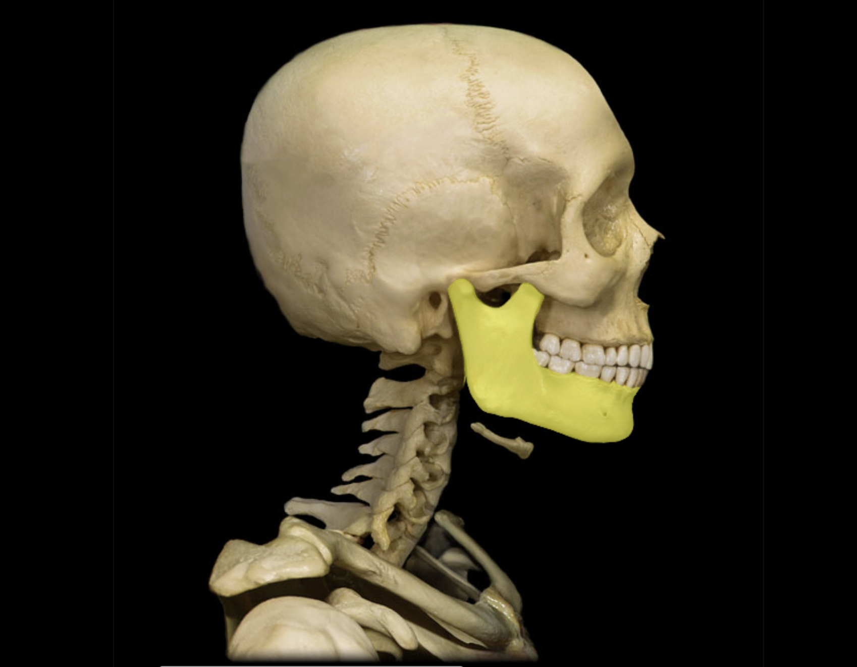
Mandible
Location:
• Skull (anterior)
Description:
U-shaped bone
Each side consists of body (horizontal) and ramus (vertical) with coronoid and condylar processes
Mental protuberance forms point of chin
Contains alveoli ("sockets") for teeth
Also known as:
• "Lower jaw"
Comment:
• Contributes to temporomandibular joint (TMJ)