RADPOS - Cranium
1/130
There's no tags or description
Looks like no tags are added yet.
Name | Mastery | Learn | Test | Matching | Spaced | Call with Kai |
|---|
No analytics yet
Send a link to your students to track their progress
131 Terms
Lateral projection
PA / PA axial projection (Caldwell Method)
AP / AP axial projection
AP axial projection (Towne Method)
PA axial projection (Haas Method)
(5) Cranium projections
SMV projection (Schuller Method)
VSM projection (Schuller Method)
(2) Cranial base
Lateral projection
AP axial projection
PA axial projection
(3) Sella turcica
22
Number of bones that make up the skull
8
Number of cranial bones in the skull
14
Number of facial bones in the skull
Calvaria
Skull dome forming the roof of the cranium
Sutures
Fibrous joints joining skull bones (except mandible)
Coronal suture
Suture between frontal and parietal bones
Sagittal suture
Suture between two parietal bones along the midline
Lambdoidal suture
Suture between occipital and parietal bones
Pterion
Skull region where several bones meet; overlies middle meningeal artery
Fontanels
Soft spots on infant skull where sutures are not yet fused
Mandible
Largest, strongest facial bone; forms lower jaw
Temporomandibular joint
Articulation of mandible and temporal bone; only movable skull joint
Sphenoid
Keystone bone articulating with all other cranial bones
Foramen magnum
Large opening for spinal cord located in the occipital bone
External auditory meatus
Ear canal in temporal bone
Auditory ossicles
Malleus, incus, and stapes bones transmitting sound in middle ear
Hyoid
Bone in neck that anchors tongue; does not articulate with other bones
Orbit
Eye socket composed of 7 bones (frontal, sphenoid, ethmoid, maxilla, zygoma, lacrimal, palatine)
Ethmoid
Bone forming part of nasal septum and medial orbital walls
Nasal conchae
Scroll-shaped bones in nasal cavity for air filtration and humidification
Tympanic membrane
Eardrum transmitting sound vibrations
Sella turcica
Pituitary gland seat in sphenoid bone
Asterion
Suture junction of parietal, occipital, and mastoid portion of temporal bone
Basal Fracture
Fracture located at the base of the skull
Blowout Fracture
Fracture of the floor of the orbit
Contre-coup Fracture
Fracture to one side of a structure caused by trauma to the other side
Depressed Fracture
Fracture causing a portion of the skull to be depressed into the cranial cavity
Leforte Fracture
Bilateral horizontal fractures of the maxillae
Linear Fracture
Irregular or jagged fracture of the skull
Tripod Fracture
Fracture of the zygomatic arch and orbital floor or rim and dislocation of the frontozygomatic suture
Mastoiditis
Inflammation of the mastoid antrum and air cells
Metastases
Transfer of a cancerous lesion from one area to another
Osteomyelitis
Inflammation of bone due to a pyogenic infection
Osteopetrosis
Increased density of atypically soft bone
Osteoporosis
Loss of bone density
Paget's Disease
Thick, soft bone marked by bowing and fractures
Polyp
Growth or mass protruding from a mucous membrane
Sinusitis
Inflammation of one or more of the paranasal sinuses
TMJ Syndrome
Dysfunction of the temporomandibular joint
Acoustic Neuroma
Benign tumor arising from Schwann cells of the eighth cranial nerve
Multiple Myeloma
Malignant neoplasm of plasma cells involving the bone marrow and causing destruction of the bone
Osteoma
Tumor composed of bony tissue
Pituitary Adenoma
Tumor arising from the pituitary gland, usually in the anterior lobe
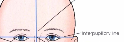
Interpupillary line
Horizontal line connecting the centers of the pupils in both eyes
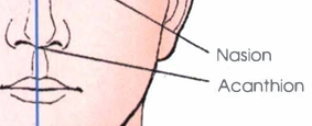
Acanthion
Midpoint at the base of the anterior nasal spine, located at the junction of the upper lip and nose
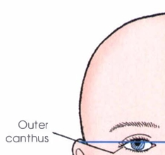
Outer canthus
Outer corner of the eye where the upper and lower eyelids meet
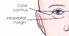
Infraorbital margin
Lower bony edge of the orbit (eye socket)
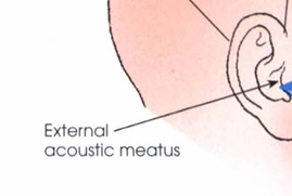
External acoustic meatus
Opening of the ear canal on the side of the head
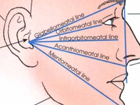
Orbitomeatal line
Line from the outer canthus of the eye to the center of the external acoustic meatus

Infraorbitomeatal line
Line from the infraorbital margin to the external acoustic meatus

Acanthiomeatal line
Line from the acanthion to the external acoustic meatus

Mentomeatal line
Line from the mental point (chin) to the external acoustic meatus
Mesocephalic
Skull shape with petrous pyramids projecting anteriorly and medially at a 47-degree angle from the midsagittal plane; represents an average or typical skull form.
Petrous pyramids
Dense, pyramid-shaped portions of the temporal bones containing the inner ear structures; their superior borders are at the base of the cranium.
Brachycephalic
Short front-to-back, broad side-to-side, and shallow vertex-to-base skull; petrous pyramids form a wider angle (~54 degrees) with the midsagittal plane; internal structures sit higher relative to the infraorbitomeatal line (IOML).
Dolichocephalic
Long front-to-back, narrow side-to-side, and deep vertex-to-base skull; petrous pyramids form a narrower angle (~40 degrees) with the midsagittal plane; internal structures sit lower relative to the IOML.
Perpendicular; external acoustic meatus (EAM)
In the lateral projection of the cranium, the central ray is directed ____________________ to enter 2 inches (5 cm) superior to the _________________.
Midsagittal; interpupillary
During the lateral skull projection, the ________________ plane of the head is placed parallel to the plane of the image receptor (IR), and the ________________ line is perpendicular to the IR.

Caudad; nasion; 15 degrees caudad
For the PA projection (Caldwell Method), the patient's forehead and nose rest on the table or upright Bucky, and the central ray is directed ___________________ exiting the _______________ at an angle of _______________.
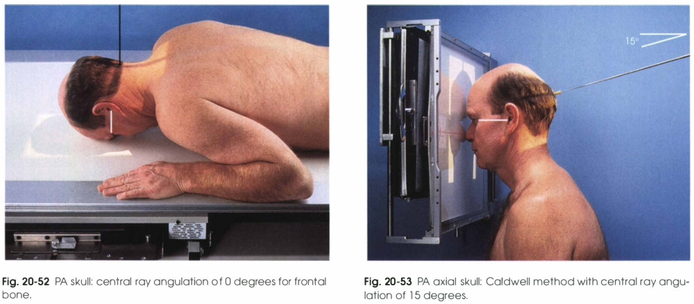
Lower third
The PA axial Caldwell method projects the petrous ridges into the _______________ third of the orbits.
Perpendicular; 15 degrees cephalad
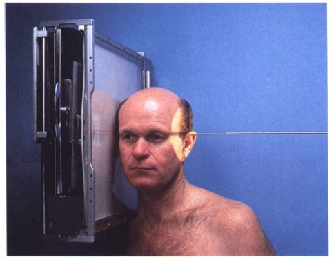
The AP skull projection is used when the patient cannot tolerate PA positioning; the central ray is ___________________ or directed to the nasion at an angle of ______________ degrees _______________.
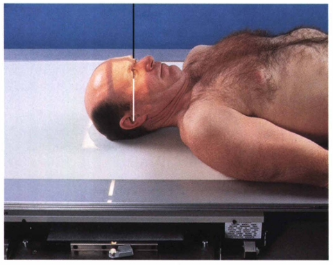
Orbitomeatal line (OML); infraorbitomeatal line (IOML); 7 degrees
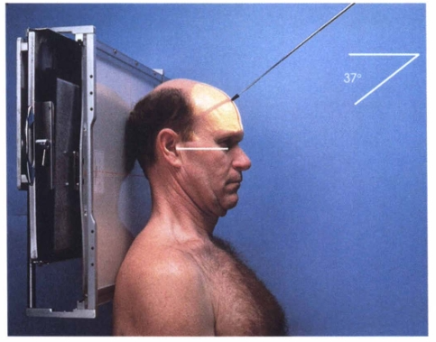
In the AP axial (Towne) projection, the patient's neck is flexed so that the ___________________ is perpendicular to the IR. If the neck cannot be flexed enough, the ________________ is made perpendicular and the central ray angulation is increased by ______________ degrees.
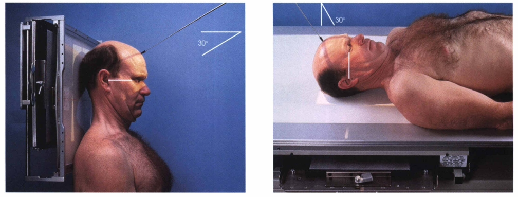
Foramen magnum; 30 degrees; 37 degrees
The central ray for the Towne method is directed through the ______________ at a caudal angle of ______________ degrees to the OML or ______________ degrees to the IOML.

Prone or seated upright; 25-degree cephalad; external occipital protuberance (inion); nasion
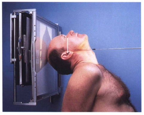
The PA axial (Haas) method is performed with the patient in the __________ or ___________ position with the OML perpendicular to the IR and the central ray directed at a __________ degree ______________ angle to enter 1 ½ inches (3.8 cm) below the ___________________ and exit 1 ½ inches (3.8 cm) superior to the ______________.

Infraorbitomeatal line (IOML); perpendicular
The submentovertical (SMV) projection requires that the _______________ line be placed as parallel as possible to the plane of the IR, and the central ray is directed ___________________ to the IOML.
Occipital
The SMV projection shows symmetric images of the petrosae, mastoid processes, foramen ovale and spinosum, carotid canals, sphenoidal and ethmoidal sinuses, and the ____________ bone.
Contraindicated; perpendicular
The verticosubmental (VSM) projection is used when the SMV projection is ________________ by the patient's condition. The central ray is directed through the sella turcica ___________________ to the IOML.
External acoustic meatus (EAM)
For the lateral projection of the sella turcica, the IR is centered 1/2 inch (1.9 cm) anterior and 1/2 inch (1.9 cm) superior to the _____________.
Dorsum sellae; posterior clinoid processes
In the AP axial projection of the sellar region, a 37-degree caudal angulation projects the ______________ and ______________ within the foramen magnum.
Glabella; cephalad
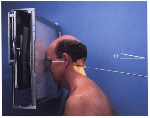
The PA axial projection central ray is directed to exit the ______________ at a 10-degree _________________ angle.
Lateral Projection (Cranium)
PA / PA Axial Projection (Caldwell Method)
AP Axial Projection (Towne Method)
PA Axial Projection (Haas Method)
SMV Projection (Schüller Method)
Lateral Projection of Sella Turcica
AP Axial Projection of Sella Turcica
PA Axial Projection of Sella Turcica
Projections that is Seated-Upright
AP / AP Axial Projection
AP Axial Projection (Towne Method)
SMV Projection (Schüller Method)
VSM Projection (Schüller Method)
Lateral Projection (Cranium)
Projections that is in Supine Position
PA / PA Axial Projection (Caldwell Method)
PA Axial Projection (Haas Method)
VSM Projection (Schüller Method)
PA Axial Projection of Sella Turcica
Projections that is in Prone Position
Lateral Projection (Cranium)
Lateral Projection of Sella Turcica
Midsagittal Plane Parallel to IR Projections
PA / PA axial (Caldwell)
AP / AP axial
Towne
Haas
PA axial sella turcica
AP axial sella turcica,
SMV
VSM
Midsagittal Plane Perpendicular to IR Projections
Lateral Projection (Cranium)
Lateral Projection of Sella Turcica
Interpupillary line perpendicular to IR Projections
PA/PA axial (Caldwell)
AP/AP axial
Towne
Haas
PA axial sella turcica
OML perpendicular to IR Projections
Lateral Cranium (front edge)
Towne (if OML can’t be)
AP axial sella turcica
IOML perpendicular to IR Projections
seated-upright; semiprone
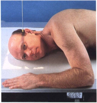
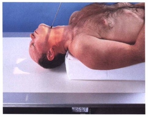
In the lateral projection of the cranium, the patient is positioned _____________ or ________________, resting on the forearm and flexed knee of the elevated side if semi prone.
parallel; perpendicular
For the lateral skull projection, the midsagittal plane of the head is placed ____________ to the image receptor (IR), and the interpupillary line is ____________ to the IR.
perpendicular; parallel
The flexion of the patient’s neck in the lateral projection should position the infraorbitomeatal line (IOML) ____________ to the front edge of the IR and ____________ to the long axis of the IR.
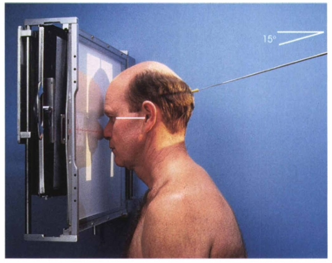
perpendicular; superior
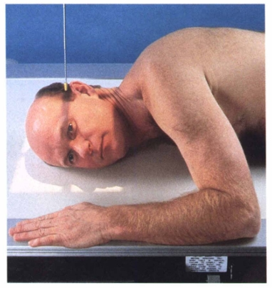
The central ray for the lateral projection of the skull is directed ________________ to enter 2 inches (5 cm) ____________ to the external acoustic meatus (EAM).

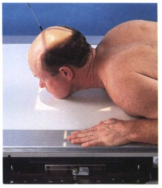
prone; seated
In the PA projection / PA axial projection (Caldwell method), the patient is positioned ___________ or ___________ with the midsagittal plane centered to the grid.
nasion; 15 degrees
For the Caldwell method, the central ray is directed to exit the ______________ at an angle of ______________ caudad.
20 to 25; caudad
To demonstrate the superior orbital fissures, the central ray is angled ______________ degrees ______________ through the midorbits.
25; 30; caudad
The central ray angle to demonstrate the rotundum foramina is ______________ to ______________ degrees ______________ directed to the nasion.
perpendicular
When the patient cannot tolerate PA positioning, the AP skull projection is performed with the patient supine and the midsagittal plane and orbitomeatal line (OML) positioned _______________ to the IR.
15 degrees; cephalad
The central ray for the AP axial projection is directed perpendicular or at an angle of ______________ degrees ______________ to the nasion.
perpendicular; perpendicular
For the AP axial (Towne) projection, the patient’s midsagittal plane is ______________ to the midline of the IR, and the neck is flexed enough to make the OML _______________ to the IR.
perpendicular; 7 degrees
If the neck cannot be appropriately flexed in the Towne method, the IOML is made ______________ to the IR, and the central ray angulation is increased by ______________ degrees.
foramen magnum; 30; 37
The central ray for the Towne method is directed through the _______________ at a caudal angle of ______________ degrees to the OML or ______________ degrees to the IOML.
2 ½ inches (6.3 cm); EAM
The AP axial projection center enters approximately ______________ inches (cm) above the glabella and passes through the level of the ______________.
prone; seated-upright; perpendicular
In the PA axial projection (Haas method), the patient is positioned ______________ or ________________, with the midsagittal plane centered and the OML positioned _______________ to the IR.
25 degree; cephalad; below; above
The central ray in the Haas method is directed at a ______________ degree ______________ angle to enter a point 1 ½ inches (3.8 cm) _______________ to the external occipital protuberance (inion) and exit approximately 1 ½ inches (3.8 cm) ______________ the nasion.
parallel; perpendicular
For the SMV projection (Schüller method), the patient’s IOML should be placed as ______________ as possible to the IR, and the central ray is directed ______________ to the IOML.
angles of the mandible; anterior
The central ray entry point for the SMV projection is the midsagittal plane of the throat between the ______________ and passes ½ inch (1.9 cm) ______________ to the level of the EAMs.
prone; perpendicular
The VSM projection is performed with the patient _____________, resting the fully extended chin on the table, and the midsagittal plane positioned ______________ to the IR.