Unit 4 - Lecture 18
0.0(0)
Card Sorting
1/109
There's no tags or description
Looks like no tags are added yet.
Last updated 9:36 PM on 4/17/23
Name | Mastery | Learn | Test | Matching | Spaced | Call with Kai |
|---|
No analytics yet
Send a link to your students to track their progress
110 Terms
1
New cards
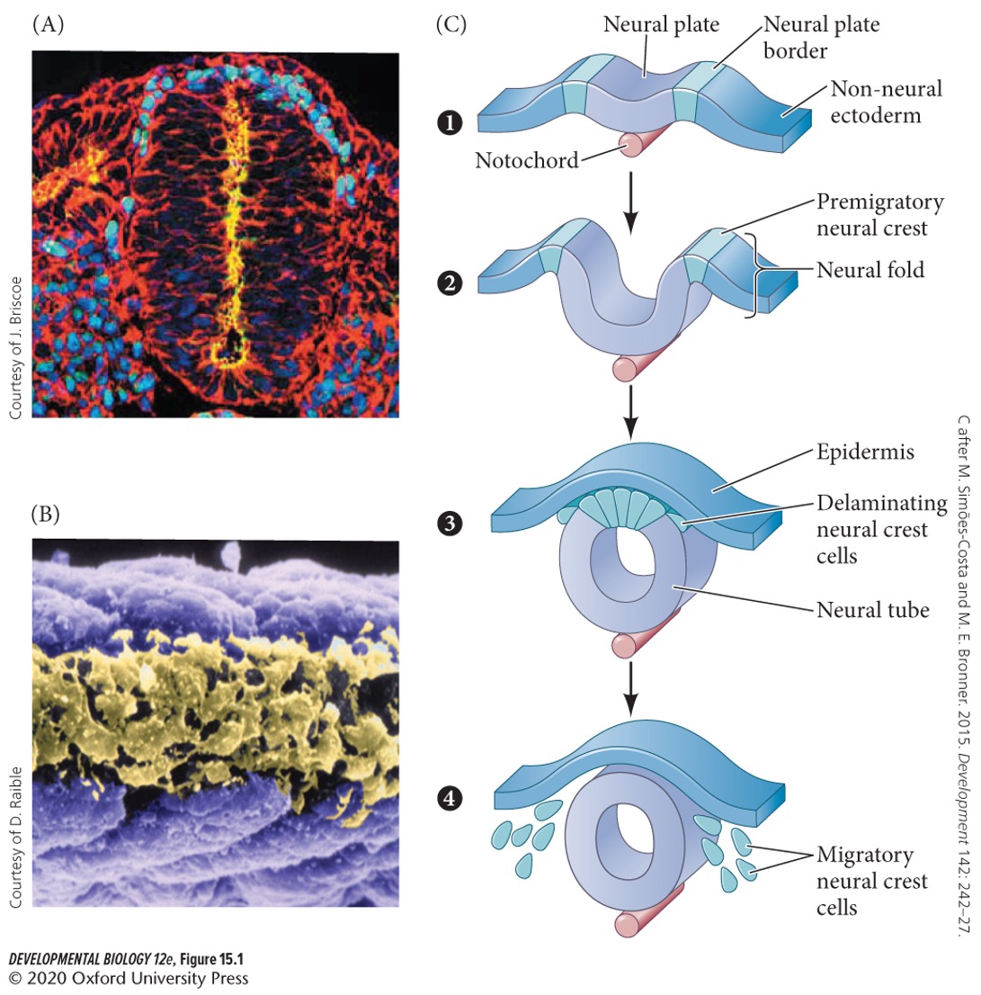
Neural crest
a collection of multipotent stem cells located at the side of the neural tube proximal to the epidermal layer after neurulation
2
New cards
The neural crest is found in:
A) only vertebrates
B) only invertebrates
C) vertebrates and invertebrates
D) neither
A) only vertebrates
B) only invertebrates
C) vertebrates and invertebrates
D) neither
A) only vertebrates
3
New cards
The neural crest is a transient structure (adults do not have a neural crest) (T/F).
True.
4
New cards
Neural crest cells originate from the
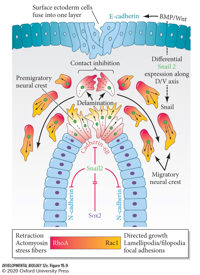
A) non-neural ectoderm
B) dorsal neural tube
C) notochord
D) neural plate
A) non-neural ectoderm
B) dorsal neural tube
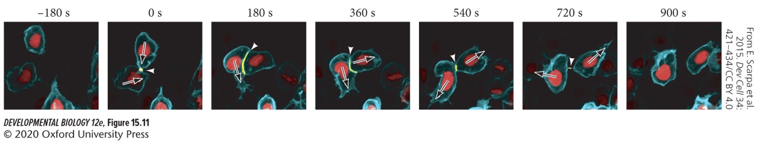
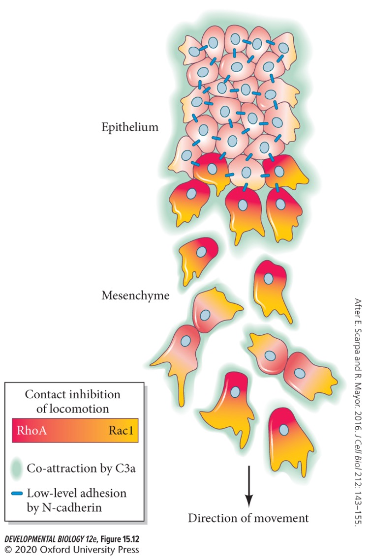
C) notochord
D) neural plate
B) dorsal neural tube (ectoderm origin)
5
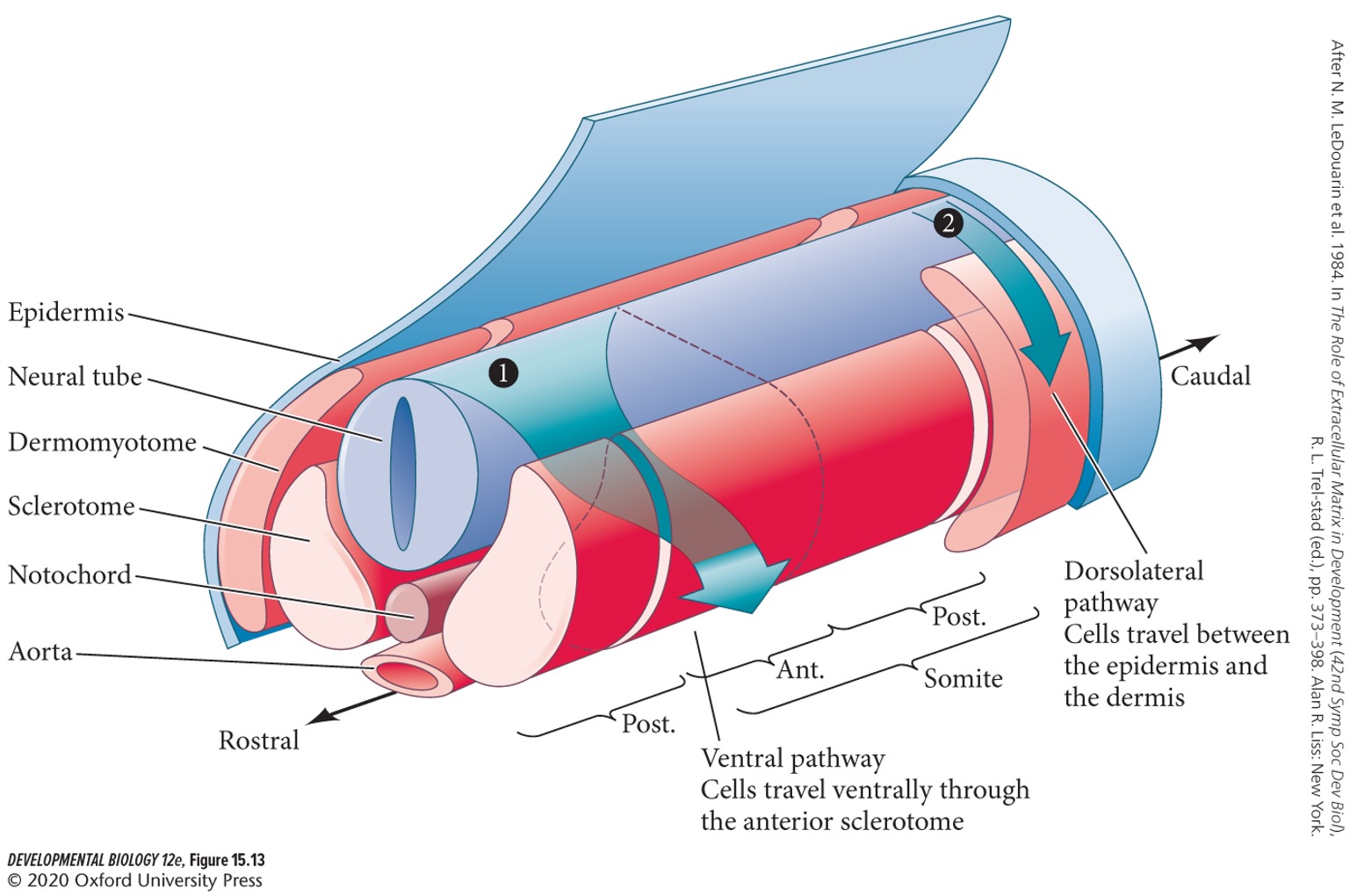
New cards
Neural crest cells undergo an ….. transition and migrate extensively to generate a huge variety of cell types.
A) mesenchymal to epithelial
B) epithelial to mesoderm
C) mesenchymal to mesoderm
D) epithelial to mesenchymal
A) mesenchymal to epithelial
B) epithelial to mesoderm
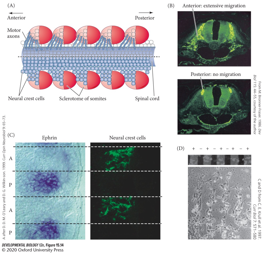
C) mesenchymal to mesoderm
D) epithelial to mesenchymal
D) epithelial to mesenchymal
6
New cards
What is used as a model system for neural crest study?
Chicks
7
New cards
**Central nervous systen** (CNS)
brain and spinal cord.
8
New cards
**Peripheral nervous system**
everything outside of the CNS, divided into:
1. **Somatic** **nervous system**
2. **Auronomic** **nervous system**
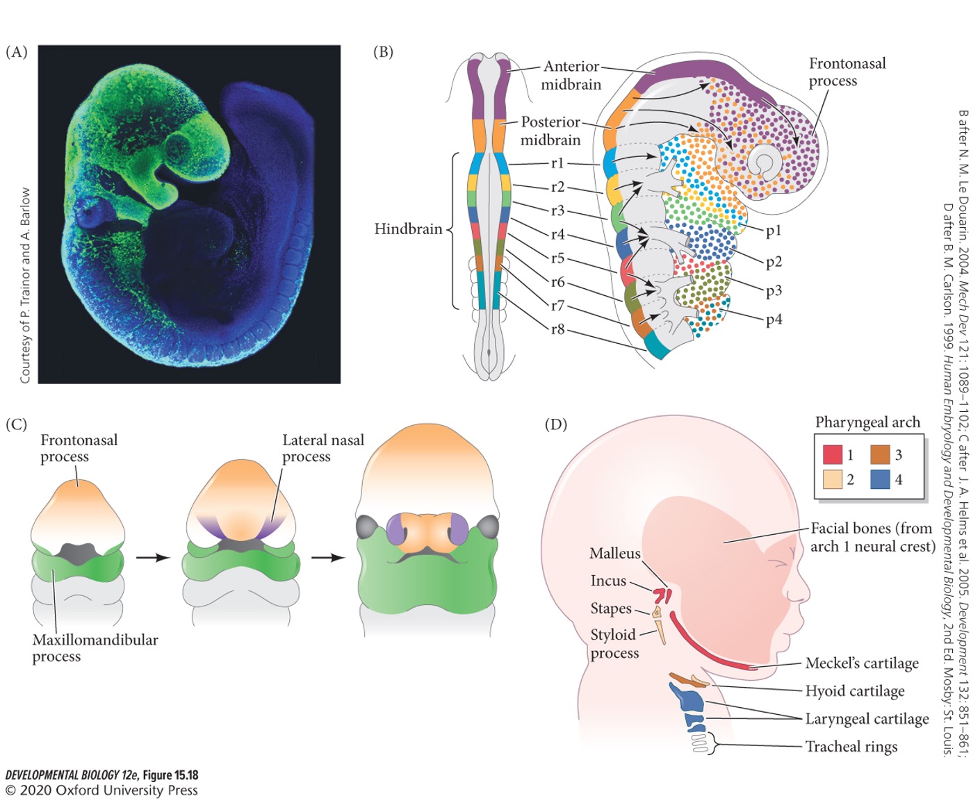
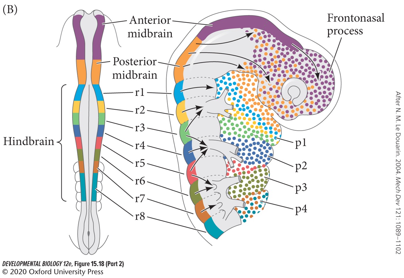
1. **Somatic** **nervous system**
2. **Auronomic** **nervous system**
9
New cards
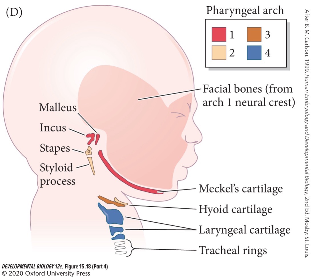
Somatic nervous system is involuntary (T/F).
False. The somatic nervous system is voluntary, the autonomic nervous system is involuntary.
10
New cards
Somatic nervous system includes which two nerves?
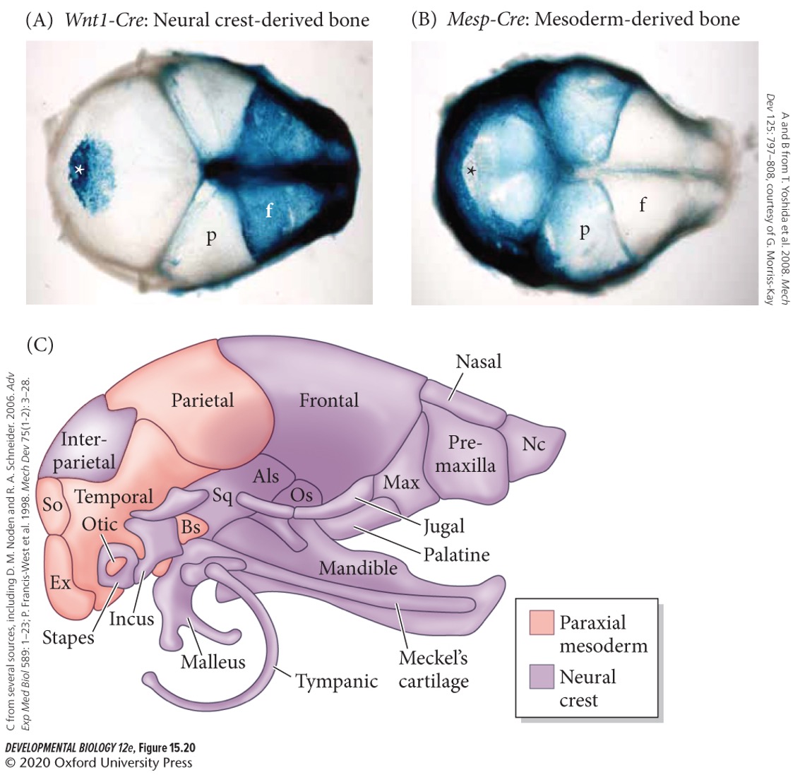
1. **Afferent** (sensory) **nerves**
2. **Efferent** (motor) **nerves**
11
New cards
The autonomic nervous system includes which three systems?
1. **Sympathetic nervous system** (fight or flight)
2. **Parasympathetic nervous system** (breed and feed, rest and digest)
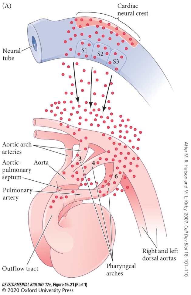
3. **Enteric nervous system** (governing gastrointestinal tract function).
12
New cards
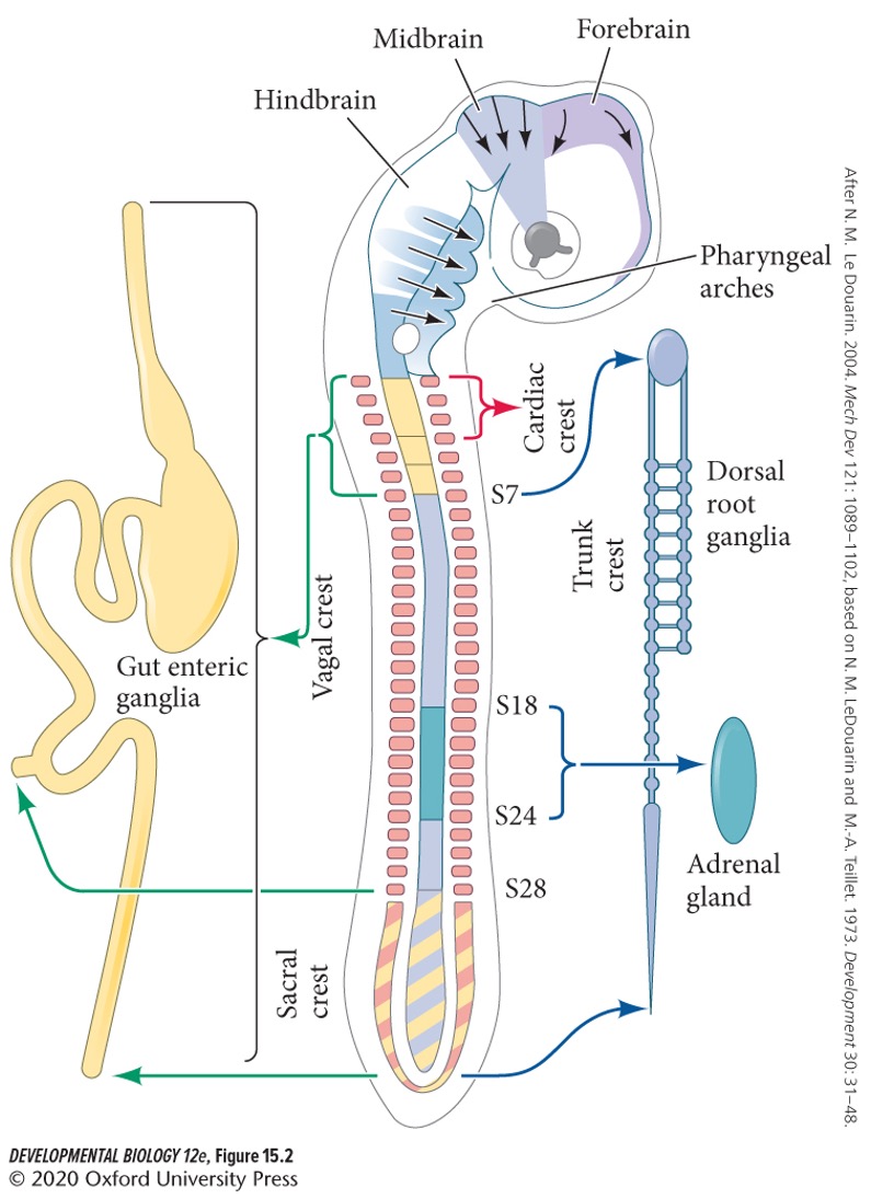
Neural crest cells can be divided into four main, overlapping anatomical regions:
1. Cranial (cephalic)
2. Cardiac
3. Trunk
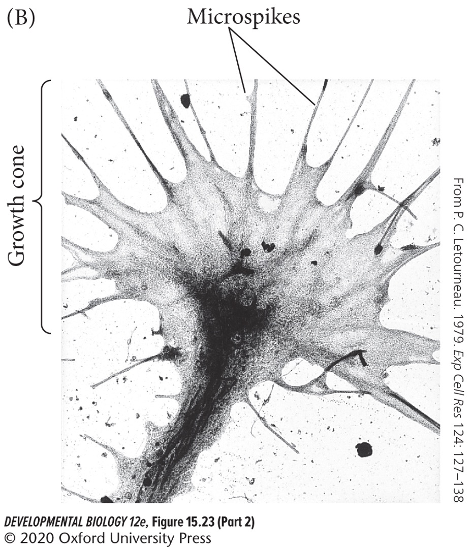
4. Vagal and Sacral

13
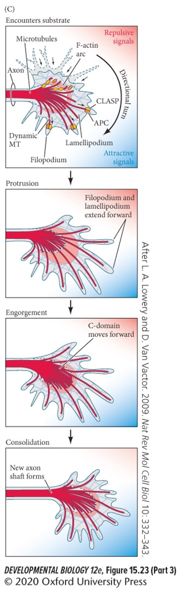
New cards
**Cranial** neural crest cells
migrate to produce the craniofacial mesenchyme, including cartilage, bone, neurons, glia, **melanocytes** (pigment cells), and connective tissues of the face.
14
New cards
**Cardiac** neural crest cells
extending from the otic (ear) places to the third somites. These contribute to the septum of the heart, form the connective tissue of the wall of the large arteries of the heart, and develop into melanocytes, neurons, cartilage, and connective tissue of the third, fourth, and sixth pharyngeal arches.
15
New cards
**Trunk** neural crest cells
lies from about stomate 6 to the tail and make the **dorsal root ganglia** containing sensory neurons, sympathetic neurons (fight or flight), the medulla part of the adrenal gland, and melanocytes.
16
New cards
**Vegal** and **Sacral** neural crest cells
form the parasympathetic (autonomous nervous system) ganglia of the gut and the enteric nervous system. The vagal (neck) neural crest overlaps that of the head and the trunk (about commits 1-7), and the sacral neural crest is posterior to somite 28.
17
New cards
Which neural crest cells enter the pharyngeal arches and pouches to give rise to thymic cells, the odontoblasts of the tooth primordial, and the bones of the middle ear and jaw?
Cranial
18
New cards
The cardiac neural crest is a subregion of the cranial neural crest (T/F).
True.
19
New cards
Nueral crest cells from different regions are *equivalent/not equivalent* in potency and specificity.
*Not equivalent*. Cranial crest cells can make cartilage, muscle, bone, and connective tissue of the cornea, but trunk cells cannot.
20
New cards
When trunk cells are transplanted to the head region, they can migrate to sites of cartilage and cornea formation and can form either structure (T/F).
False. They __cannot__ form either structure.
21
New cards
Expression of which gene helps determine specificity of the trunk neural crest?
A) Wnts
B) Nodal
C) Hox gene
D) Sox9
A) Wnts
B) Nodal
C) Hox gene
D) Sox9
C) Hox gene
22
New cards
Most neural crest cells behave as multipotent stem cells even after they begin migrating (T/F).
True. Like other stem cells, paracrine factors specify more and more committed cell fates.
23
New cards
Multipotent stem cells
cells that have the capacity to self-renew by dividing and to develop into multiple specialised cell types present in a specific tissue or organ.
24
New cards
1\. Which of the following is __**TRUE**__regarding the neural crest?
A) Neural crest cells have mesodermal origin
B) Neural crest cells undergo a mesenchymal to epithelial transition
C) The neural crest is a transient structure
D) The neural crest is found in both vertebrates and invertebrates
E) Neural crest cells are stationary
A) Neural crest cells have mesodermal origin
B) Neural crest cells undergo a mesenchymal to epithelial transition
C) The neural crest is a transient structure
D) The neural crest is found in both vertebrates and invertebrates
E) Neural crest cells are stationary
C) The neural crest is a transient structure
25
New cards
Specification of neural crest cells begins when?
During early gastrulation.
26
New cards
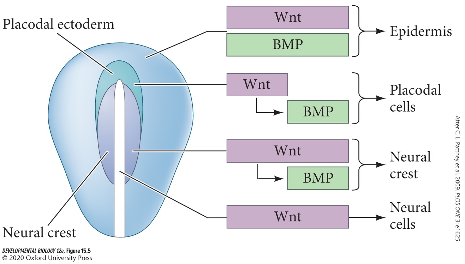
…. and …. activate a sequential series of transcription factors that progressively specify neural crest cell fate. Reconstitution of these pathways allows the reprogramming of other cell types into neural crest cells.
Wnts and BMPs
27
New cards
*Epidermis* is specified by:
A) BMP
B) Wnt
C) BMP and Wnt
D) Wnt expression early, BMP expression later.
E) Wnt expression later, BMP expression early.
F) Wnt expression persistence, BMP expression later.
A) BMP
B) Wnt
C) BMP and Wnt
D) Wnt expression early, BMP expression later.
E) Wnt expression later, BMP expression early.
F) Wnt expression persistence, BMP expression later.
C) BMP and Wnt
28
New cards
*Neural* is specified by:
A) BMP
B) Wnt
C) BMP and Wnt
D) Wnt expression early, BMP expression later.
E) Wnt expression later, BMP expression early.
F) Wnt expression persistence, BMP expression later.
A) BMP
B) Wnt
C) BMP and Wnt
D) Wnt expression early, BMP expression later.
E) Wnt expression later, BMP expression early.
F) Wnt expression persistence, BMP expression later.
B) Wnt
29
New cards
*Placodes* is specified by:
A) BMP
B) Wnt
C) BMP and Wnt
D) Wnt expression early, BMP expression later.
E) Wnt expression later, BMP expression early.
F) Wnt expression persistence, BMP expression later.
A) BMP
B) Wnt
C) BMP and Wnt
D) Wnt expression early, BMP expression later.
E) Wnt expression later, BMP expression early.
F) Wnt expression persistence, BMP expression later.
D) Wnt expression early, BMP expression later.
30
New cards
*Neural crest* is specified by:
A) BMP
B) Wnt
C) BMP and Wnt
D) Wnt expression early, BMP expression later.
E) Wnt expression later, BMP expression early.
F) Wnt expression persistence, BMP expression later.
A) BMP
B) Wnt
C) BMP and Wnt
D) Wnt expression early, BMP expression later.
E) Wnt expression later, BMP expression early.
F) Wnt expression persistence, BMP expression later.
F) Wnt expression persistence, BMP expression later.
31
New cards
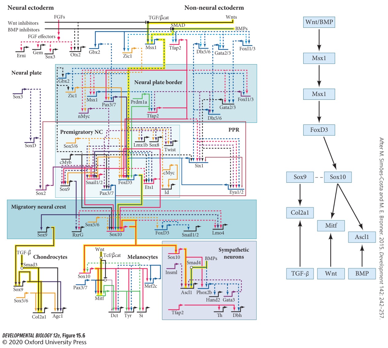
Which is the key specifier of neural crest?
A) Sox10
B) Sox9
C) Snail
A) Sox10
B) Sox9
C) Snail
A) Sox10
32
New cards
Which factors are required for the epithelia to mesenchymal transition?
A) Sox10
B) Sox9
C) Snail
A) Sox10
B) Sox9
C) Snail
B + C) Sox9 and Snail
33
New cards
Neural crest cells must migrate. What effects their migration?
Environmental cues. Including cells around them, local adhesive cues, lang range secreted factors, and tracks.
34
New cards
Do neural crest cells migrate collectively or individually?
Both collectively and individual.
35
New cards
What signal initiate migration (**epithelial-mesenchymal transition** (EMT))?
Activation of Wnt genes by BMP.
36
New cards
Where are BMPs made?
Dorsal region of the neural tube (below neural crest tissue).

37
New cards
When BMP inhibitor concentration from the … is reduced, BMPs can become active.
notochord.
38
New cards
What happens when the Wnt genes are activated?
This primes the cytoskeleton for cell migration and dissociates the cadherin connections.
39
New cards
Which genes are activated by Wnt signaling to prime the cytoskeleton?
A) PAX3; SP1
B) Rho; Rac GTPases
C) FGFs; SOX2
D) Sox9;Snail
E) GATA5; HEY
A) PAX3; SP1
B) Rho; Rac GTPases
C) FGFs; SOX2
D) Sox9;Snail
E) GATA5; HEY
B) Rho; Rac GTPases
40
New cards
Which proteins dissociate the cadherin connections (repress N-cadherin and E-cadherin domain)?
A) BMPs
B) Sox9
C) Nodal
D) Snail-2
A) BMPs
B) Sox9
C) Nodal
D) Snail-2
D) Snail-2
41
New cards
Neural crest cells display **contact inhibition of locomotion**, this causes what?
Dispersal of cells.
42
New cards
Which two signals cause contact inhibition of locomotion?
1. Non-canonical Wnt signaling
2. RhoA
43
New cards
Certain neural crest cells display **collective migration**. How is this mediated?
1. Low level N-cadherin
2. **C3a**
44
New cards
After early specification and delamination from the dorsal neural tube, neural crest cells migrate along different paths to their specific locations for final differentiation (T/F).
True.
45
New cards
What are the two major pathways for trunk neural crest migration?
1. **Ventral pathway**
2. **Dorsolateral pathway**
46
New cards
Which pathway do early trunk cells follow?
Ventral pathway.
47
New cards
What do the cells following the ventral pathway form?
* Sensory neurons
* Autonomic neurons
* Adrenomedullary cells
* Schwann cells
* Autonomic neurons
* Adrenomedullary cells
* Schwann cells
48
New cards
**Sclerotome**
Place where the Schwann cells travel through (part of the somite that will form the vertebrae).
49
New cards
Later cells follow the dorsolateral pathway. What do they become?
* Melanocytes (travel between the dermis and epidermis).
50
New cards
Ventrally migrating trunk cells can move either between the somites (to form the sympathetic ganglia of the aorta) or through the somites (T/F).
True.
51
New cards
Early trunk cells go between somites, but this pathway is soon blocked by a protein that repels neural crest cells, called?
**Semaphorin-3F**
52
New cards
The receptor for semaphorin-3F on the neural crest cells is?
**Neuropilin-2**.
53
New cards
Only the extracellular matrix of the posterior of each somite allows migration (T/F).
False. Only the extracellular matrix of the __anterior__.
54
New cards
The posterior portion of each somite contains a protein which also repel neural crest cells, called?
**Ephrins**.
55
New cards
The receptor for ephrins on neural crest cells is?
**Eph**
56
New cards
This patterning of neural crest cell migration generates the overall segmented character of the *peripheral nervous system/central nervous system.*
peripheral nervous system
57
New cards
Ventrally-migrating trunk cells that differentiate within the sclerotome become what?
**Dorsal root ganglia.**
58
New cards
Dorsal root ganglia
contain the sensory neurons that relay information to the central nervous system, and glia cells.
59
New cards
Ventrally-migrating cells that continue migrating past the sclerotome become part of:
A) Parasympathetic autonomous nervous system
B) Sympathetic autonomous nervous system
C) Enteric autonomous nervous system
D) A + B
A) Parasympathetic autonomous nervous system
B) Sympathetic autonomous nervous system
C) Enteric autonomous nervous system
D) A + B
D) A + B
60
New cards
Trunk cells that will colonize the gut (enteric nervous system) are attracted to the digestive tube by which paracrine factor?
Glial-derived neurotrophic factor (GDNF).
61
New cards
Glial-derived neurotrophic factor (GDNF)
activate cell division and induce neural differentiation.
62
New cards
The receptor for GDNF on neural crest cells is?
**Ret**
63
New cards
**Melanoblasts** (precursors to **melanocytes**, the pigment cells) migrate via the *ventrally pathway/dorsolateral pathway*.
dorsolateral pathway.
64
New cards
The transcription factor that specifies melanoblasts is?
MITF
65
New cards
MITF
activates genes responsible for pigment production, specifies travel along the dorsolateral pathway into the skin, and prevents apoptosis of migrating cells.
66
New cards
Mutations in MITF lead to defects. What is an examples?
Dalmatians and American paint horses heterozygous for MITF have spots due to random death of melanoblasts. Melanocytes also induce blood formation in the inner ear, so they have a high frequency of deafness.
67
New cards
Other neural crest-expressed genes lead to defects too. A mutation in the **KIT** gene for example results in what?
Piebaldism
68
New cards
Piebaldism
reduced proliferation of neural crest cells as well as germ cell and blood cell precursors.
69
New cards
2\. _____________ and ________________ are required for the epithelia to mesenchymal transition of neural crest cells.
A) PAX3; SP1
B) Wnts; Nodal
C) FGFs; SOX2
D) Sox9; Snail
E) GATA5; HEY2
A) PAX3; SP1
B) Wnts; Nodal
C) FGFs; SOX2
D) Sox9; Snail
E) GATA5; HEY2
D) Sox9; Snail
70
New cards
Which cells are largely responsible for the formation of the head?
Cranial
71
New cards
Cranial neural crest cells migrate from the midbrain and hindbrain anterior to:
A) Frontonasal process
B) Lateral nasal process
C) rhombomere 8
D) Pharyngeal arches
A) Frontonasal process
B) Lateral nasal process
C) rhombomere 8
D) Pharyngeal arches
C) rhombomere 8
72
New cards
Rhombomere 8 migrates into the:
A) Frontonasal process
B) Lateral nasal process
C) Pharyngeal arches
D) A + C
A) Frontonasal process
B) Lateral nasal process
C) Pharyngeal arches
D) A + C
D) A + C
73
New cards
What do the pharyngeal arches and the frontonasal process form?
Face.
74
New cards
What are the three major pathways of cranial cell migration?
1. Neural crest cells from the midbrain and rhombomeres 1 and 2 of the hindbrain migrate to the first pharyngeal arch (the mandibular arch).
2. Here they form the jawbones, the incus and malleus bones of the ear, and various nerves.
3. Additional cells enter the head and form the frontonasal process, which is composed of the forehead, middle of the nose, and primary palate.
75
New cards
Rhombomere 4
populate the second pharyngeal arch, which forms;
* the upper hyoid cartilage of the neck
* the stapes bone of the middle ear
* additional nerves
* the upper hyoid cartilage of the neck
* the stapes bone of the middle ear
* additional nerves
76
New cards
Rhombomeres 6-8
Migrate into the third and fourth pharyngeal arches to form:
* Lower hyoid cartilage
* Contribute to the thymus, parathyroid, and thyroid glands
* Contribute to the aorta and pulmonary arteries.
* Lower hyoid cartilage
* Contribute to the thymus, parathyroid, and thyroid glands
* Contribute to the aorta and pulmonary arteries.
77
New cards
Rhombomeres 3 and 5
Join streams from rhombomeres 2, 4 and 6.
78
New cards
Cranium
vertebrate skull.
79
New cards
Neural crest cells form most of the **cranium** (T/F).
True.
80
New cards
Intramembranous bones
Bones created by laying down calcified spicules directly in connective tissue without a cartilaginous precursor.
81
New cards
Are cranial bones intramembranous?
Yes.
82
New cards
The front of the head is derived from:
A) Neural crest
B) Mesoderm
C) Neural crest and mesoderm
A) Neural crest
B) Mesoderm
C) Neural crest and mesoderm
A) Neural crest
83
New cards
The back of the skull is derived from:
A) Neural crest
B) Mesoderm
C) Neural crest and mesoderm
A) Neural crest
B) Mesoderm
C) Neural crest and mesoderm
C) Neural crest and mesoderm
84
New cards
Facial muscles are derived from:
A) Neural crest
B) Mesoderm
C) Neural crest and mesoderm
A) Neural crest
B) Mesoderm
C) Neural crest and mesoderm
C) Neural crest and mesoderm
85
New cards
Rates and directions of cranial neural crest cell divisions determine what our face looks like (T/F).
True.
86
New cards
Where does the heart originally form?
Neck region.
87
New cards
**Cardiac neural crest**
In the caudal (posterior) part of the cranial neural crest and gives rise to the endothelium (inner lining) of the aortic arch arteries and the septum between the aorta and the pulmonary artery.
88
New cards
Cardiac neural crest cells are attracted to the region by which gene?
**Fgf8**.
89
New cards
Cardiac cells also form other neck structures, including:
* Thyroid parathyroid
* Thymus glands
* Carotid body
* Thymus glands
* Carotid body
90
New cards
Which gene do cardiac cells express in mice?
Pax3
91
New cards
How do congenital heart defect often occur?
Defects in the neck structures.
92
New cards
Neurons can generate extremely complex neural networks (T/F).
True.
93
New cards
Axons grown via?
Growth cones.
94
New cards
Growth cones
the locomotion apparatus of an axon.
95
New cards
How do growth cones migrate?
via environmental sensing using the same types of signals as migrating neural crest cells.
96
New cards
Growth cones move by pointed filopodia called?
Microspikes.
97
New cards
The navigation of axons to their appropriate targets depends on guidance molecules in the extracellular environment. What are the major regulator of actin filament dynamics that drive movement.
Rho GTPases
98
New cards
Pioneer nerve fibers
go ahead of other axons and serves as guides. They migrate while embryonic distances are still short and embryonic tissue is relativist uncomplicated.
99
New cards
All pioneer neurons die after follow up neurons reach their destination (T/F).
False. Some die.
100
New cards
The specificity of neuronal connections depends on which three steps?
1. Pathway selection
2. Target selection
3. Address selection