Unit 2: Textbook/slide definitons
1/107
There's no tags or description
Looks like no tags are added yet.
Name | Mastery | Learn | Test | Matching | Spaced |
|---|
No study sessions yet.
108 Terms
Cell theory
(what are the three parts?)
All living things are made of cells.
The cell is the fundamental unit of life
All cells come from pre-existing cells
Basic unit of life
Cells
Prokaryotic cells
Cells that lack a nucleus
EX: Bacteria, Archaea
Eukaryote cells
Cells that have a nucleus
EX: Plants, Animals, Fungi & Protists
Nucleoid
Found in prokaryotic cells (where DNA is found)

Flagella
Structures that extend that allow them to move
Organelles
Small structures inside a cell that perform specific functions to keep the cell alive
Transcription
Making mRNA from a DNA template
✅ Happens in the nucleus
✅ DNA → mRNA (message to build a protein)
Translation
Where ribosomes read mRNA to build a protein using amino
Happens in the cytoplasm
✅ Uses mRNA, tRNA, and ribosomes
✅ Each 3-letter mRNA code = 1 amino acid
Process of transcription/translation in Eukaryote cells
Transcription in nucleus > translation later in cytoplasm
Process of transcription/translation in Prokaryote cells
Translation occurs as soon as mRNA is transcribed from the DNA template
The main component of cell membranes
Lipids
Their properties form a barrier in an aqueous (watery) environment
(creating the cell membrane)
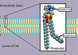
Major types of lipids found in cell membranes
Phosolipids
Amphipathic
Hydrophilic and Hydrophobic groups being in the same molecule
Cell membrane (aka plasma membrane)
Thin, flexible outer layer that surrounds the cell
It controls what goes in and out of the cell
Made mostly of phospholipids (a type of lipid)
Found in ALL cells
Phosolipid
Type of lipid with a hydrophilic head and two hydrophobic tails
Shapes of structures phospholipids can form
Micelles
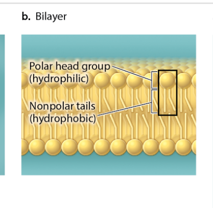
Bilayer
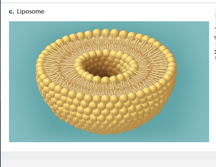
Liposomes
These are the shapes a cell membrane can form into
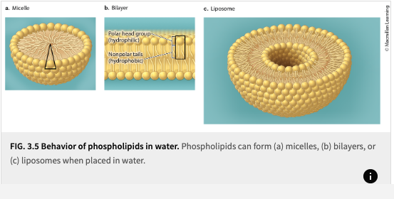
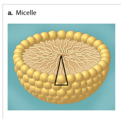
Micelles
Wedge-shaped and packed into spherical structures
(Bulky heads and a single hydrophobic tail are packed)
Bilayer
Structure of two layers where the phospholipid heads are on the outside and the tails are in the inside
Liposome
A cell like structure sphere made up of phospholipid bilayers
Two features that affect the membranes fluidity
Length/number of carbon-carbon double bonds in fatty acid tails
The longer the fatty acid tails
Explanation:
The longer the fatty acid tails, the less fluid in the membrane
The shorter the fatty acid tails, the more fluid in the membrane
Longer fatty acid tails have more surface area, so they stick together more through Van der Waals forces, making the membrane less fluid
also: the less carbon-carbon double bonds, the less fluid in the membrane
How do saturated and unsaturated fatty acids affect membrane fluidity?
Saturated fats → less fluid (tails pack tightly)
Unsaturated fats → more fluid (tails prevent tight packing)
Major component of animal cell membranes
Cholestreol
(also amphipathic)
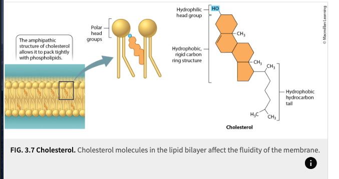
What chemical group is cholesterol in?
Hydroxyl group (-OH)
How does temperature affect cholesterol’s membrane fluidity?
Reduces fluidity at moderate temperatures.
Prevents solidification at low temperatures.
💡 Think of cholesterol as a buffer:
It helps the membrane stay just right — not too fluid, not too rigid — across temperature changes.
Lipid rafts
Small, organized patches in the cell membrane made of tightly packed lipids and proteins.
They gather special lipids that help stabilize and organize parts of the membrane
They help with cell signaling and membrane organization.
FACT: Most membranes contain proteins as well as lipids`
Membrane proteins serve different functions
main functions of membrane proteins are?
Transport
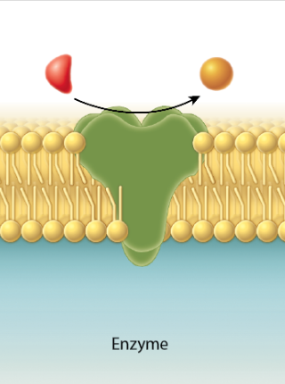
Enzymatic activity
Signal transduction
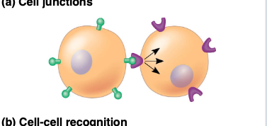
Cell-cell recognition
Intercellular joining
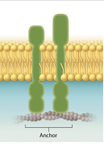
Attachment to the cytoskeleton and extracellular matrix (ECM)
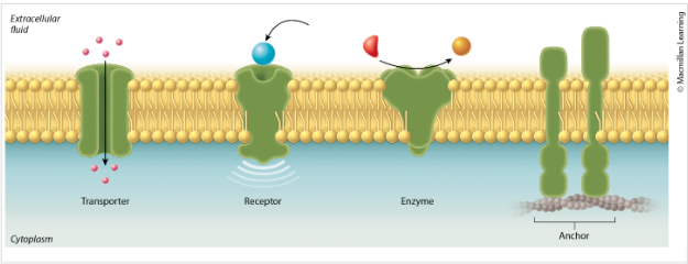
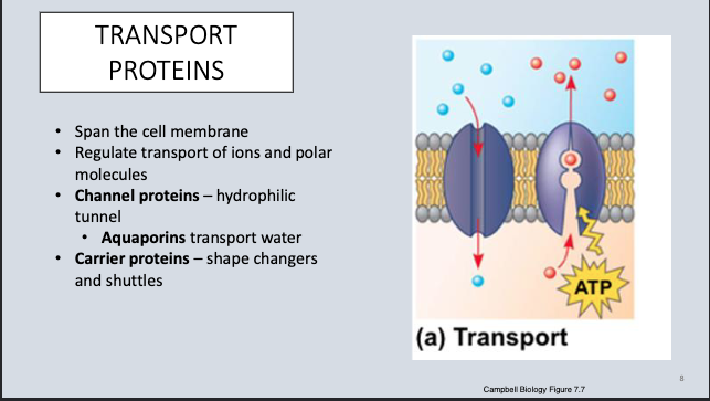
Transporter proteins
Moves Ions or molecules across the membrane
Channel proteins- Hydrophilic tunnel
Aquaporins- Transport water
Carrier proteins- Shape changers and shuttles
(May be passive or active)

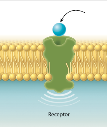
Receptors
Allows the cells to recieve signals form the environment
Enzymes
Catalyze (speed up) chemical reactions
Anchors
Attaches to other protiens and help maintain cell structure + shape
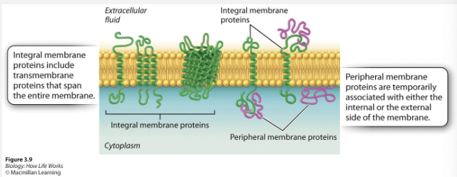
Intergral membrane proteins
Proteins that are permanently embedded in the membrane and can’t be removed without damaging it
Partially or fully inside membrane

Peripheral membrane proteins
Proteins temporarily attached to the membrane, easily removed, and involved in signaling or support

Transmembrane protein
A TYPE of integral protein that spans the entire lipid bilayer with both hydrophilic and hydrophobic regions (Fully crosses from one side to the other)
Key Function:
Transfers signals or materials from one side of the membrane to the other
What does FRAP show about membrane proteins?
That they are mobil and can move within the membrane
FRAP: is a technique use to test whether proteins moved around in the cell membrane
How it works:
Proteins in the membrane are tagged with fluorescent dye.
A laser bleaches one section so it loses its glow.
Over time, if unbleached (glowing) proteins move into the bleached spot, it proves proteins are mobile.
Main takeaway:
FRAP shows that membrane proteins are not locked in place — they can move within the membrane.
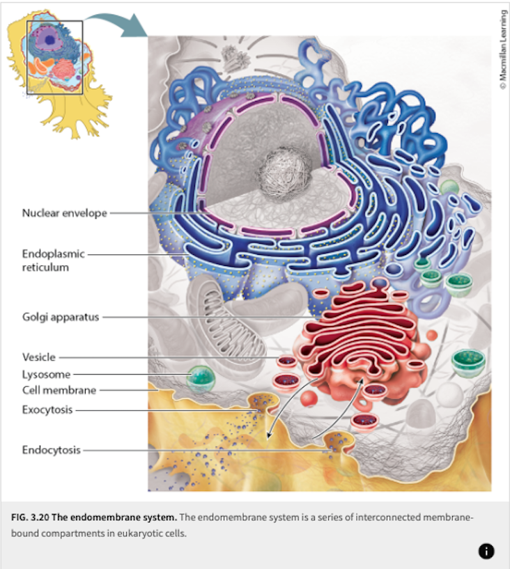
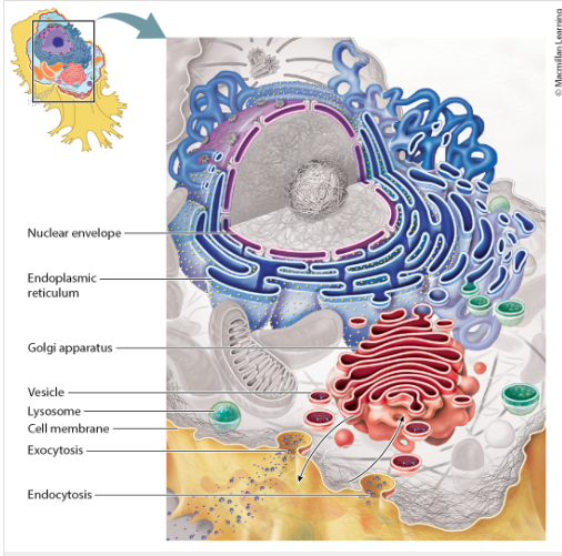
KEY POINT: Endomembrane system
A specific group of membrane-bound organelles in eukaryotic cells that:
Make, modify, and move proteins and lipids
Use vesicles to transport materials
Work together as a connected team through membranes
(Found in eukaryotes (not common in prokaryotes)
Examples: Rough/Smooth ER, Golgi apparatus, Lysosomes, plasma membrane, etc.
(Inside/functions within cells)

Nuclear envelope (double membrane)
Double membrane surrounding the nucleus
The nuclear envelope has two phospholipid bilayers
(inner membrane + outer membrane)It’s like the nucleus is wearing two jackets, not just one
Thats why we say double membrane
Nucleus function
Stores DNA and controls gene expression
Nuclear pores
Controls what leaves/enters the nucleus
mRNA leaves through pores to go to ribosomes for protein synthesis
Endoplasmic Recticulum (ER)
It makes proteins (rough ER) and lipids (smooth ER), and helps with detox and calcium storage
Connected to the nuclear envelope
Rough ER
Makes proteins, helps fold and modify them, send them to Golgi in vesicles
Has ribosomes attached (that’s why it looks “rough”)
Smooth ER
Makes lipids, Detoxifies harmful substances, Stores calcium
Has no ribosomes
Golgi apparatus
Modifies and ships proteins to the cell membrane
Functions:
1⃣ Modifies proteins and lipids received from ER.
2⃣ Sorts and packages them into vesicles for delivery.
3⃣ Adds carbohydrates to proteins/lipids (glycosylation) to form glycoproteins & glycolipids.
4⃣ Directs proteins to:
Cell membrane
Lysosomes
Other organelles
Secretion outside the cell

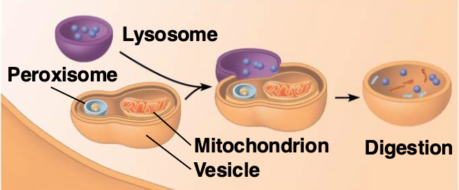
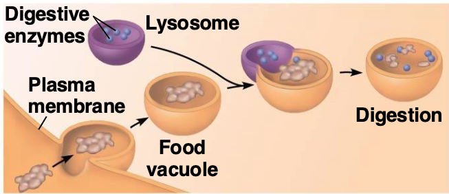
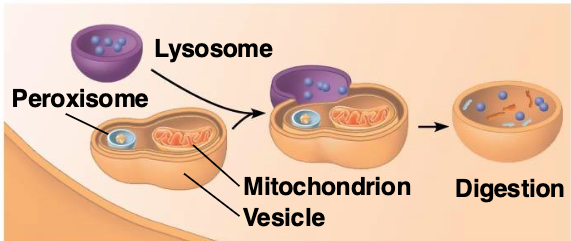
Lysosomes
Break down waste, damaged cell parts, and other materials using digestive enzymes

Vesicles
Transport materials between organelles or to/from ER > Golgi > its other destinations
Vesicle Transport = Tiny workers carrying boxes inside the store (cell)
Vesicles are like delivery trucks or employees
They move materials between departments (organelles)
Or carry stuff to/from the "storefront" (cell membrane)
🔁 What vesicles do:
Pick up stuff from the ER
Drop it off at the Golgi
Then deliver it to the membrane, lysosomes, or storage areas
They never float randomly — they move on tracks (cytoskeleton) or fuse with membranes on purpose.

What is the role of the cell membrane in the endomembrane system?
It’s the outer boundary that helps regulate what enters and exits the cell; part of the system's transport and signaling
Cell wall
Provides structure and support to the plant cell and protects against damage
(found in plants)

Large vacuole (stores water)
Stores water, nutrients and wast; helps maintain turgor pressure
Turgor pressure is the pressure of the water pushing against the inside of a plant cell’s cell wall.
It happens when the large central vacuole fills with water, causing the cell to swell slightly and stay firm.
Why it matters:
Turgor pressure helps keep plants upright
Without it (like in drought), plants get wilted and floppy
(found in plants)
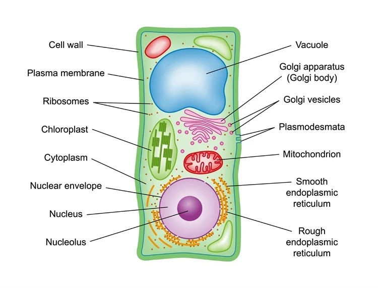
Chloroplasts (photosynthesis)
Chloroplasts convert sunlight into chemical energy through photosynthesis
By chemical energy take away:
They take light energy from the sun
And turn it into energy stored in molecules (aka chemical energy)
Specifically:
They make glucose (sugar), which is a chemical energy source the plant can later use to make ATP through cellular respiration.

Plasmodesmata
Channels that connect plant cells and allow molecules like proteins and mRNA to pass between them

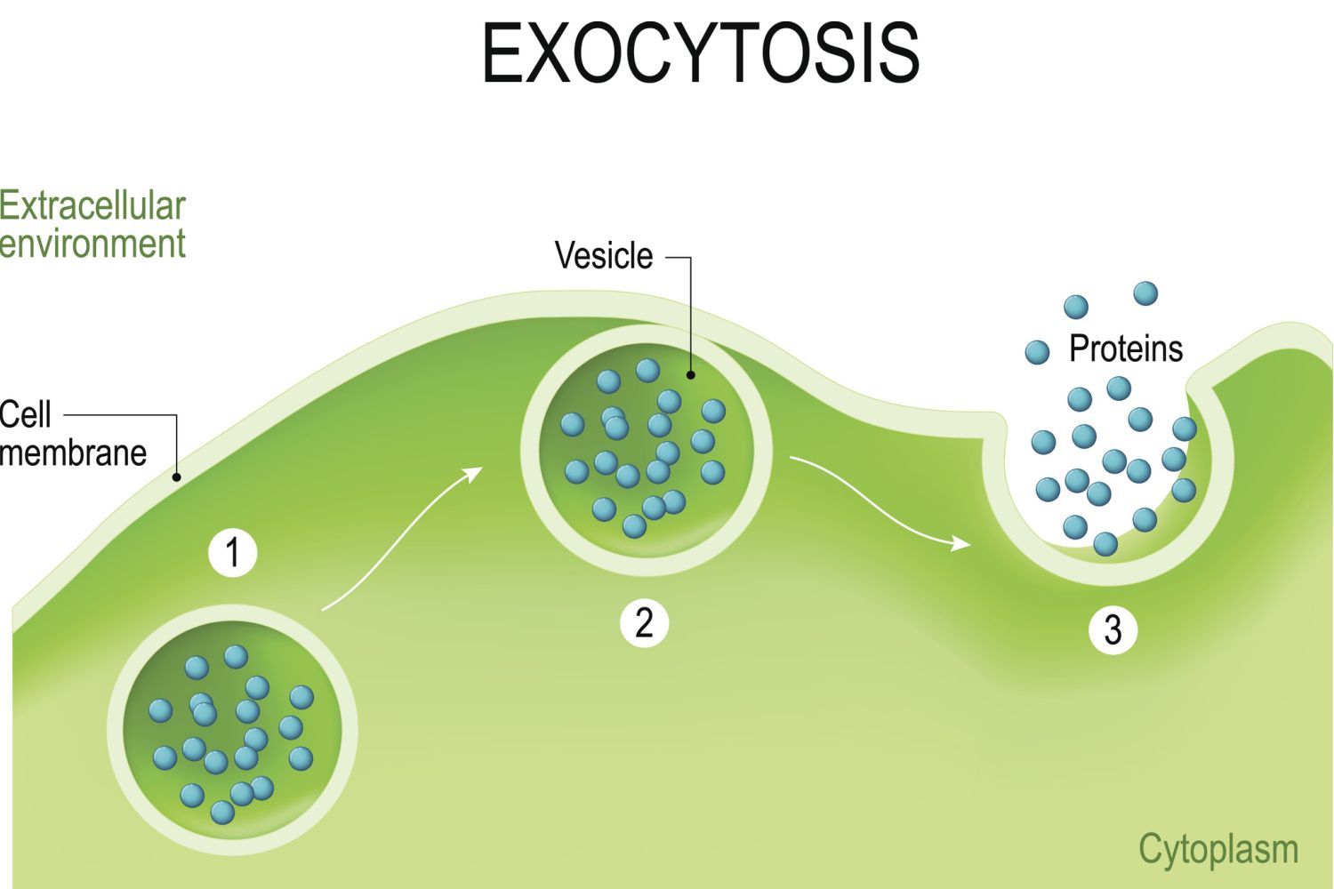
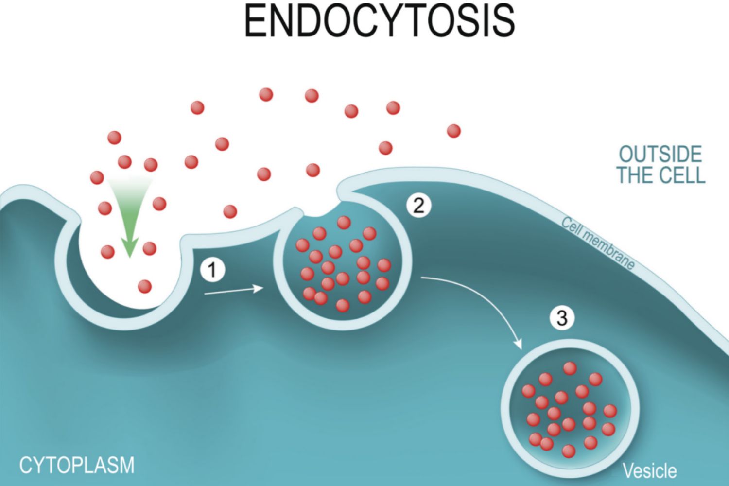
Exocytosis
vesicle fuses with the membrane to release contents outside the cell
Endocytosis
vesicle buds inward from the membrane to bring things inside
the membrane itself folds inward to create the vesicle.
Physical separation in cells
Inside organelles
Cytosol (fluid outside organelles but inside the cell)
Molecules are restricted to specific areas unless moved by vesicles
Cytosol
Fluid outside organelles but inside the cell
(In both Prokaryote and Eukaryote cells)
What is the cytosol vs cytoplasm?
Cytosol = fluid portion; Cytoplasm = cytosol + all organelles except the nucleus
Glycosylation
As proteins and lipids pass through the Golgi → enzymes attach sugars to them.
This creates:
Glycoproteins (protein + sugar)
Glycolipids (lipid + sugar)
Apoptoisis
Programmed cell death where the cell safely destroys itself to protect the organism
Chromosomes
carry genetic info

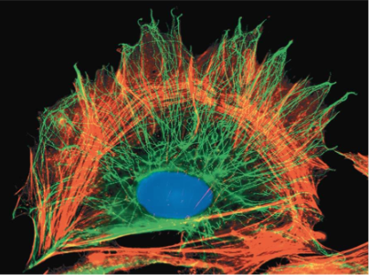
Cytoskeleton
Network of fibers inside cystoplasm
Organizes the cell’s structures and activities, anchoring many organelles
plays a role in:
shape
cell motility
regulation of biochemical activities
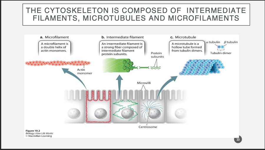
Cystoskeleton components
Microfilament
double helix of acting monomers
Shape, movement, muscle contraction
Intermediate filament
A strong fiber composed of intermediate filament protein subunits
Strength, anchor nucleus
Microtubule
A hallow tube formed from tubulin dimers
Movement, shape, chromosome separation
Cristae
Inner membrane folds inside mitochondria
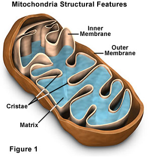
Matrix
Fluid-filled space inside mitochondria
Stroma
Fluid-filled space inside chloroplast
Thylakoid
Disc-like sacs inside chloroplast where light reactions happen
Granum
Stack of thylakoids (Disc-like sacs inside chloroplast where light reactions happen)
Autophagy
Process where lysosomes break down damaged organelles
Phagocytosis
Phagocytosis = “cell eating.”
It’s a type of endocytosis where the cell engulfs large particles (like bacteria, dead cells, or food).
A type of endocytosis where the cell engulfs large particles by surrounding them and bringing them into the cell.
Step-by-step:
1⃣ Cell extends its membrane outward → forms pseudopodia ("arms").
2⃣ Surrounds the particle.
3⃣ Membranes fuse and form a vesicle (phagosome) inside the cell.
4⃣ The vesicle often fuses with a lysosome to break down whatever was eaten.
Peroxisomes
Organelles that detox & break down fatty acids
Hydrolytic enzymes
Enzymes that break down molecules using water
Found in lysososmes
Motor Proteins
Proteins that move things along cytoskeleton tracks
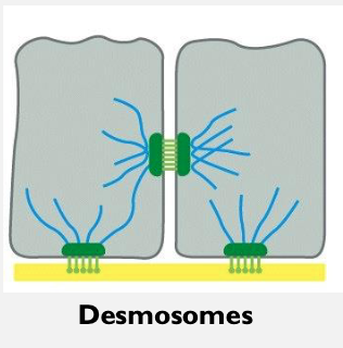
3 Types of cell junctions
Desmosomes
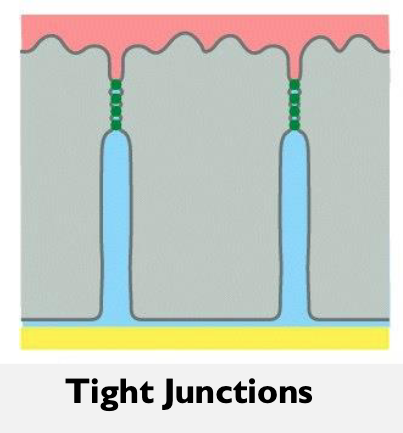
Tight junctions
Gap junction
Desmosomes
Anchor cells together using strong proteins > gives tissues strength & flexibility
Example:
Skin, heart muscle → tissues that stretch or handle mechanical stress.
Tight junctions
Structures that connect cells together or allow communication
Example:
Intestines → keeps food/waste in gut from leaking into body.
Gap junctions
Form channels (pores) between cells → allows small molecules, ions, and signals to pass directly from one cell to another.
Example:
Heart muscle → allows electrical signals to pass quickly for synchronized beats.
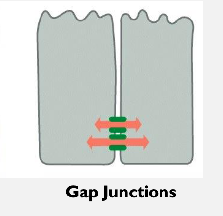
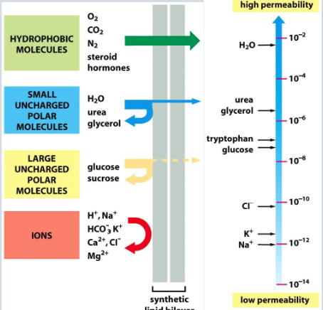
Chart of permeability
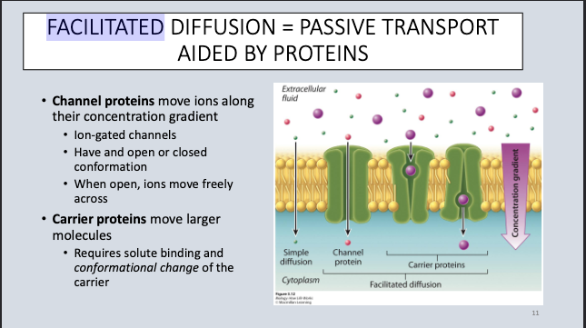
Channel proteins (type of transport protein)
Act like a tunnel/pore for molecules to pass through.
Usually for small ions or molecules.
Hydrophilic tunnel → allows polar/charged molecules to move through membrane.
Aquaporins (a type of transport protein)
Special type of channel protein.
Specifically for water transport.
Allow rapid movement of water across membrane.
Carrier proteins (a type of transport protein)
Bind to molecules.
Change shape to shuttle molecules across the membrane (conformational change).
Work for slightly larger or specific molecules.
Types of transport
Passive transport (req no energy)
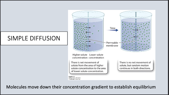
Simple diffusion (small, hydrophobic molecules)
Facilitated diffusion (Large, polar, or charged molecules) (They use transport proteins for this)
Osmosis (Facilitated diffusion of water across selectively permeable membrane)
Simple diffusion
Small, hydrophobic molecules
Molecules move down their concentration gradient to establish equilibrium
Facilitated diffusion = PASSIVE transport aided by proteins
Channel prteins
Carrier proteins
Tonicity
The ability of a surrounding solution to cause a cell to gain or lose water

membrane potential
Established by having different concentrations of ions on each side of the membrane
(create potential energy for active transport)
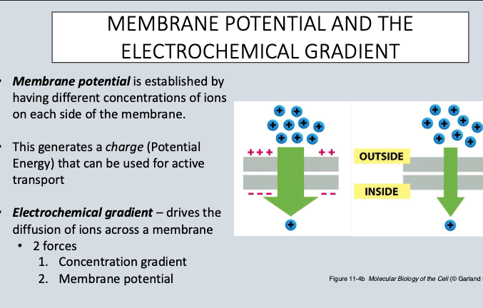
Electrochemical gradient
Drives the diffusion of ions across a membrane
2 forces:
Concentration gradient
Membrane potential
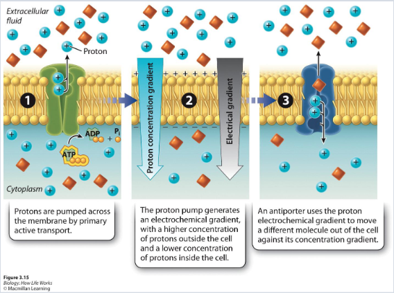
Electrogenic pump (this establishes membrane potential)
Transport protein that generates voltage across a membrane; help store energy that can be used for cellular work
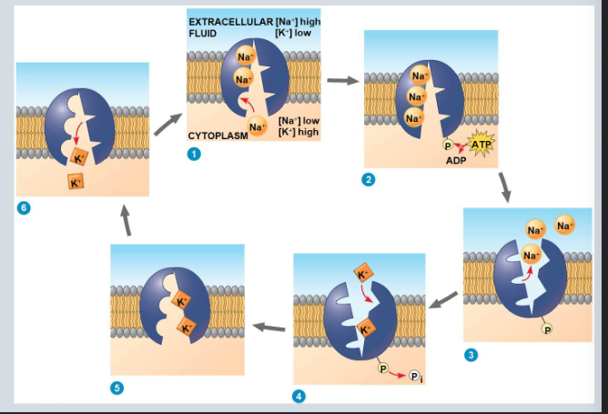
Major electrogenic pump in animal cells = sodium-potassium pump
Major electrogenic pump in plants, bacteria and fungi = proton pump
Sodium potassium pump (Primary active transport)
Establishes membrane potential in resting cells
Secondary Active Transport (Cotransport)
Uses energy from one molecule’s gradient to move another molecule.
Ex: H⁺ gradient helps move other substances.
Bulk transport
For moving large molecules (proteins, polysaccharides)
Requires energy.
1⃣ Exocytosis:
Vesicle fuses with membrane → releases contents outside.
2⃣ Endocytosis:
Membrane folds inward to bring material in:
Phagocytosis ("cell eating"): engulfs large particles.
Pinocytosis ("cell drinking"): engulfs fluids.
Receptor-mediated endocytosis: highly specific intake using receptors.
Cell Junctions |
allows
passage of signaling
molecules w/out crossing
plasma membrane
Cell-Cell Recognition
Direct contact between membrane-bound molecules.
Paracrine Signaling
Local regulators affect nearby cells (ex: growth factors).
Synaptic Signaling
Nerve cells release neurotransmitters to target nearby cells.
Autocrine Signaling
Cell signals to itself.
Endocrine signaling (Long-distance signaling)
Hormones travel via bloodstream to distant cells
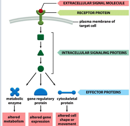
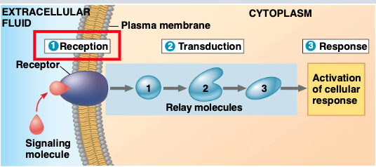
3 stages of signal transduction
1⃣ Reception:
Signal (ligand) binds to receptor protein.
Binding is highly specific.
Receptor shape changes (conformational change).
2⃣ Transduction:
Signal relayed through proteins (relay molecules).
Signal amplified like falling dominoes.
3⃣ Response:
Cellular activity changes (e.g. movement, gene expression, division, death).
Stages of cell signal image
Receptors
Receptors are proteins in or on the cell membrane (or sometimes inside the cell).
They receive signals from outside the cell — these signals are called ligands (like hormones, neurotransmitters, etc).
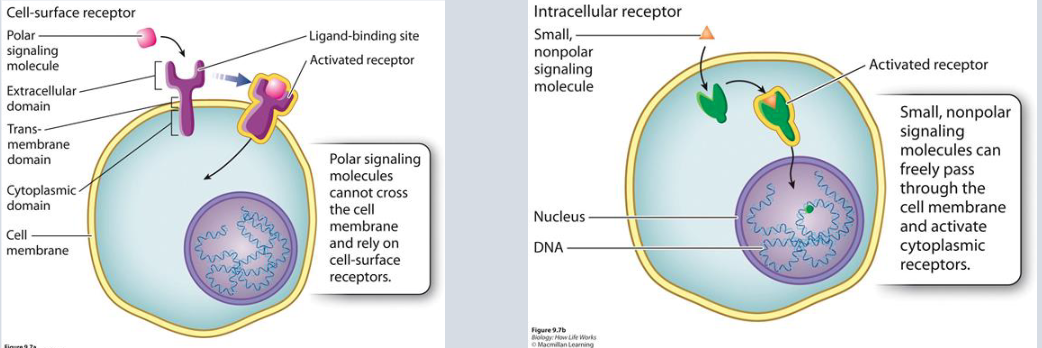
Types of receptors
Receptor Type | Function |
|---|---|
G Protein-Coupled Receptors (GPCRs) | Most common; 7-pass membrane proteins that activate G proteins. |
Ion Channel Receptors | Act as gates for ions. |
Intracellular Receptors | Inside cell; activated by small/hydrophobic ligands (ex: hormones). |
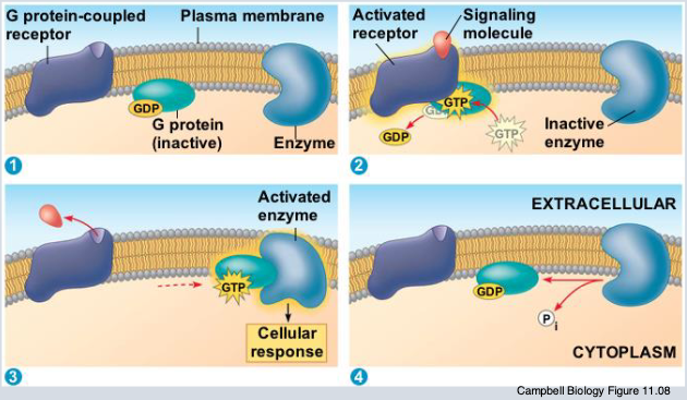
G Protein-Coupled Receptors (GPCRs)
What it is:
The most common type of receptor in your body.
Found in the cell membrane.
What does "7-pass transmembrane protein" mean?
The protein crosses (passes through) the membrane 7 times like a snake weaving back and forth.
Basically, parts of the protein stick out both inside & outside the cell.
How it works:
A ligand (signal molecule) binds to the outside part of the receptor.
The receptor changes shape.
This activates a G protein inside the cell.
The G protein triggers other molecules inside to create a bigger response (like enzymes, second messengers, etc).
Purpose:
Start many important processes: smell, taste, adrenaline response, etc.