Bio Book Important Book Terms and Images
1/13
Earn XP
Description and Tags
For Bio Final Dec 9
Name | Mastery | Learn | Test | Matching | Spaced | Call with Kai |
|---|
No analytics yet
Send a link to your students to track their progress
14 Terms
Explain this image
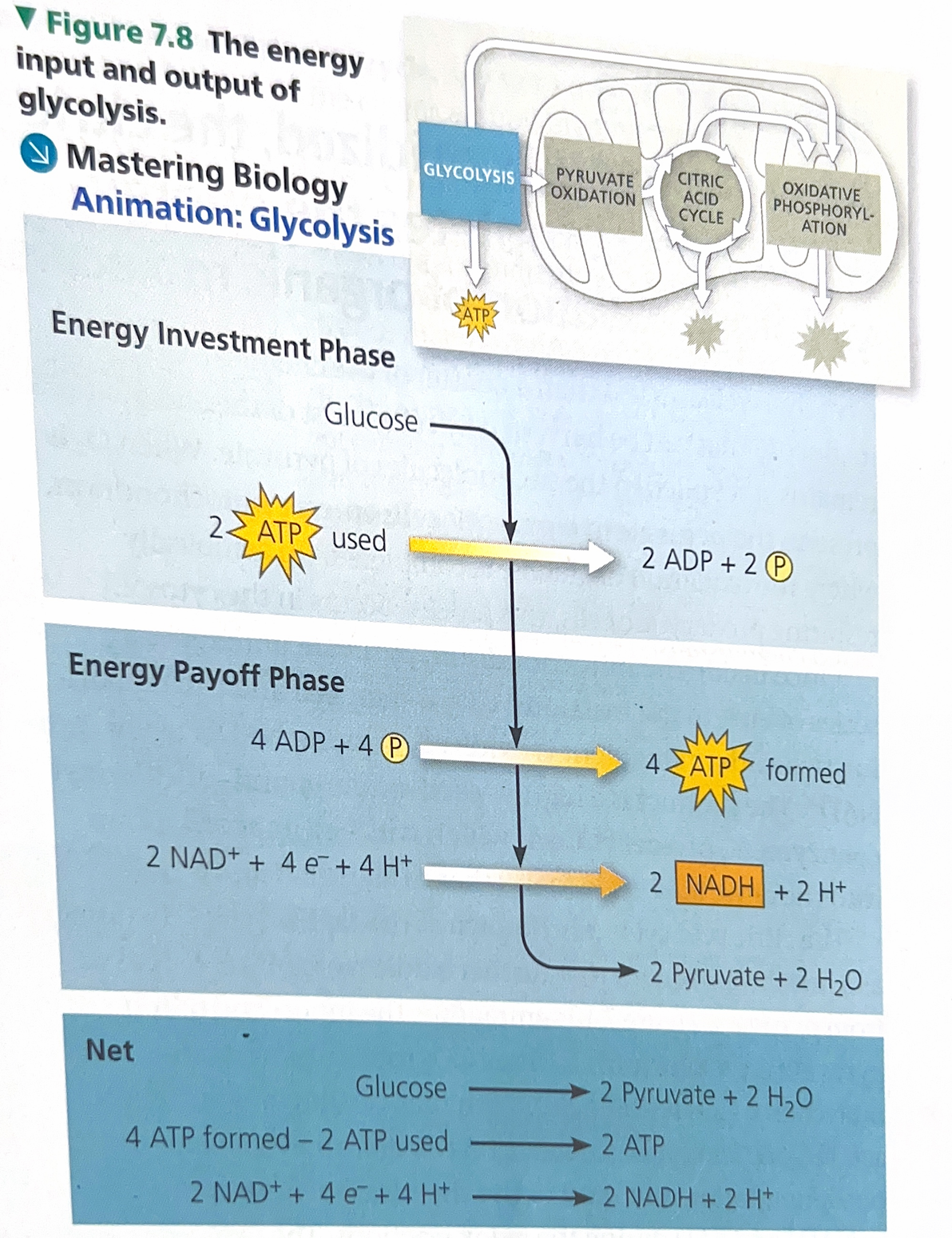
Glycolysis can be divided into two phases: energy investment and energy payoff. During the energy investment phase, the cell actually spends ATP. This investment is repaid with interest during the energy payoff phase, when ATP is produced by substrate-level phosphorylation and NAD+ is reduced to NADH by electrons released from the oxidation of glucose. The net energy yield from glycolysis, per glucose molecule, is 2 ATP plus 2 NADH.
Study this image
These are the 10 steps of glycolysis.
All of the carbon originally present in glucose is accounted for in the two molecules for pyruvate; no carbon is released as CO2 during glycolysis. Glycolysis occurs whether or not O2 is present. However, if O2 is present, the chemical energy stored in pyruvate and NADH can be extracted by pyruvate oxidation, the citric acid cycle, and oxidative phosphorylation.
Explain this image
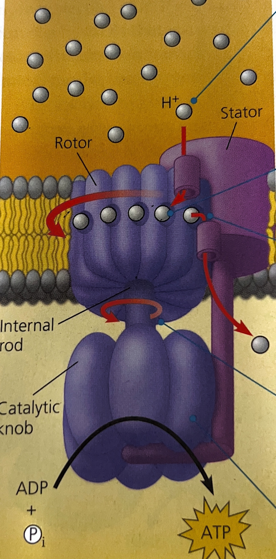
H+ ions flowing down their gradient enter a channel in a stator, which is anchored into the membrane.
H+ ions enter binding sites within a rotor, changing the shape of each subunit so that the rotor spins within the membrane.
Each H+ ion makes one complete turn before leaving the rotor and passing through a second channel in the stator into the mitochondrial matrix.
Spinning of the rotor causes an internal rod to spin as well. This rod extends like a stalk into the knob below it, which is held stationary by part of the stator.
Turning of the rod activates catalytic sites in the knob that produce ATP from ADP and inorganic phosphate.
ATP synthases, a molecular mill
Explain this image
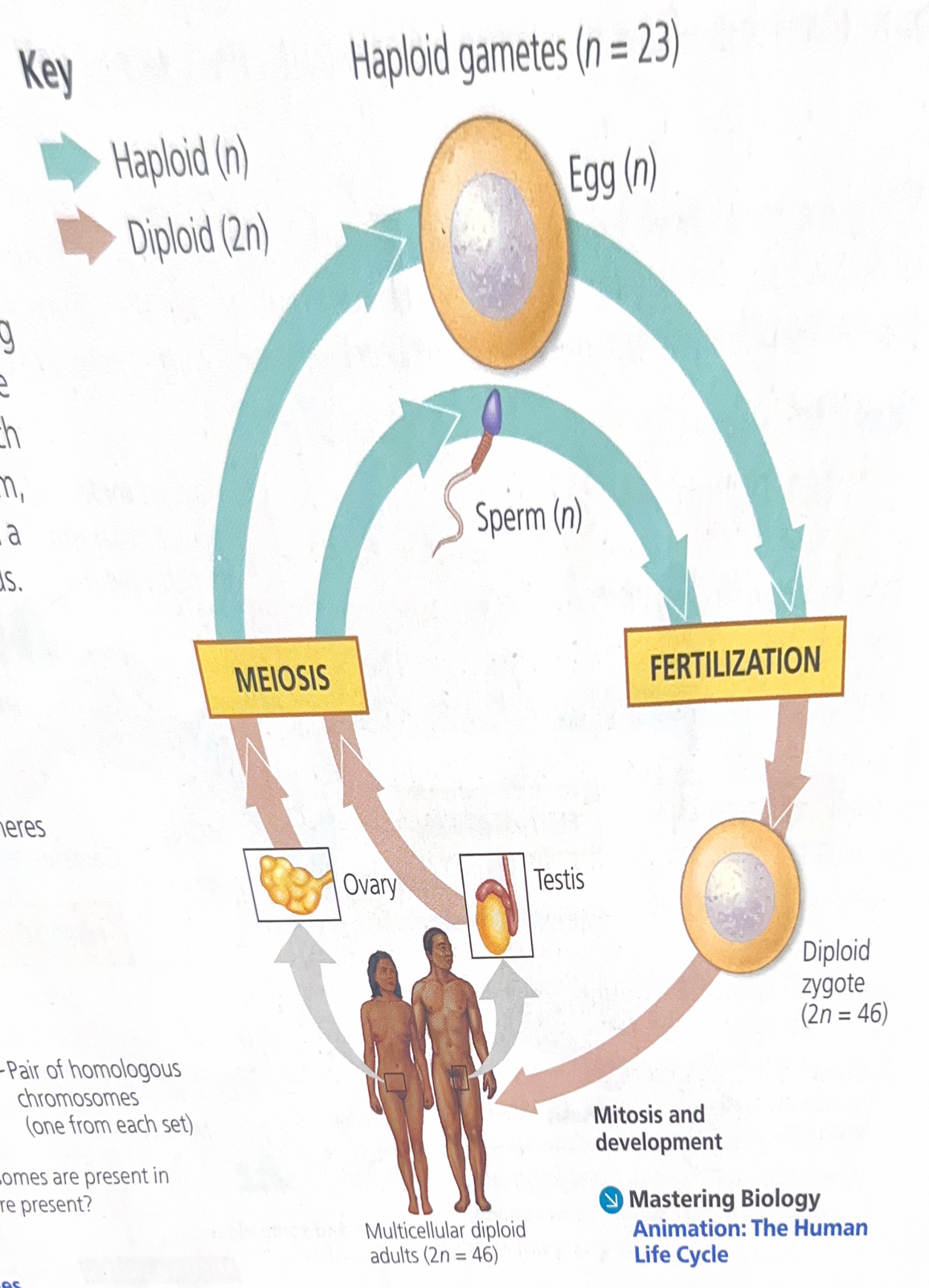
The human life cycle
In each generation, the number of chromosomes sets is halved during meiosis but doubles at fertilization. For humans, the number of chromosomes in a haploid cell is 23, consisting of one set (n=23); the number of chromosomes in the diploid zygote and all somatic cells arising from it is 46 consisting of two sets (2n=46)
Explain this image
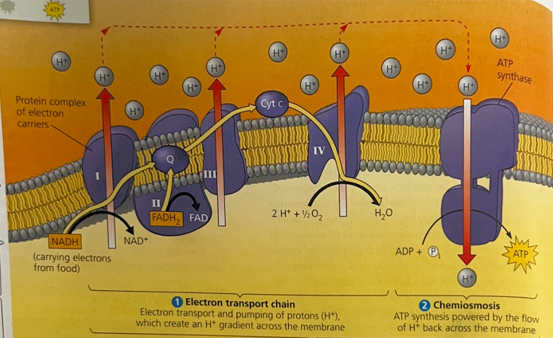
The chain is an energy converter that uses the exergonic flow of electrons from NADH and FADH2 to pump H+ across the membrane, from the mitochondrial matrix into the intermembrane space. The H+ has a tendency to move back across the membrane, diffusing down its gradient. And the ATP synthases are the only sites that provide a route through the membrane for H+. The passage of H+ through ATP synthase uses the exergonic flow of H+ to drive the phosphorylation of ADP. Thus, the energy stored in an H+ gradient across a membrane couples the redox reactions of the electron transport chain to ATP synthesis.
Explain this image
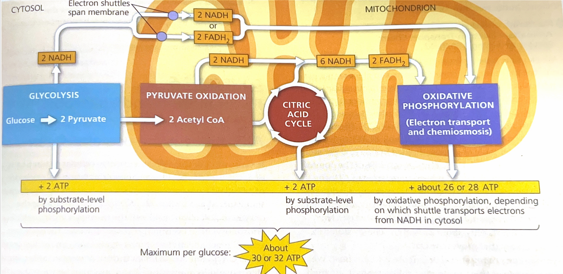
A detailed accounting of the ATP yield per glucose molecule oxidized. The tally adds the 4 ATP produced directly by substrate-level phosphorylation during glycolysis and the citric acid cycle to the many molecules of ATP generated by oxidative phosphorylation. Each NADH that transfers a pair of electrons from glucose to the electron transport chain contributes enough to the proton motive force to generate a maximum of about 3 ATP.
Explain this image
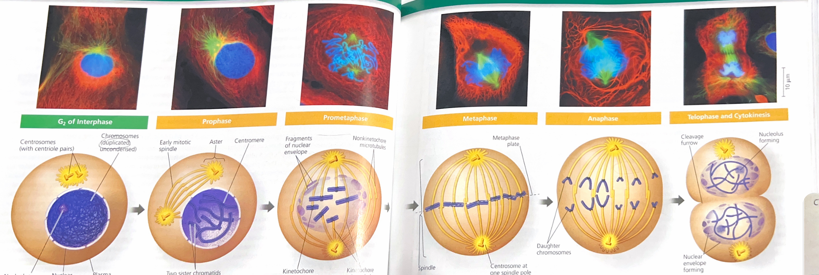
Mitosis is broken down into 5 stages.
P- prophase
The chromatin fibers become more tightly coiled condensing into discrete chromosomes. The nucleoli disappear. Each duplicated chromosome appears as two identical sister chromatids joined at their centromeres and, in some species, all along their arms by sister chromatid cohesion. The mitotic spindle begins to form, and it composed of the centrosomes and the microtubules that extend from them.
PM-prometaphase
The nuclear envelope fragments. The microtubules extending from each centrosome can now invade the nuclear area. The chromosomes have become more condensed. A kinetochore has now formed at the centromere of each chromatid.
M- metaphase
The centrosomes are now at opposite poles of the cell. The chromosomes have all arrived at the metaphase plate, a plane that is equidistant between the spindle’s two poles. The chromosomes’ centromeres lie at the metaphase plate.
A- anaphase
Anaphase begins when the cohesion protein are cleaved. This allows the two sister chromatids of each pair to par suddenly. Each chromatid thus becomes an independent chromosome. The two new daughter chromosomes begin moving toward opposite ends of the cell as their kinetochore microtubules shorten. The cell elongates as the nonkitnetochore microtubules lengthen.
T- Telophase
Two daughter nuclei form in the cell. Nuclear envelopes arise from the fragments of the parent cell’s nuclear envelope and other portions of the endomembrane system. Nucleoli reappear. The chromosomes become less condenses.
Cytokinesis
The division of the cytoplasm is usually well under way by late telophase, so the two daughter cells appear shortly after the end of mitosis.
Explain this image
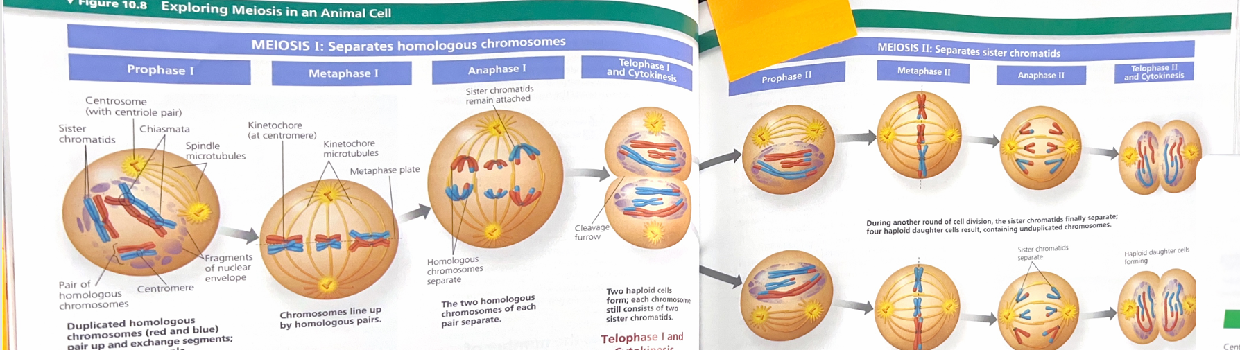
Detail the stages of the two divisions of meiosis for an animal cell.
PI- Prophase I
Centrosome movement, spindle formation, and nuclear envelope breakdown occur as in mitosis. Chromosomes condense progressively throughout prophase I. During early prophase I, before the stage shown above, each chromosome pairs with its homolog, aligned gene by gene, and crossing over occurs: The DNA molecules of nonsister chromatids are broken and are rejoined to each other. At the stage shown above, each homologous pair has one or more X-shaped regions called chiasmata, where crossovers have occurred. Later in prophase I, microtubules from one pole or the other attach to the kinetochores, one at the centromere of each homolog.
MI- Metaphase I
Pairs of homologous chromosomes are now arranged at the metaphase plate, with one chromosome of each pair facing each pole; Each pair has lined up independently of other pairs (independent assortment). Both chromatids of one homolog are attached to kinetochore microtubules from one pole; the chromatids of the other homolog are attached to microtubules from the opposite poles.
AI- Anaphase I
Breakdown of proteins that are responsible for sister chromatids cohesion along chromatid arms allowing homologs to separate. The homologs move towards opposite poles, guided by the spindle apparatus. Sister chromatids cohesion persists at the centromere, causing chromatids to move as a unit towards the same pole.
TI- Telophase I and Cytokinesis
When Telophase I begins each half of the cell has complete haploid set of duplicated chromosomes. Each chromosome is composed of two sister chromatids; one or both include regions of nonsister chromatids DNA. Cytokinesis usually occurs simultaneously with telophase I, forming two haploid daughter cells. NO CHROMOSOME DUPLICATION OCCURS BETWEEN MEIOSIS 1 AND 2.
PII- Prophase II
A spindle apparatus forms. In late prophase II, chromosomes, each still composed of two chromatids associated at the centromere, are moved by microtubules towards the metaphase II plate.
MII- Metaphase II
The chromosomes are positioned at the metaphase plate as in mitosis.
Because of crossing over in meiosis I, the two sister chromatids of each chromosome are not genetically identical.
AII- Anaphase II
Breakdown of proteins holding the sister chromatids together at the centromere allows the chromatids to separate. The chromatids move towards opposite poles as individual chromosomes.
TII- Telophase II and Cytokinesis
Nuclei form, the chromosomes begin decondensing and cytokinesis occurs. The meiotic division of one parent cell produces FOUR daughter cells, each with a haploid set of unduplicated chromosomes. The four daughter cells are genetically DISTINCT from one another and from the parent cell.
Explain this image
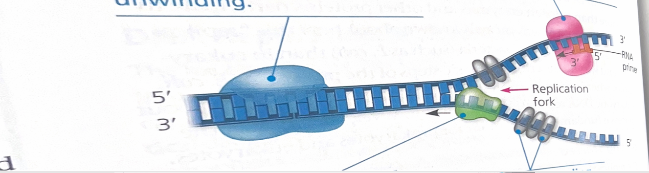
At each end of a bubble is a replication fork, a Y shaped region where the parents strand of DNA is being unwound. Several kinds of proteins participate in the unwinding. Helicases are enzymes that untwist the double helix at the replication forks, separating the two parental strands and making them available as template strands. After the parental strands separate, single- strand binding proteins bind to the unpaired DNA strands, keeping them from re-pairing. The untwisted of the double helix causes tighter twisting and strain ahead of the replication fork. Topoisomerase is an enzyme that helps relieve this strain by breaking, swiveling, and rejoining DNA strands.
Explain this image
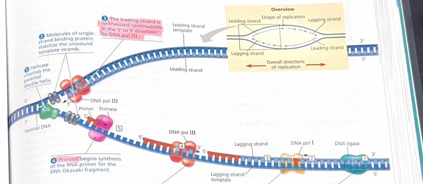
The helicase unwinds the parents’ double helix
Molecules of single strand binding protein stabilize the unwound template strands
The leading strand is synthesized continuously in the 5’ to 3’ direction by DNA pol III
Primase begins synthesis of the RNA primer for the fifth Okazaki fragment.
DNA pol III is completing synthesis of fragment 4. When it reaches the RNA primer on fragment 3, it will detach and begin adding DNA nucleotides to the 3’ end of the fragment 5 primer in the replication fork
DNA pol I removes the primer from the 5’ end of fragment 2, replacing it with DNA nucleotides added one by one to the 3’ end of fragment 3. After the last addition, the backbone is left with a free 3’ end.
DNA ligase joins the 3’ end of fragment 2 to the 5’ end of fragment 1
This is a summary of DNA replication.
Explain this image
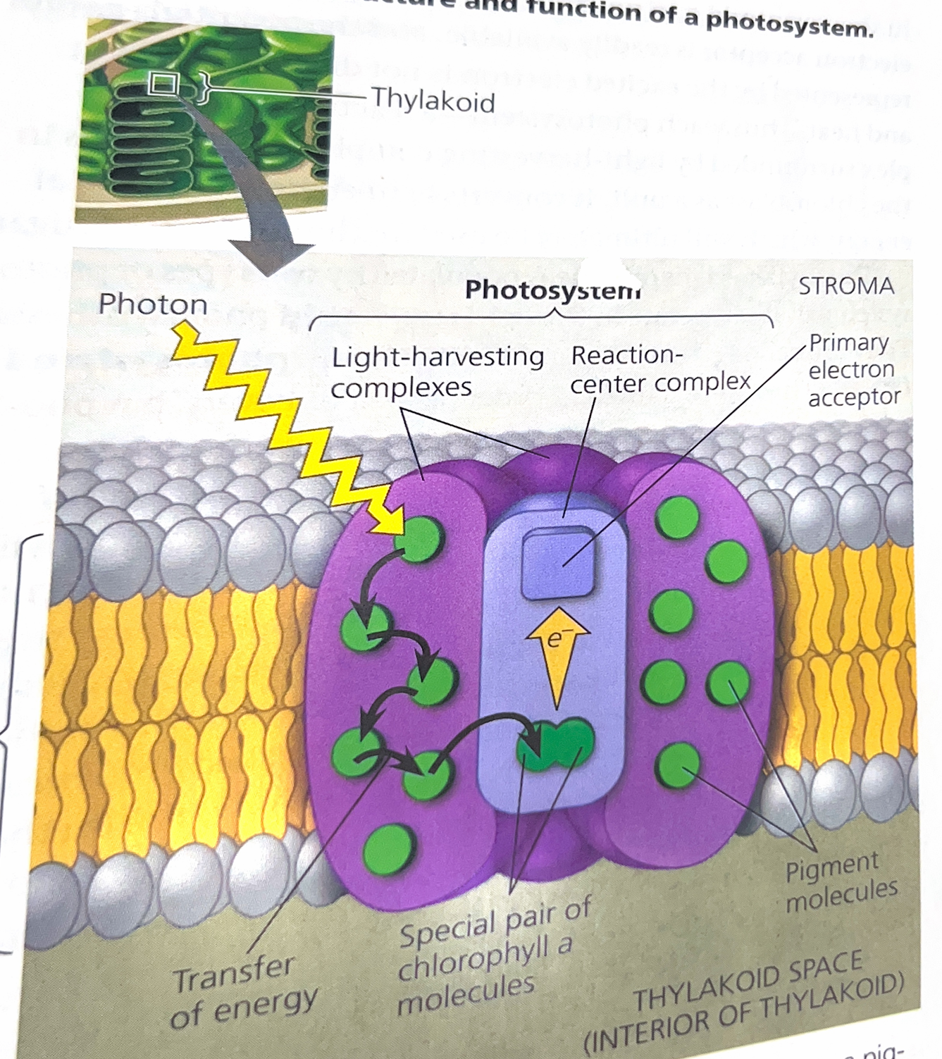
When a photon strikes a pigment molecule in a light-harvesting complex, the energy is passed from molecule to molecule until it reaches the reaction-center complex. Here, an excited electron from the special pair of chlorophyll a molecules is transferred to the primary electron acceptor.
Explain this image
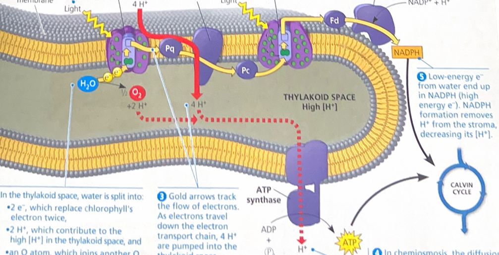
This explains light reactions. Electrons flow pushes electrons from water, where they are at a state of low potential energy, ultimately o NADPH, where they are stored at a state of high potential energy. The light-driven electrons flow also generates ATP. Thus, the equipment of the thylakoid membrane converts light energy to chemical energy stored in ATP and NADPH (O2 is produced as a by-product)

Explain this image
In the chloroplasts, the thylakoid membranes are the sites of the light reactions, whereas the Calvin cycle occurs in the stroma. The light reactions use solar energy to make ATP and NADPH, which supply chemical energy and reducing power, respectively to the Calvin cycle. The Calvin cycle incorporates CO2 into organic molecules, which are converted to sugar.
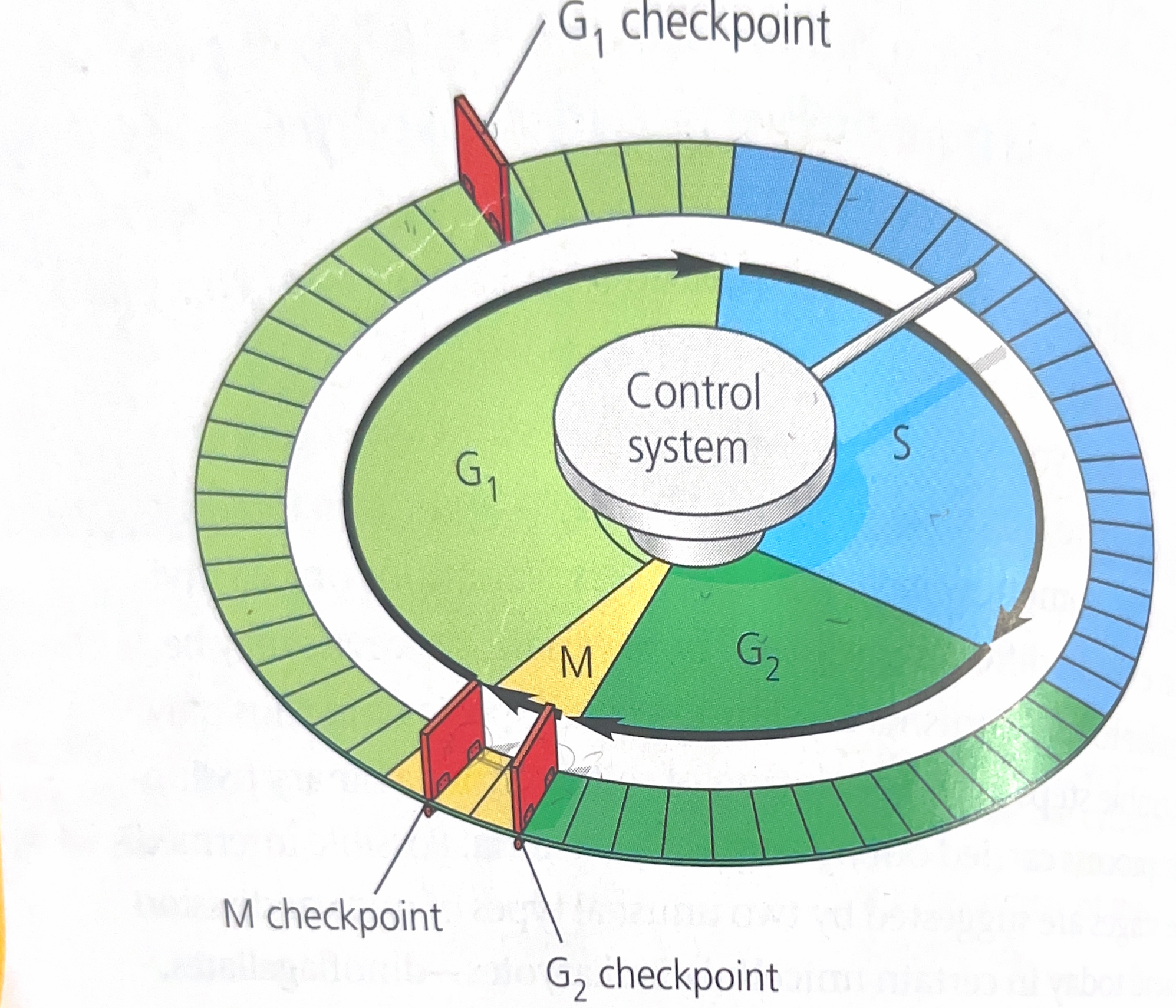
Explain this image
The flat “stepping stones” around the perimeter represent the sequential events. Like the control device of a washing machine, the cell cycle control system proceeds on its onw, driven by a built in clock. However, the system is subject to internal and external regulation at various checkpoints; 3 important checkpoints. These checkpoints come from a cellular surveillance mechanism inside the cell. These signal report whether crucial cellular processes that should have occurred by the point have in face been completed correctly and thus whether or not the cell cycle should proceed.