PS231: Hormones and the Brain III
1/19
There's no tags or description
Looks like no tags are added yet.
Name | Mastery | Learn | Test | Matching | Spaced |
|---|
No study sessions yet.
20 Terms
The major vertebrae endocrine glands
Hypothalamus: control of hormone secretions
Pituitary gland
Anterior pituitary: hormone secretion by thyroid, adrenal cortex and gonads; growth
Posterior pituitary: water balance; salt balance
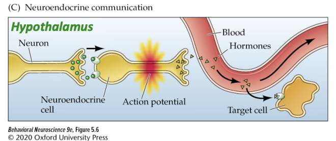
Neuroendocrine communication
In the hypothalamus
Neuroendocrine cells fire action potentials to release hormones directly into the blood
Then affect cells in the anterior pituitary
Level I: the hypothalamus
Small peptide hormones that increase or decrease the release of hormones from the anterior pituitary gland (left: little green triangles)
Can be thought of as a special class of neurotransmitters
Released by neurosecretory cells at the base of the hypothalamus
These neurons act as endocrine cells, but look like conventional neurons, firing action potentials that cause the release of “releasing hormones”
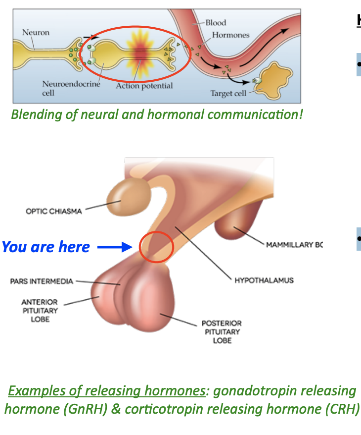
Level II: the pituitary gland
Latin for “mucus”
Sits directly below the hypothalamus, encased in the “sella tursica” (Turkish saddle) bone
Made up of two anatomically distinct parts:
Anterior pituitary (glandular tissue)
Posterior pituitary (neural tissue)

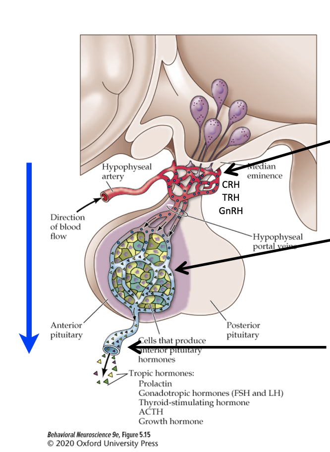
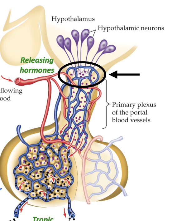
IIa: the anterior pituitary
Hypothalamic neurons release neurohormones (or “releasing” hormones) into capillaries of the portal vessels (closed blood circuit linking the hypothalamus with the anterior pituitary)
Releasing hormones from the portal vessels stimulate or inhibit the release of tropic hormones from distinct anterior pituitary endocrine cells
Anterior pituitary “tropic” hormones leave the pituitary gland into the general circulation to act on peripheral endocrine organs (e.g. adrenal glands, thyroid gland, gonads, etc.)
The anterior pituitary contains cells that secrete hormones—it’s an endocrine gland
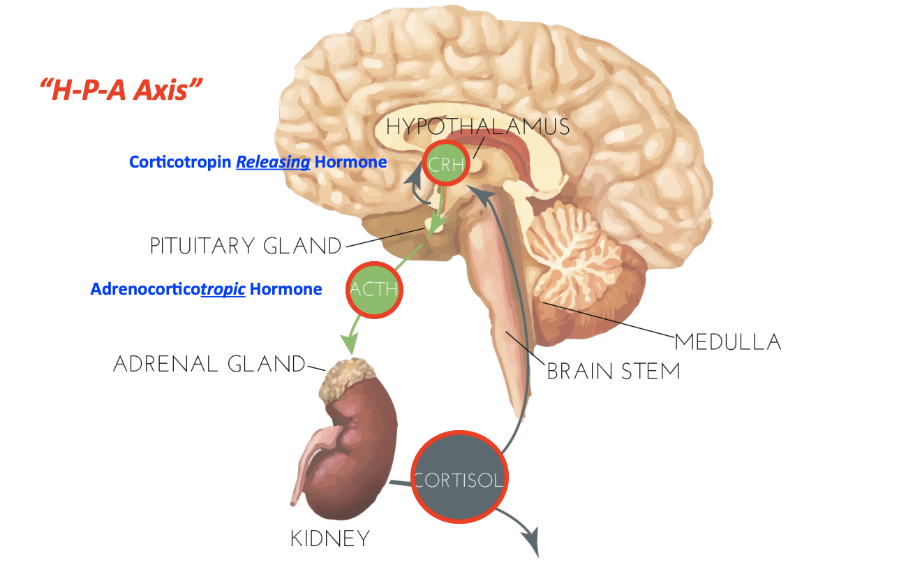
HPA axis
blood pressure/stress
HPT axis
metabolic rate
HPG axis
reproducition
Anterior pituitary gland associated hormones
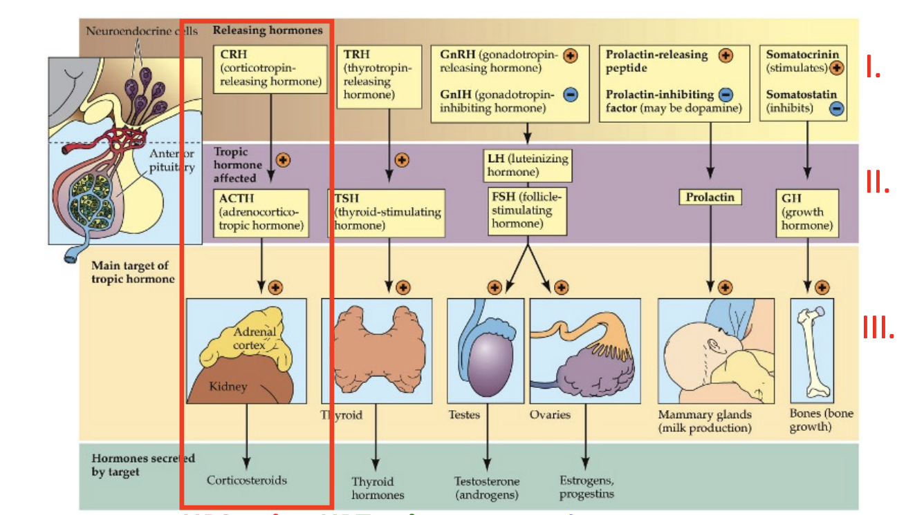
Stress response system
CRH → ACTH → Glucocorticoids (e.g. cortisol)
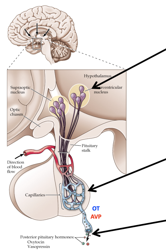
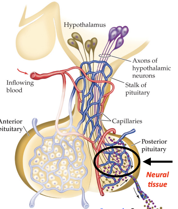
IIb: the posterior pituitary
Magnocellular (large) neurons in the hypothalamus (PVN/SON) synthesize oxytocin (OT) and vasopressin (AVP) and transport them to their axon terminals, located in the posterior pituitary—no portal system!
Oxytocin and vasopressin are directly released from axon terminals in the posterior pituitary and diffuse into capillaries
Oxytocin and vasopressin then leave the posterior pituitary via the bloodstream into the general/systemic circulation to act on peripheral tissues
Posterior Pituitary Gland-Associated Hormones: Oxytocin & Vasopressin
9 amino acid peptides (“nonapeptides”)
Differ by only two amino acids
Very different functions
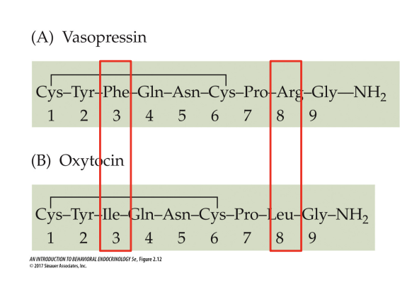
Vasopressin
Water retention
Vasoconstriction
Oxytocin
Uterine contractions
Milk ejection
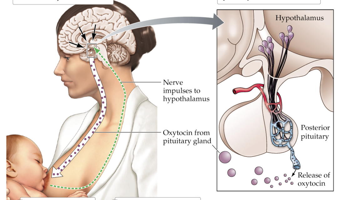
Oxytocin and Milk Letdown
Stimulation of the mother’s nipple by the infant’s suckling response produces brain activity in the mother
This brain activity stimulates hypothalamic cells to release oxytocin from the posterior pituitary
The oxytocin causes the cells of the mammary glands to contract, thereby releasing milk
The baby, rewarded with milk, continues suckling until sated
Anterior pituitary
Produces and secretes its own hormones
Glandular tissue
Connected by network of blood vessels
Tropic hormones
Posterior pituitary
Stores and releases hormones made in the hypothalamus
Neural tissue
Connected by nerve fibers that extend from the hypothalamus
Oxytocin and vasopressin
Compare and contrast neural vs. hormonal communication. Explain how neuroendocrine cells at the base of the hypothalamus “blend” neuronal and neuroendocrine mechanisms. Do these neurons fire action potentials? Do they release anything? If so, what do they release and where do they release it?
Neural communication uses electrical impulses (action potentials) and chemical neurotransmitters through specific neural pathways. They are fast, precise, and very short-lived.
Hormonal communication uses chemical messengers (hormones) released into the bloodstream and are relatively slow but with long-lasting effects.
Neuroendocrine receive neuronal signals and fire action potentials but they also release hormones directly into the blood, affecting cells in the anterior pituitary.
How, specifically, does the hypothalamus communicate with the anterior pituitary gland? Which types of hormones are involved, and what do anterior pituitary cells do in response to receiving a hormonal signal from the hypothalamus?
Neurosecretory cells at the base of the hypothalamus release small peptide hormones that either increase or decrease the release of hormones from the anterior pituitary gland
Receiving hormones are involved in the process and it leads to either the increase or decrease of the synthesis and secretion of their own hormones
What are oxytocin and vasopressin? What role do each play in the body? What role do they play in the brain? From where are they released into the bloodstream?
Oxytocin:
Role in the Body (Peripheral Actions):
Labor and Childbirth: Stimulates powerful contractions of the uterine muscles.
Milk Ejection: Causes the contraction of myoepithelial cells in the mammary glands, leading to the "let-down" of milk for breastfeeding.
Role in the Brain (Central Actions):
Social Bonding: Promotes pair bonding, maternal-infant bonding, and other social attachments.
Emotional Connection: Often called the "love hormone" or "cuddle hormone," it is associated with trust, empathy, and sexual arousal.
Stress Reduction: Can have anti-anxiety and calming effects.
Vasopressin (also called Antidiuretic Hormone or ADH):
Role in the Body (Peripheral Actions):
Water Retention: Its primary role is to regulate water balance in the body. It acts on the kidneys to increase water reabsorption, thereby concentrating the urine and conserving water. This is crucial for preventing dehydration.
Vasoconstriction: At very high concentrations (e.g., during severe blood loss), it causes constriction of blood vessels, which helps to increase blood pressure.