CTL Anatomy 30% Liver, GB, Pancreas
1/141
There's no tags or description
Looks like no tags are added yet.
Name | Mastery | Learn | Test | Matching | Spaced |
|---|
No study sessions yet.
142 Terms
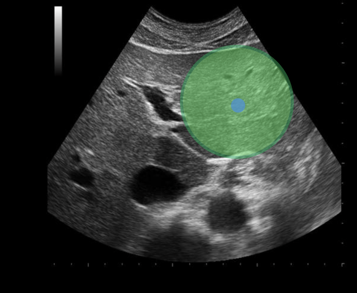
Use your mouse to place your cursor over the center of the lateral left lobe of the liver and click to mark the structure
The caudate lobe occupies much of the ___________ surface of the liver
Posterior superior |
.The medial segment of the left lobe lies between:
|
In a normal fasting adult, blood sugar levels should not exceed __________ of blood.
100 mg/100ml |
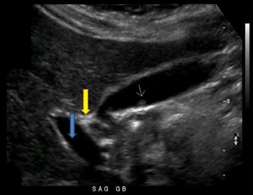
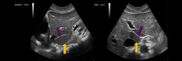
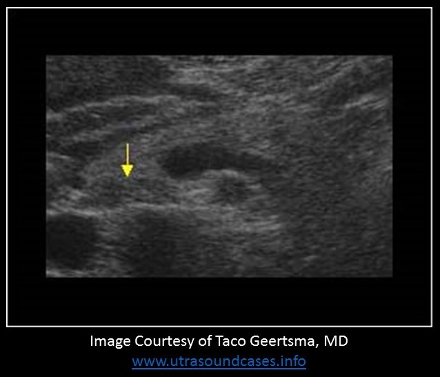
What structure is indicated by the yellow arrow?
main lobar fissure |
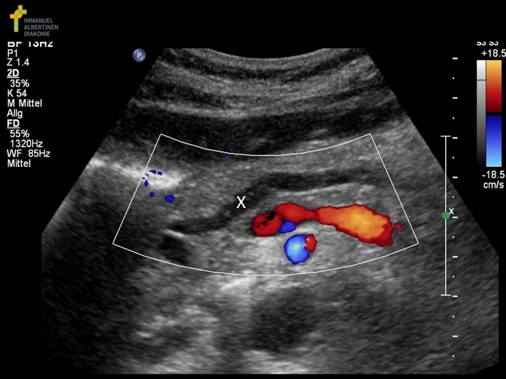
What structure is marked with the X on the image?
|

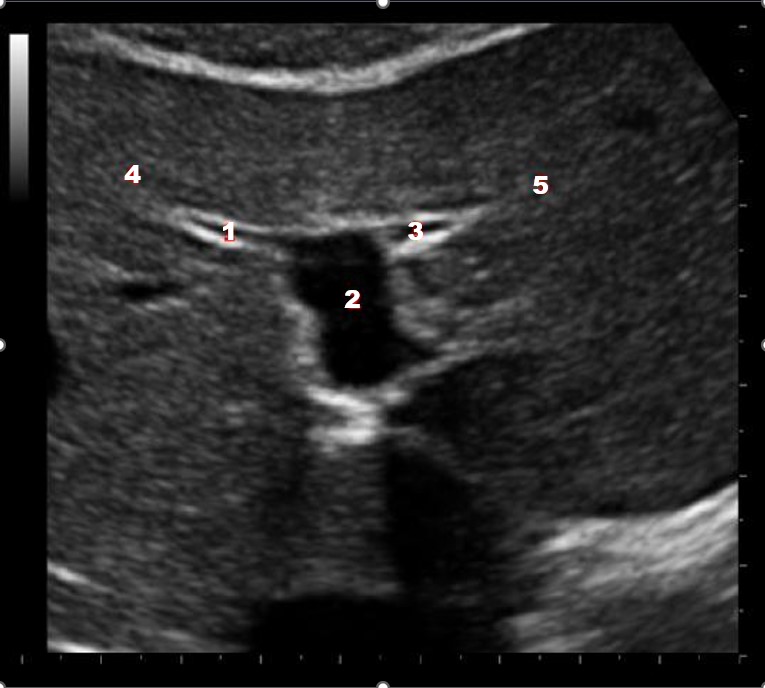
Which of the following structures is labeled #5?
lateral left lobe |
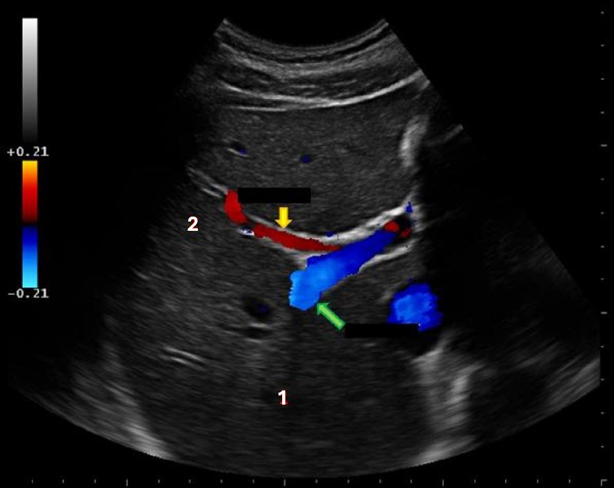
What lobe of the liver is indicated by #1?
posterior right lobe
Which of the following hormones is responsible for causing the gallbladder to contract?
|
The yellow arrow on the images represents which of the following structures?
|
The ______________ can be identified anterolateral to the pancreas tail.
|
What organ produces sodium bicarbonate?
|
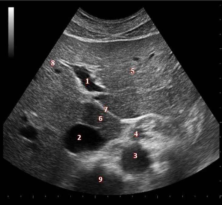
What structure/vessel is indicated by #8?
medial left lobe |
The ___________________ is located within the inferior margin of the falciform ligament.
|
The caudate lobe lies between what two structures?
|
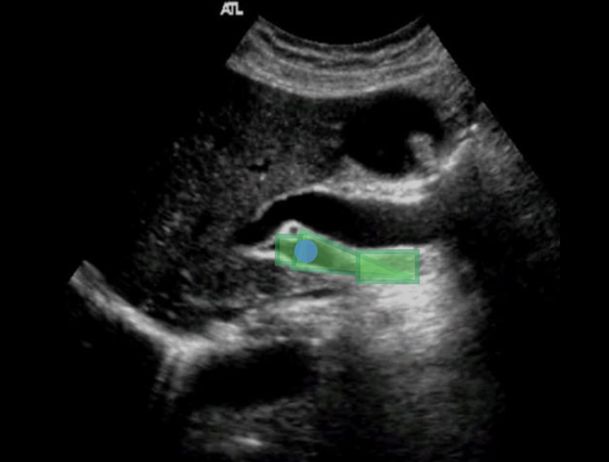
During an abdominal ultrasound, a 4mm circular anechoic structure is identified at the posterior portion of the pancreas head. Color flow is not identified in the structure. What is it?
common bile duct

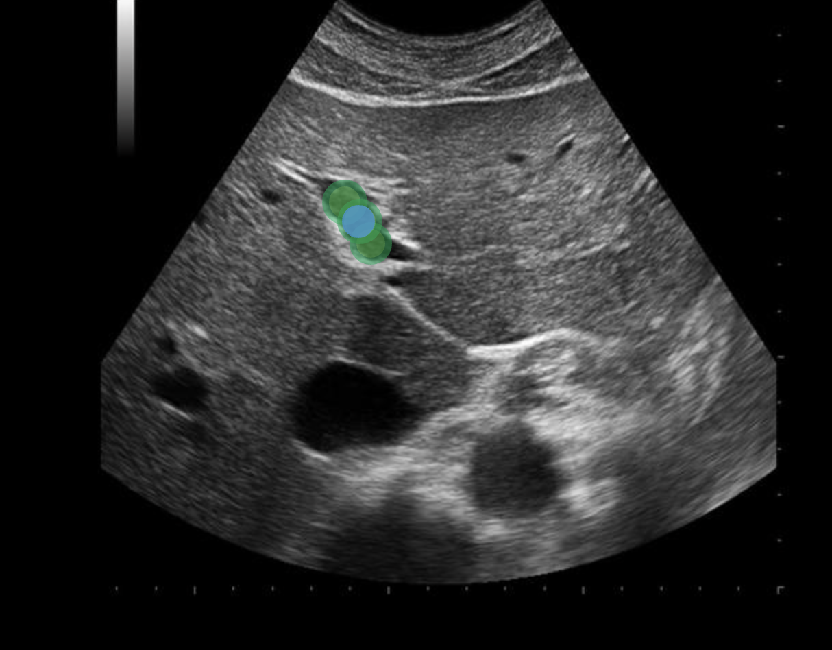
What liver vessel is indicated by the green arrow?
posterior right portal vein |
Use your mouse to place your cursor over the ligamentum venosum and click to mark the structure.
The liver's exocrine function includes producing:
Bile |

What structure/vessel is indicated by #5?
|
The abdominal organ that produces the majority of alkaline phosphatase is?
|
Which of the following terms can be used to describe the normal flow in the portal vein?
continuous |
Branches of which of the following vessels supply the pancreas with blood?
|

What lobe of the liver is indicated by #2?
anterior right lobe |
All of the following laboratory tests are used to evaluate the liver function, EXCEPT
Sodium bicarbonate |
The cystic artery originates at the _____________________ and the cystic vein empties into the __________________________.
|
The common bile duct is formed by:
Common hepatic duct + the cystic duct |
A patient presents with a history of Reidel lobe. What are the expected findings on the ultrasound exam?
The right lobe of the liver will have a tongue-like extension that extends inferior to the lower pole of the right kidney.
What vessels drain the blood from the caudate lobe?
|

Which of the following structures is labeled #1?
|
The pancreatic duct should normally be less than _______ in diameter in children and young adults
|
Which of the following correctly describes the pediatric pancreas?
|
Which of the following statements is true regarding the image displayed?
|
Which of the following statements correctly describes imaging of the dome of the liver?
Deep inspiration is required to evaluate this portion of the liver. |
The majority of the liver is covered by a thick capsule composed of fibrous and elastic elements called _________________.
|
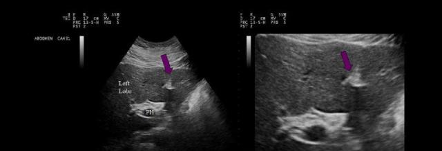
The purple arrows point to which of the following liver structures?
ligamentum teres |
While discussing the medical history with a patient, he tells you that the doctor recommended that he increase his intake of vitamin K due to some recent abnormal lab results. Which of the following lab values was abnormal?
|
An ERCP is commonly performed to evaluate:
the Ampulla of Vater |
Use your mouse to place your cursor over the main lobar fissure and click to mark the structure.

Which of the following structures is labeled #7?
anterior right lobe |
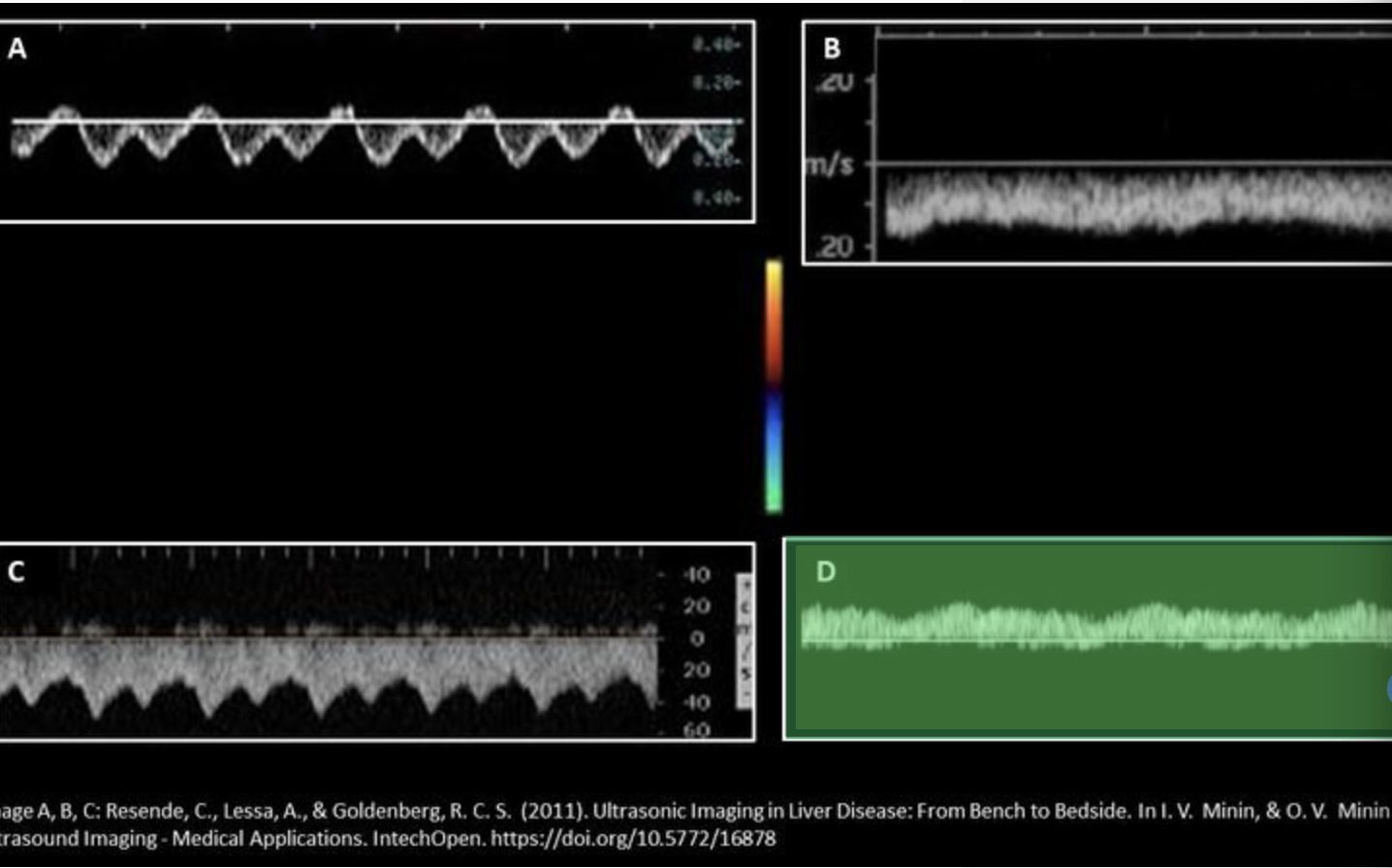
Which waveform represents a normal portal vein waveform? Use your mouse to position your cursor over the correct waveform and click to set the answer.
***the color map displayed applies to all displayed Doppler tracings.
Which of the following structures is labeled #2?
gastroduodenal artery |
Which abdominal vessel is the best landmark to identify the body and tail of the pancreas?
|
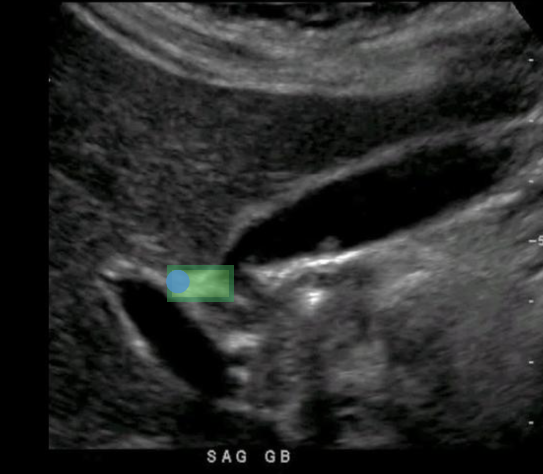
Use your mouse to place your cursor over the superior mesenteric vein and click to mark the vessel. If the vessel is not demonstrated on the image, mark the purple box that says "Not demonstrated on the image".

What structure/vessel is indicated by #9?
|
Which of the following correctly lists the structures found in an intrahepatic portal triad?
|
What structure/vessel is indicated by #4?
|

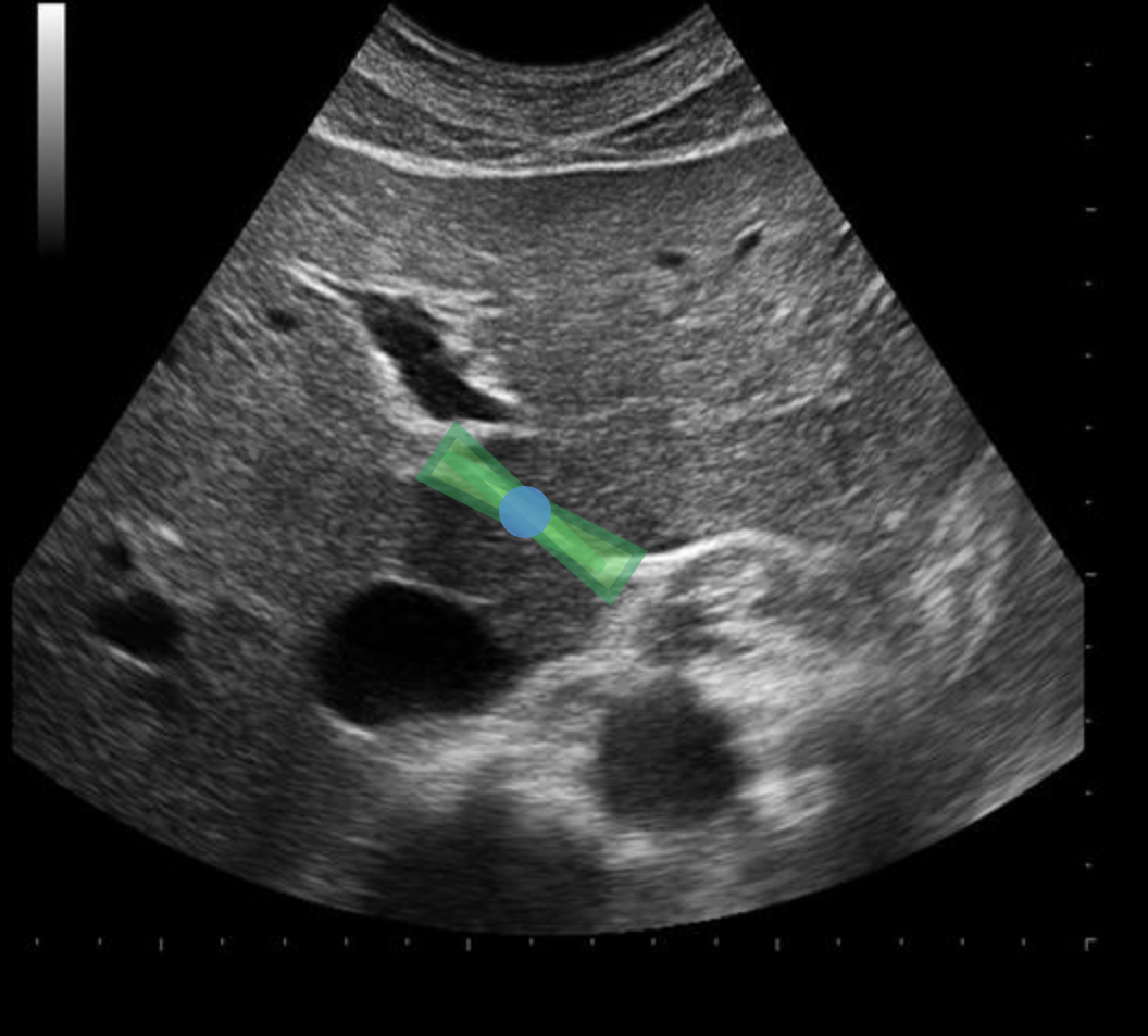
What structure is indicated by the blue arrow?
right portal vein |
Use your mouse to place your cursor over the left portal vein and click to mark the structure.
Use your mouse to place your cursor over the main portal vein and click to mark the vessel.
The right lobe of the liver is divided into ____________________ segments, while the left lobe is divided into ________________ segments.
anterior and posterior, medial and lateral |
Normal hepatic venous flow will demonstrate:
|
Which of the following produces insulin?
|
Which of the following hepatic ligaments separate the medial and lateral left lobes of the liver?
|
The _______________________ lobe of the liver is located between the right and middle hepatic vein.
|
Which portions of the gallbladder and/ or biliary tree are involved in the formation of a phrygian cap?
|
In the normal liver, which of the following correctly describes the changes in flow in the hepatic artery after eating?
|
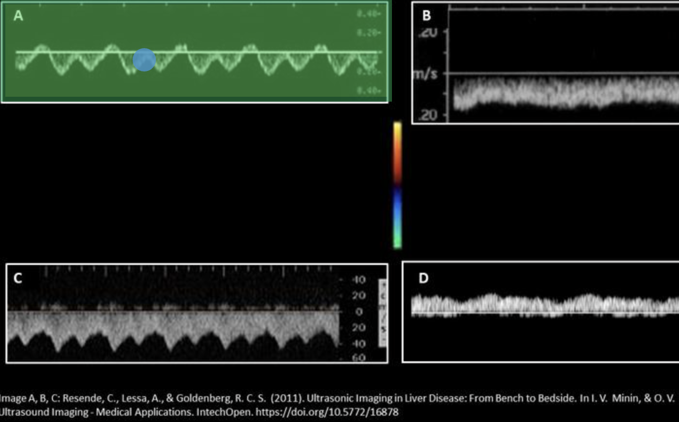
Which waveform represents a normal inferior vena cava waveform? Use your mouse to position your cursor over the correct waveform and click to set the answer.
***the color map displayed applies to all displayed Doppler tracings.
The superior mesenteric vein
|
In the porta hepatis, what structure is anterior to the portal vein and medial to the common hepatic duct?
Points: 1 / 1
proper hepatic artery |
In the porta hepatis, what structure is anterior to the portal vein and lateral to the proper hepatic artery?
|
The IVC is located posterior to the pancreatic __________________.
|
Islets of Langerhans secrete ___________________ into ___________________.
insulin, bloodstream |

What structure/vessel is indicated by #4?
|

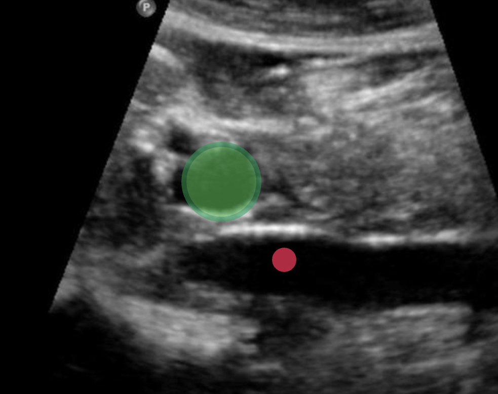
What structure is indicated by the green arrow?
Main portal vein |
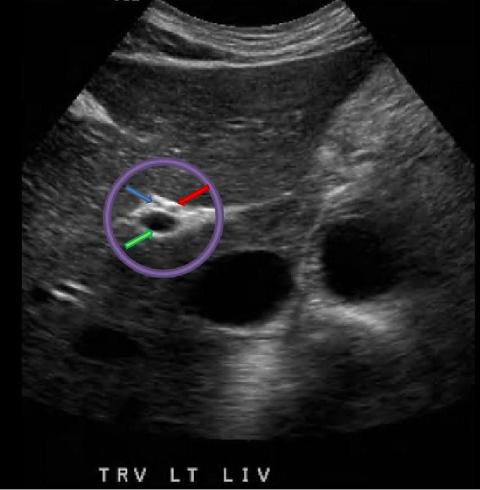
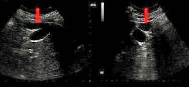
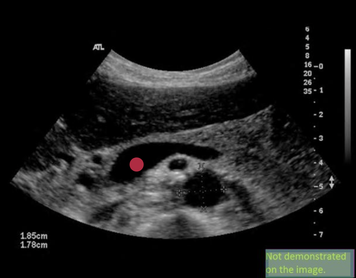
What structure is indicated by the red arrow?
|

Use your mouse to place your cursor over the hepatic artery and click to mark the vessel.

What structure/vessel is indicated by #12?
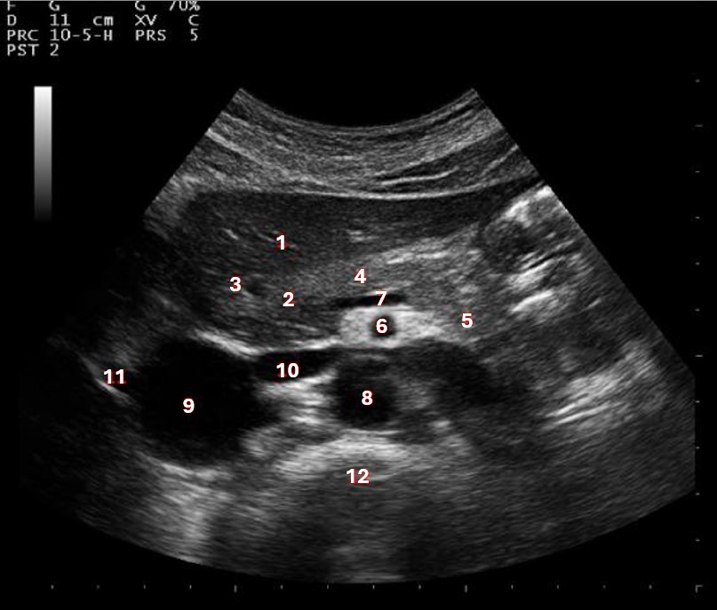
spine

What structure/vessel is indicated by #2?
|
What causes the appearance of this normal variant of the uncinate process?
|

Which of the following structures is labeled #6?
medial left lobe |
The right and left hepatic ducts come together to form the common hepatic duct:
Inside the liver, near the porta hepatis |

Which of the following structures is labeled #3?
IVC
Use your mouse to place your cursor over the main portal vein and click to mark the vessel.
Which of the following ligaments separates the medial and lateral left lobes of the liver?
|
All of the following are true regarding Couinaud liver segmentation except:
|
The hepatoduodenal ligament contains which of the following structures?
|
A patient presents for an abdominal sonogram due to a history of hepatic congestion. What structure(s) should be closely evaluated for related findings?
|

What structure/vessel is indicated by #2?
left portal vein
The formation of a Hartmann pouch usually occurs in what portion of the gallbladder?
neck |
The normal gallbladder is usually less than __________ in transverse diameter.
|
The main lobar fissure:
|
During an abdominal ultrasound, a small circular anechoic structure is identified at the anterior portion of the pancreas head. Color flow is identified in the structure. What is it?
celiac axis |
Which lab value is associated with the development of jaundice?
bilirubin |

What structure/vessel is indicated by #6?
|

What structure/vessel is indicated by #7?
|
What structure prevents free fluid in Morison pouch from moving into the subphrenic space?
right coronary ligament |

Use your mouse to place your cursor over the left medial superior segment of the liver and click to mark the structure.
In a patient with complete situs inversus, the liver will be:
|
A small rounded prominence on the anteroinferior aspect of the normal caudate lobe is called
distal papillary process |
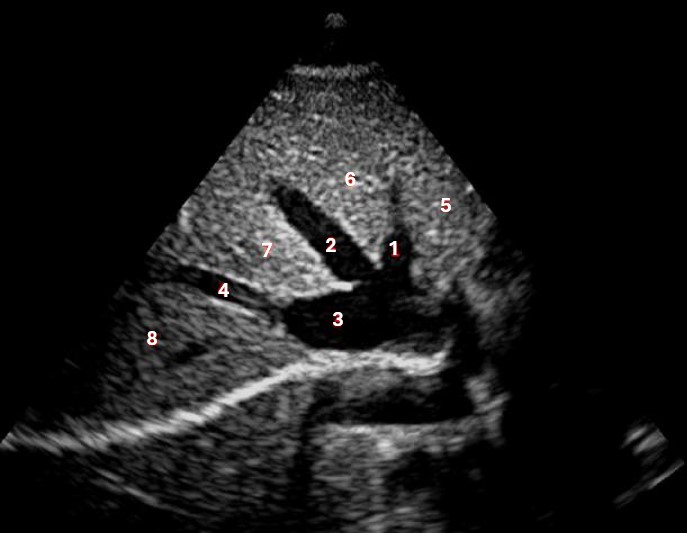
Which of the following structures is labeled #8?
posterior right lobe |

Which of the following structures is labeled #2?
|
A normal portal vein will:
increase in diameter by more than 20% with deep inspiration |
On a transverse sonogram, the CBD enters the _________________ aspect of the head of the pancreas and lays ____________ to the IVC.
|
The Rex-Cantlie line is an imaginary line that extends from the ____________ in the liver.
|

What structure is indicated by the purple circle?
portal triad |
The liver is divided into superior and inferior segments by the:
branching of the portal veins |
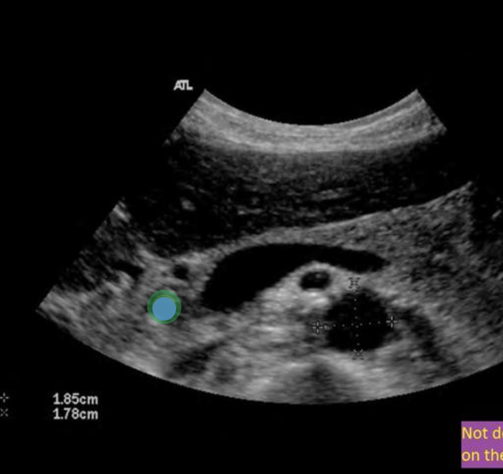
Use your mouse to place your cursor over the common bile duct and click to mark the vessel. If the vessel is not demonstrated on the image, mark the purple box that says "Not demonstrated on the image".
What structure/vessel is indicated by #3?
|

What structure is indicated by the blue arrow?
|