ANAT321 post midterm notes
1/208
There's no tags or description
Looks like no tags are added yet.
Name | Mastery | Learn | Test | Matching | Spaced |
|---|
No study sessions yet.
209 Terms
How is movement conveyed from the brain?
Movement starts with an internal representation (the goal
of the intended movement) and translates it into the
appropriate motor commands to achieve the goal
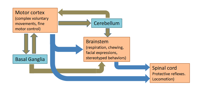
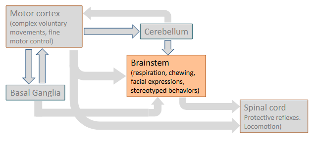
What are the 2 ways the motor system is organized?
The motor system has both hierarchical (blue)
and parallel (green) organization.
Prefrontal cortex function?
Executive decisions: not directly involved in movement
Frontal lobe function? How is it organized?
The frontal lobe is involved in action. It is functionally organized along a rostral-caudal gradient. The abstract aspects of action (e.g., “I’m going to make a sandwich.”) are rostral, whereas the specific action required to achieve the abstract goal (e.g., the movements involved in slicing bread) are cauda
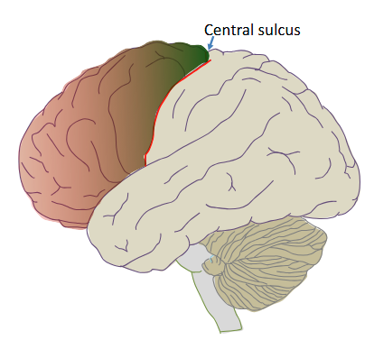
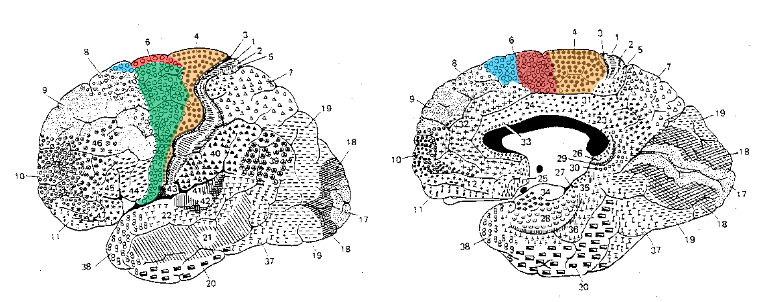
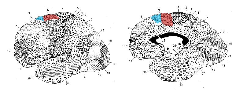
What does the motor cortex comprise?
Motor cortex comprises area 4 (primary motor cortex, M1) and area 6 (lateral premotor cortex, supplemental motor area (SMA) and pre-SMA).
What would be the consequence of a lesion in the primary motor cortex?
Lesions of primary motor cortex result in contralateral paralysis and increased muscle tone (spasticity). Movements requiring dexterity are especially affected
What would be the consequence of a lesion in the premotor cortex or the supplemental motor area?
lesions to premotor cortex or SMA affect the organization and control of movements
Which part of the cortex is most directly involved with movement?
Primary motor cortex is most directly connected to movement.
Does the primary motor cortex contain a somatotopic map?
Early work by Wilder Penfield and others showed that primary motor cortex contained a map of the body musculature that paralleled the somatotopic map in primary somatic sensory cortex. There are extensive connections between these two maps
When do neurons in the primary motor cortex fire when executing a movement?
Neurons in primary motor cortex fire before and during voluntary movements of contralateral muscles.
What mediates sensory-motor transformations?
Reciprocal connections between the parietal lobe (sensory + visual) and premotor cortex mediate sensory-motor transformations, the computations that enable sensory information to guide interactions with objects in the environment
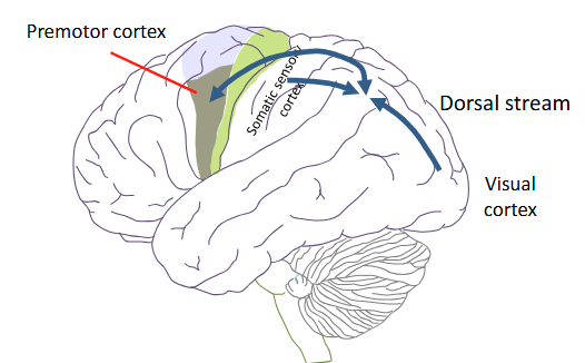
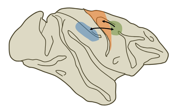
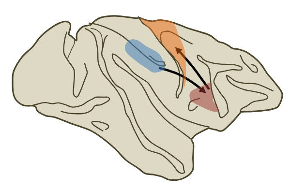
What’s the network that directs arm movements towards objects based on visual information?
A network comprising the parietal lobe <-> dorsal premotor cortex → primary motor cortex
Parietal (blue) connects to the premotor cortex (green), which then connects to the motor cortex (orange.)
Dorsal pathway
Parietal lobe function in spatial recognition?
Many neurons in the parietal lobe respond to both tactile (physical) and visual stimuli with receptive fields that are spatially in register. These neurons are thought to be involved construction of a peripersonal spatial map used to guide goal- directed movements
Neurons that fire if there’s a visual or physical stimulus near a region of the face
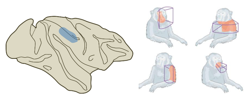
Peripersonal spatial map
Identifies “what is in my reach”

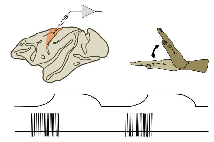
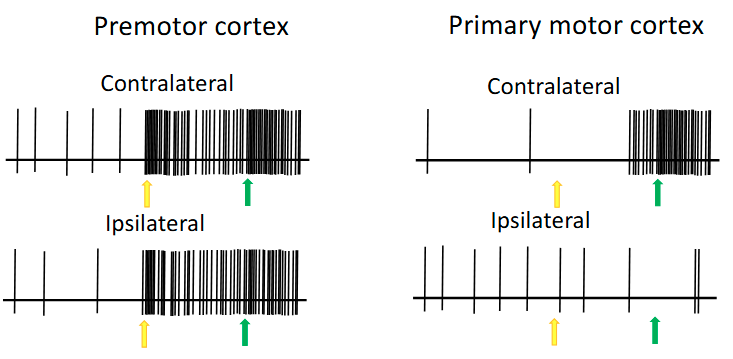
When do premotor neurons fire? What are they involved in?
Premotor neurons fire both during movements and during an imposed delay prior to the movement, suggesting they are involved in the planning and preparation to move.
Active when thinking about what/how movement they will make
(Note: M1 ONLY fires during movement - contralateral)
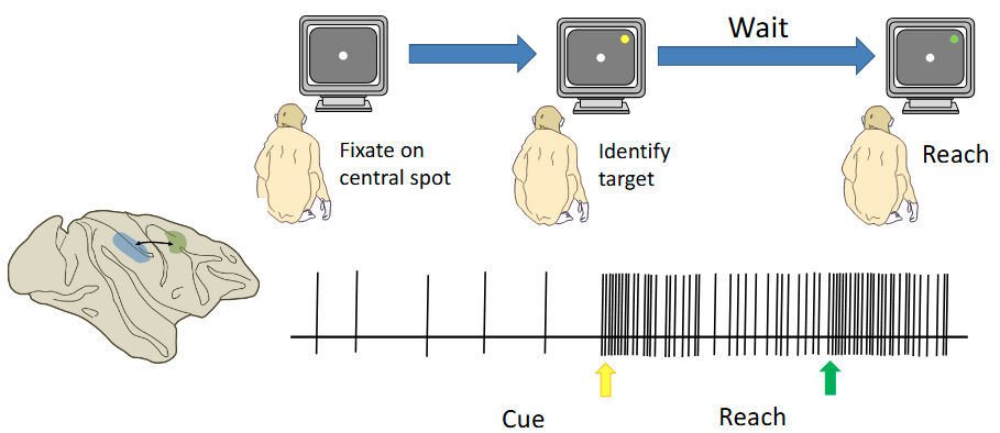
Premotor vs primary motor neuron
The premotor neuron on the left is active in preparation for and during the execution of an arm (either) movement toward a target, regardless of which arm is used. In contrast, the neuron in primary motor cortex is active only during the execution phase and only for the contralateral arm
Yellow means don’t touch
Green means touch
What’s the network responsible for shaping your hand to grasp objects?
A network comprising the parietal lobe → ventral premotor cortex → primary motor cortex is involved in shaping the hand to grasp an object
Ventral pathway
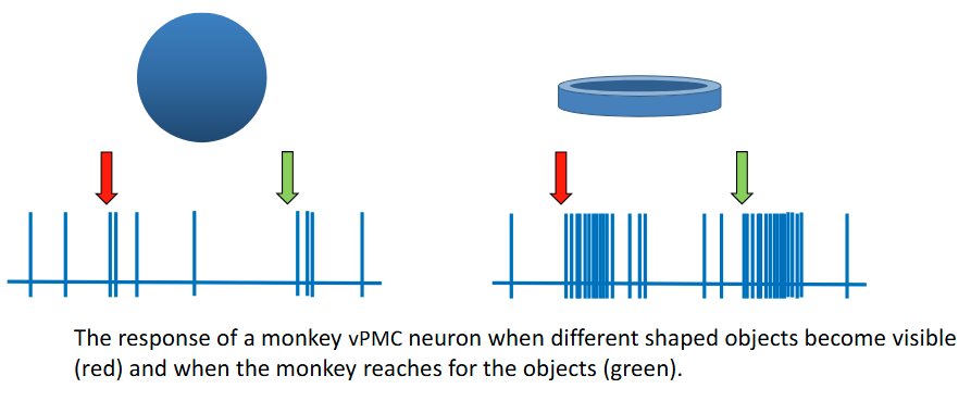
How do ventral premotor neurons differ from other premotor neurons?
Many ventral premotor neurons respond to preferred shapes. This neuron prefers a ring and responds strongly both when the stimulus is presented and when the monkey reaches for the object. It responds much less strongly to a sphere.
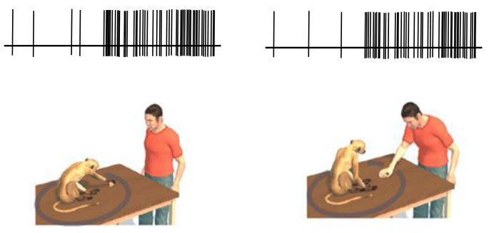
Mirror neurons
Mirror neurons were first discovered in premotor cortex. Mirror neurons respond when the monkey reaches for an object and when he watches the experimenter reach for the object. They do not respond to the object alone or to non-goal-directed movements of the experimenter’s arm
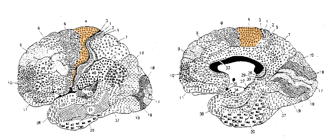
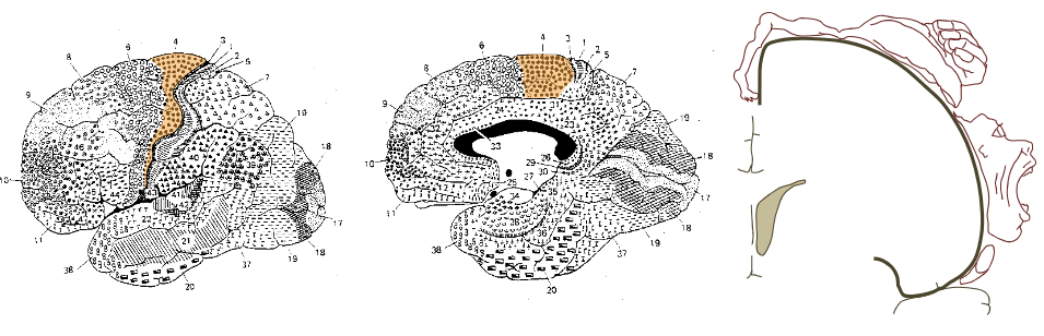
Supplementary motor area function?
The SMA has been proposed to be involved in internally generated (i.e “free-willed”) movements (not stimuli), especially learned complex movement sequences
Organizes a complex sequence of movements
Responsible for purposeful movements
What would happen if there were a lesion in the Supplementary motor areas?
lesions to SMA and pre-SMA can cause paradoxical effects on volitional movement, including alien limb syndrome, or, conversely, loss of spontaneous movement

Alien limb syndrome
Action of random free-willed movements (unbuttoning the shirt) uncoupled from the SMA
Movements of the arms’ free will (not yours)
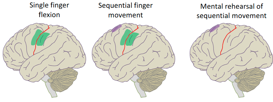
When will the Supplementary motor areas be active?
SMA (purple) is active during complex learned movement sequences, even when they are just imagined
Green is M1
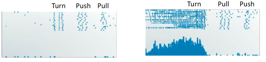
How can Supplementary motor areas be selective?
SMA neurons respond to selective components of learned movements sequences. This monkey SMA neuron is active prior to the turning motion in a sequence of movements, but only when the turn is followed by a pull and not a push movement
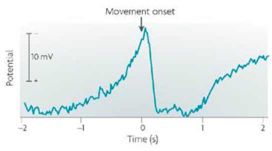
readiness potential
The readiness potential (Berietschaftspotential) is an EEG signal that is recorded from the medial frontal lobes (~ SMA) of humans, starting around 1 second before voluntary movements
EEG was active in SMA before the movement
Is the primary motor cortex the final common motor output from the cortex (i.e. do the other regions of the cortex ONLY feed it information to send to the spinal cord?)
No, M1 is indeed most directly connected to movement; however, it contributes only around 1/3 of cortical motor output to the brainstem and spinal cord. The remainder comes largely from premotor cortex, SMA and S1
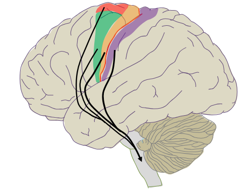
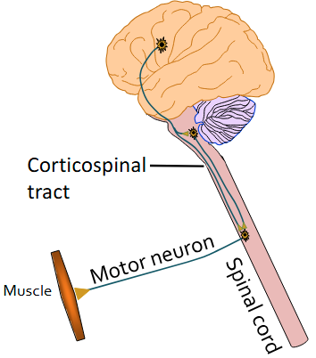
corticospinal tract
Projections (from layer V) from the motor cortex to the spinal cord form the corticospinal tract
Passes through the pryamids

corticobulbar tract
Projections (from layer V) from the motor cortex to the brainstem form the corticobulbar tract - then to the spinal cord
Doesn’t pass through the pryamids
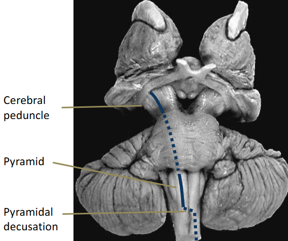
Explain the pathway of how motor fibres end up in the brainstem or spinal cord
Motor fibers pass within the internal capsule and then form the cerebral peduncles and the pyramids. Some fibers (corticobulbar) end in the brainstem. Others (corticospinal) project to the spinal cord (contralateral as it moves down.)
Travel through pons
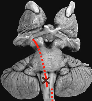
What would happen if there were a lesion in the pyramids? Why?
Monkeys with lesions to the pyramids, after a period of recovery, exhibit almost normal movement, except for loss of control of individual fingers (motor cortex → spinal cord severed)
The monkeys could still move because the corticobulbar doesn’t pass through the pyramids (motor cortex → brainstem → spinal cord not severed, rubrospinal tract allows them to move)
Brainstem function?
The brainstem controls stereotyped movements of the head (e.g. facial expressions, chewing, gag reflex). In addition, through descending connections with the spinal cord it contributes to the control of voluntary movements of the body
corticobulbar relation to cranial nerves?
Some corticobulbar projections control cranial nerves involved in voluntary movements in the head (e.g. facial expressions, tongue movements, jaw movements, and eye movements.)

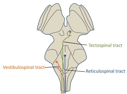
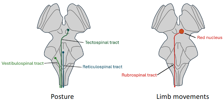
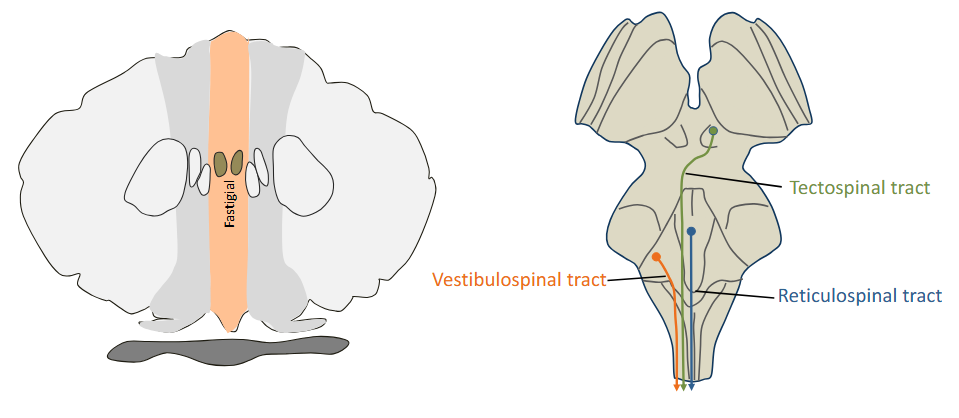
Medial brainstem pathway
Medial brainstem pathways innervate axial muscles that control posture and balance. These fibers travel down the ventral spinal cord white matter and terminate in ventromedial regions of the cord gray matter
Without them, you would just tip over
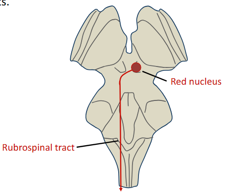
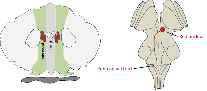
rubrospinal tract
The main lateral pathway is the rubrospinal tract, which originates in the red nucleus of the midbrain. Rubrospinal fibers descend in the contralateral dorsolateral column of the spinal cord and terminate in the dorsolateral gray matter. The rubrospinal tract contributes to control of voluntary limb movements.
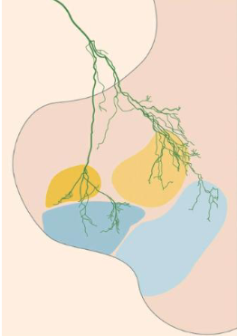
How does the primary motor cortex make precise connections (i.e. to control fingers)?
This axon (directly) from primary motor cortex makes many divergent synaptic connections, both directly onto motor neurons (blue regions) and onto spinal cord interneurons (yellow regions)
basal ganglia function?
• Movement preparation
• Action selection
• Scaling amplitude and velocity
• Movement sequencing
• Motor learning, especially learning of action-outcome
and stimulus-response relationships
They also have functions related to cognition and motivation.
What are the basal ganglia?
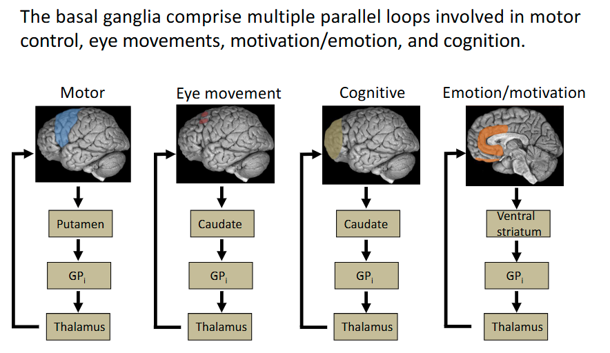
The basal ganglia are four interconnected subcortical nuclei, involved in voluntary movement, cognition and emotion
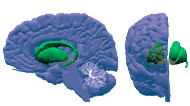
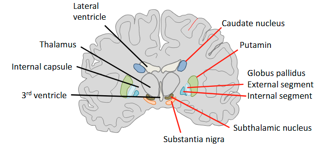
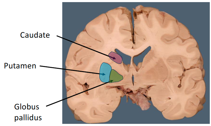
What is the basal ganglia comprised of?
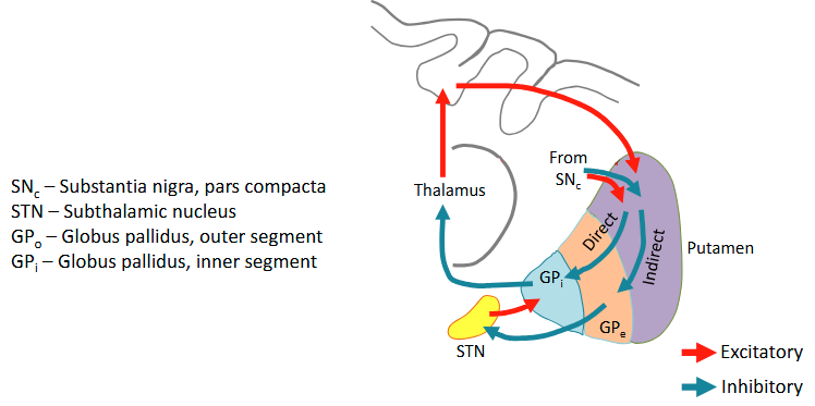
The basal ganglia comprise the striatum (the caudate, and putamin), the globus pallidus (GPe & GPI), the substantia nigra and the subthalamic nucleus.
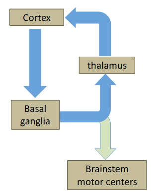
Basal ganglia projections
The basal ganglia forms a loop with the thalamus and cerebral
cortex. Basal ganglia output also projects to the brainstem
What are the input structures of the basal ganglia?
Caudate and putamen (striatum)
The caudate forms the sides of the lateral ventricle
What are the two pathways of the basal ganglia?
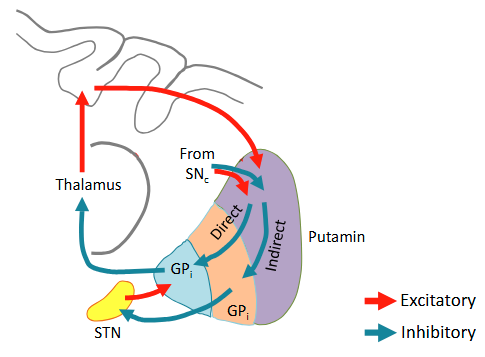
The basal ganglia modulate the activity of the thalamus and brainstem through direct and indirect pathways.
The direct pathway facilitates movement. The indirect pathway inhibits movement.
Dopaminergic input from the substantia nigra to the putamen facilitates the direct pathway and inhibits the indirect pathway
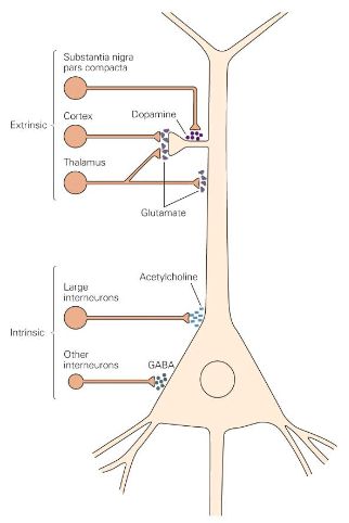
medium spiny neurons
The GABAergic projection neurons of the striatum (putamen) are called medium spiny neurons (MSNs). They make up 95% of striatal neurons. Of the remaining neurons, 3% are GABAergic interneurons and the remaining 2% are large, cholinergic interneurons
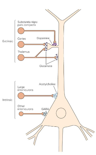
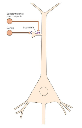
How do the medium spiny neurons get excitatory input?
Spines of MSNs receive excitatory glutamatergic inputs from the cerebral cortex. Dopaminergic neurons innervate spine shafts, which makes them well located to influence excitatory input from the cortex
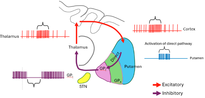
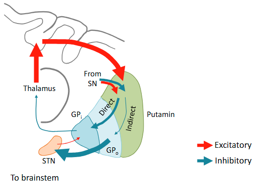
Explain the mechanism of the direct pathway
In the absence of movement, Gpi neurons are spontaneously active, whereas putamen neurons are silent. For the direct pathway, movement commands from the cortex (and thalamus?) activate MSNs in the putamen, inhibiting Gpi neurons, which relieves inhibition on thalamic and brainstem neurons, facilitating movement.
Selects movements you want to make
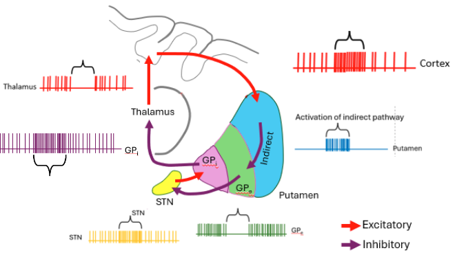
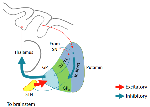
Explain the mechanism of the indirect pathway
Activation of indirect pathway neurons in the putamen inhibits Gpe. This relieves inhibition on excitatory STN neurons, resulting in increased firing of Gpi and inhibition of movement
Selects movements you don’t want to make
What are the 2 different dopamine receptors of the MSNs? How do these functions differ?
Dopamine from substania nigra projections increases excitability of direct pathways MSNs (through activation of D1 receptors) and decreases excitability of indirect pathway MSNs (through activation of D2 receptors). Thus, dopamine facilitates movement
Explain the mechanism for parkinson’s disease
In Parkinson’s disease, loss of dopaminergic input from the substantia nigra results in reduced activity in the direct pathway and accentuated activity in the indirect pathway. Parkinson’s patients exhibit a paucity of spontaneous movement.
How can we treat parkinson’s disease?
Destroy a part of the subthalamic nucleus

Explain the mechanism for hungtinton’s disease
reduced activity in the indirect pathway reduces the normal inhibition on the thalamus and brainstem, resulting in excessive, unwanted movements
Descending projections of the brainstem function?
Descending projections from the brainstem control posture and contribute to limb movements
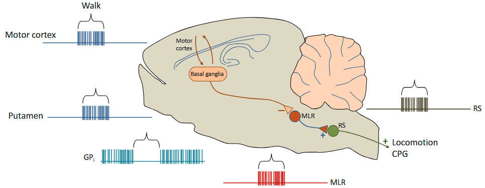
Explain the activation pathway of locomotion (to walk)
Tonic output from the basal ganglia (GPi) inhibits neurons in the mesencephalic locomotion region (MLR). Cortical motor commands for walking cause relief of this inhibition (activate the putamen). MLR neurons activate descending medullary reticulospinal neurons which activate locomotion central pattern generators in the spinal cord
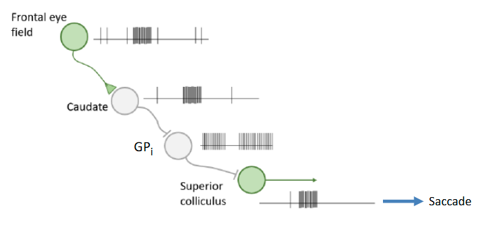
Explain the activation mechanism for saccades (rapid eye movements)
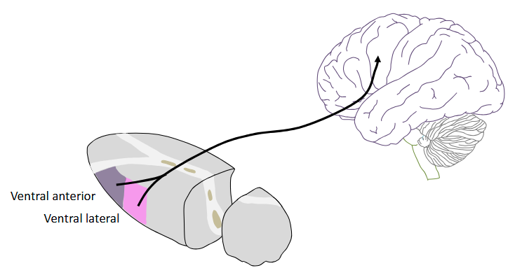
Where do the GPi project to in the thalamus? Why?
The Gpi projects to the ventral anterior and ventral lateral nuclei specific relay nuclei of the thalamus to create a loop between the basal ganglia and the cerebral cortex
What are the parallel loops of the basal ganglia?
All may be affected if the basal ganglia is damaged
What’s responsible for learning the relationship between actions and outcomes?
The basal ganglia are involved in the learning of relationships between actions and outcomes. This function is related to their role in action selection.
The basal ganglia play a role in stimulus-response learning and the transformation of consciously-mediated, goal-directed responses into automated habits
e.g. after a while, we know if we turn right, there will be water (non-conscious action when thirsty)
How does dopamine impact the direct and indirect pathways
Activation of dopaminergic synapses on the spine shafts of MSN dendrites promote long-term potentiation of glutamatergic synapses on direct pathway neurons and long-term depression of synapses on indirect pathway neurons
Increase/decrease dopamine on certain movements
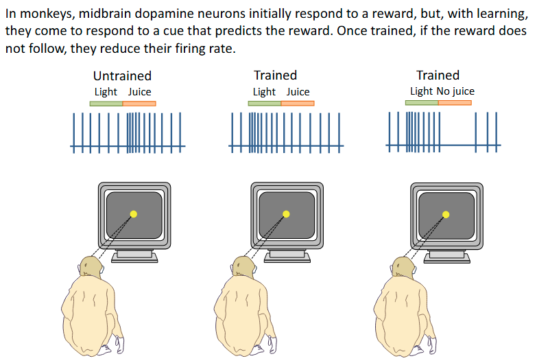
Light-juice experiment on monkeys
After some time, the dopamine fires in response to the light
Habit learning, not declarative
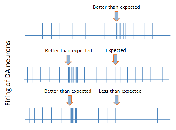
Reinforcement Learning Models
Dopamine firing encodes a prediction-error signal, which informs other brain regions of the deviation between experience and internally represented goals.
Basal ganglia function in habit learning?
The basal ganglia play a role in stimulus-response learning and the transformation of consciously-mediated, goal-directed responses into automated habits
to relieve the stress of the brain
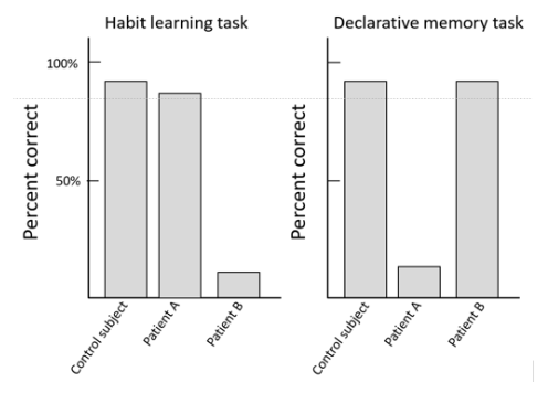
Damage in the medial temporal lobe leads to? How about basal ganglia?
Patients with damage to the medial temporal lobes show deficits in a declarative memory task, but not a habit learning task: Patient A.
Parkinson’s patients exhibit the opposite deficits: Patient B (damage to the basal ganglia)
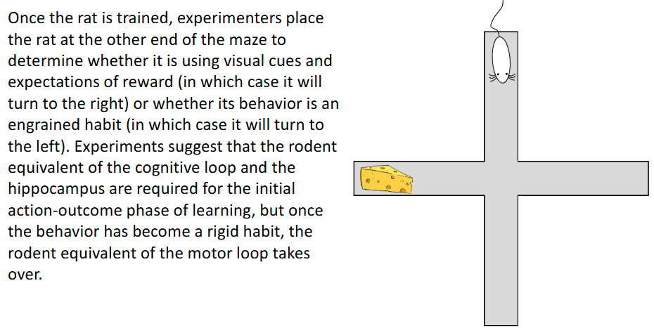
What’s responsible for the initial phases of habit learning? What about after?
Hippocampus then basal ganglia:
Hippocampus for learning (i.e. mouse WILL know where cheese is, regardless of where it’s placed)
Basal ganglia for autopilot (i.e. mouse will only turn left due to habit)
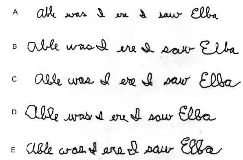
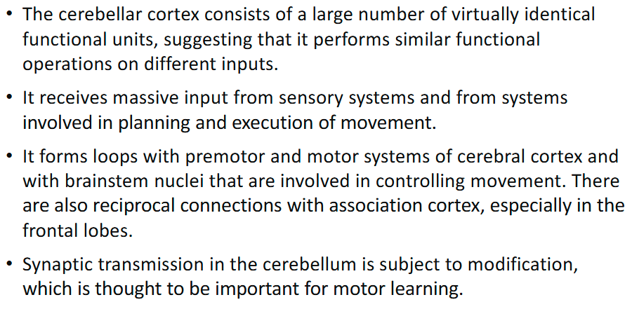
Cerebellum function?
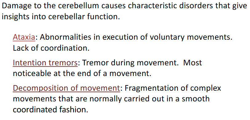
What would happen if the cerebellum is damaged?
Cannot hold finger out and then touch nose:
tries again but overshoots the boundaries
Rest tremors are caused by?
Damage to the basal ganglia
Does cerebellar lesions cause paralysis?
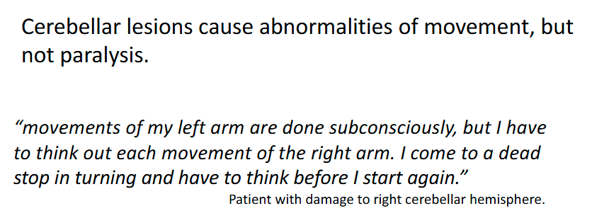
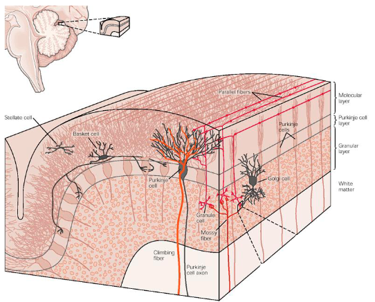
Folia
The surface of the cerebellum is covered with folds called folia. The folia greatly increase the surface area of the cerebellar cortex
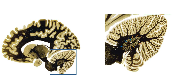
What does the cerebellum comprise?
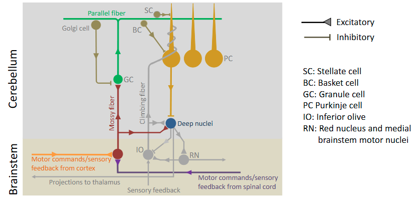
The cerebellum comprises a cortex and deep nuclei.
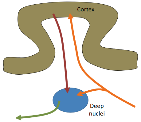
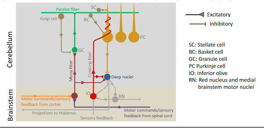
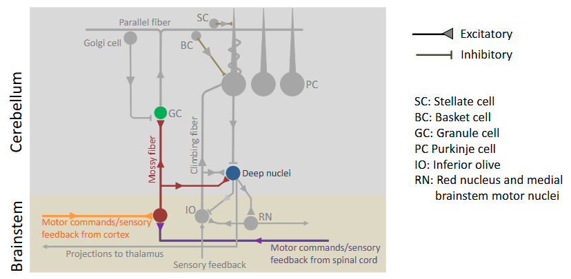
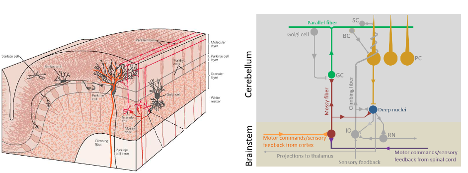
How does the cerebellum handle inputs and outputs?
Inputs to the cerebellum project (mossy fibers) to both the cortex and the deep nuclei. Outputs from the cortex project to the deep nuclei, which, in turn, form the outputs of the cerebellum
2 copies of the same information: deep nuclei → cerebellum & cortex → deep nuclei → cerebellum

How is the cerebellum connected to the brainstem?
The cerebellum is connected to the brainstem through three large fiber tracts called cerebellar peduncles.

Superior cerebellar peduncle
Outputs to the brainstem and thalamus
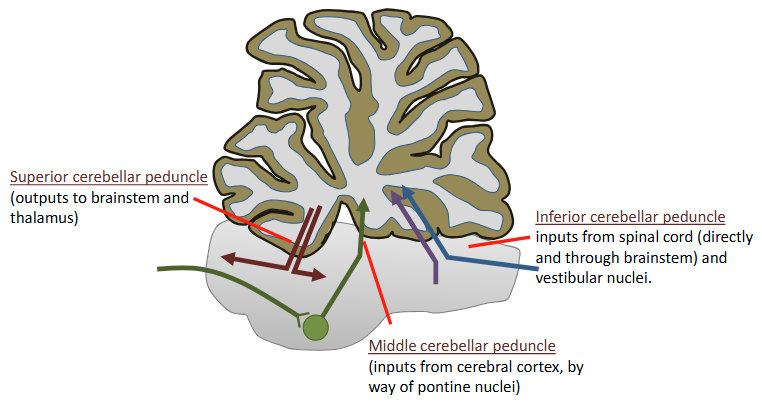
Inferior cerebellar peduncle
Inputs from spinal cord (directly and through brainstem) and vestibular nuclei

Middle cerebellar peduncle
inputs from cerebral cortex, by way of pontine nuclei (Descending pathways → synapse in pons → Middle cerebellar peduncle)

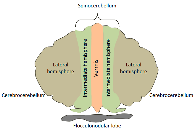
Functional Domains of the Cerebellar Cortex
Lateral hemispheres = lateral cerebellum gets all inputs from cerebral cortex
The spinocerebellum gets most inputs from spinal cord
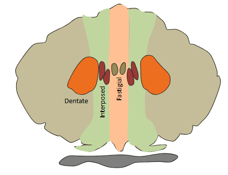
What are the 3 deep nuclei of the cerebellum?
There are three deep cerebellar nuclei, called fastigial, interposed and dentate.
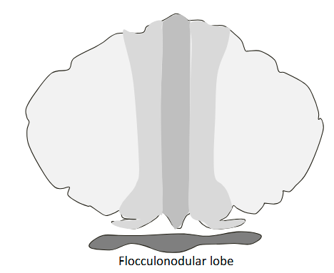
vestibulocerebellum function?
The vestibulocerebellum comprises the flocculonodular lobe. It receives inputs from the semicircular canals, the otolith organs, the visual system and somatic sensory input from the head and neck. It is involved in balance and eye movements.
outputs go directly back into brainstem: doesn’t go through deep nuclei
Input: Vestibular system - Output: brainstem
spinocerebellum consists of?
vermis and the intermediate hemispheres

spinocerebellum function?
It receives massive somatic sensory input (mainly proprioceptive) as well as efference copy of motor commands, through mossy fibers, coming either directly from the spinal cord or from the cord by way of the brainstem reticular formation
Coordination of the limbs
Output: brainstem
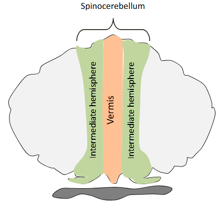
vermis function?
The vermis projects to the fastigial nucleus, which in turn projects to the vestibular nuclei and the reticular formation. These projections are mainly involved in posture and axial musculature
Intermediate hemispheres function?
The Intermediate hemispheres project through the interposed nuclei to primary motor cortex (via the ventral lateral thalamic nucleus) and to the red nucleus, which sends descending projections involved in control of limb movements.
How is the cerebrocerebellum formed?
The lateral hemispheres form the cerebrocerebellum
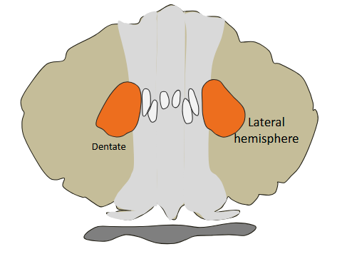
cerebrocerebellum function?
This region gets all its input from the cerebral cortex and sends its output through the dentate nucleus to the frontal lobes and to the red nucleus. The cerebrocerebellum is much larger in humans than in other primates. It is thought to be involved in the planning, programming and timing of complex, precisely coordinated movement sequences (e.g. finger movements).
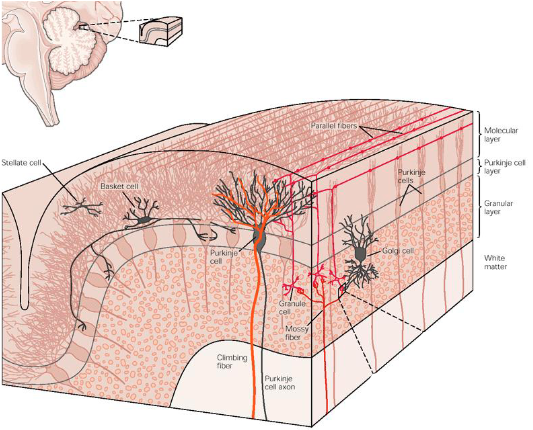
How is the cerebellar cortex organized?
The cerebellar cortex consists of five cell types, arranged in three layers: the molecular layer (dendrites & synapses), the Purkinje cell layer (1 cell layer) and the granule cell layer (lots of cell bodies).
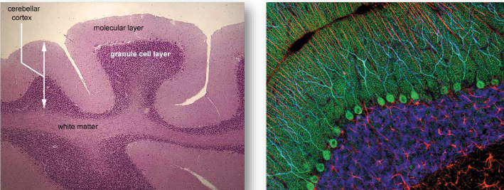
What are the 5 cell types of the cerebellum organized?
The five cell types of neurons in the cerebellar cortex are arranged in a highly ordered, repeating architecture
The most numerous inputs to the cerebellum are…
Mossy fibers
Mossy fibers function?
They convey copies of motor commands (efference copies) and sensory feedback from the cerebral cortex and from the spinal cord
Mossy fibers make excitatory synapses on granule cells and on cells in the deep nuclei
In cerebellum
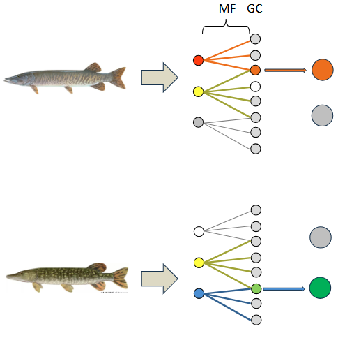
Granule cells function? Why are there so many?
Pattern generalization: Similarities between 2 things
Pattern separation: Distinction between similar things
Explain the mechanism for Pattern separation
In the sparse coding model, granule cells (GC) only spike in response input from multiple mossy fibers (MS). As a result, overlapping mossy fiber input is reencoded as nonoverlapping output from granule cells.
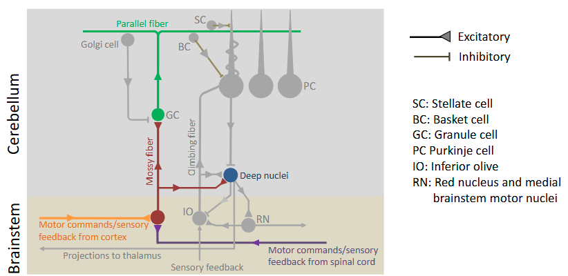
Where do the axons of granule cells project to?
The axons of granule cells ascend to the molecular layer and then bifurcate to form parallel fibers that run along the length of the folia.
Each parallel fiber makes excitatory synapses with hundreds of Purkinje cells along the length of the folia. Purkinje cells in turn project to the deep nuclei where they make inhibitory synapses
How are Purkinje cells organized in the folia?
Purkinje cells are lined up in rows along the folia. Parallel fibers run along the folia passing through the dendrites of Purkinje cells
What are the 5 cell types in the cerebral cortex?
Stellate cells
Basket cells
Granule cells
Purkinje cells
Golgi cells
How are parallel fibres organized in the cerebral cortex?
Parallel fibers are densely packed in the molecular layer
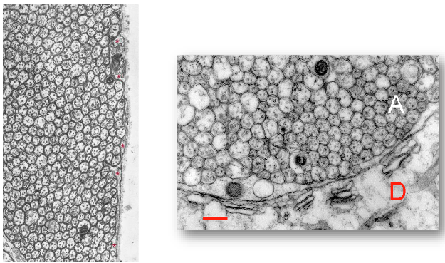
What do parallel fibres create excitatory synapses with?
A single parallel fiber spans around 5 mm and synapses with hundreds of Purkinje cells. A single Purkinje cell receives excitatory input from hundreds of thousands of parallel fibers
Runs through purkinje cells
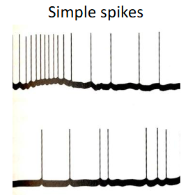
What are the spikes created by Mossy fibers?
Mossy fiber activity produces a steady stream of simple spikes in Purkinje cells, at rates of up to several hundred simple spikes per second. The frequency of simple spikes is strongly modulated by sensory stimuli and voluntary movements
Needs a lot of these spikes to fire action potentials
Spike patterns are important
What are the 3 inhibitory interneurons of the cerebral cortex?
stellate cells, basket cells and Golgi cells

What do parallel fibers create inhibitory synapses with?
Basket cells make inhibitory synapses on the cell bodies of Purkinje cells in adjacent rows.
Golgi cells provide feedback inhibition of granule cells
Basket cells function?
Basket cells make inhibitory synapses on the cell bodies of Purkinje cells in adjacent rows.

Stellate cells function?
Stellate cells make inhibitory synapses on the distal dendrites of nearly Purkinje cells.

Golgi cells function?
Golgi cells provide feedback inhibition of granule cells

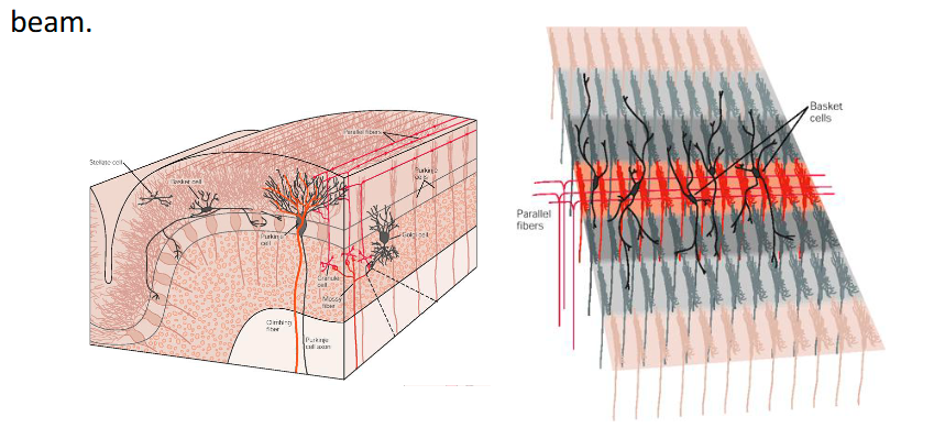
How do parallel cells activate basket cells for lateral inhibition?
Bundles of parallel fibers, called beams, run transversely and excite the dendrites of Purkinje cells, stellate cells, and basket cells. The basket cells inhibit the Purkinje cells flanking the parallel fiber beam.
Red active, grey inactive
Red stands out more
What are the 2 input pathways into the cerebellum?
1.) Mossy fibers + parallel fiber
2.) Climbing fibers