BIO 13: Exam 2
1/175
There's no tags or description
Looks like no tags are added yet.
Name | Mastery | Learn | Test | Matching | Spaced |
|---|
No study sessions yet.
176 Terms
Respiration
gas exchange (oxygen and CO2), talking about capillaries because gas exchange can only occur between capillary walls
Ventilation
moving air in and out of the lungs
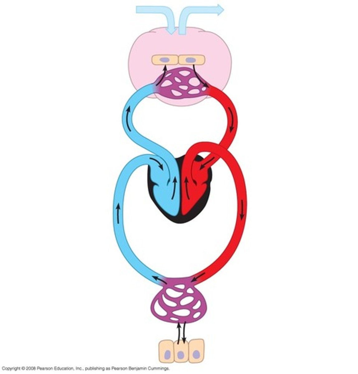
external respiration
pulmonary capillaries: pick up oxygen from lungs and remove CO2, breathing circuit, oxygen moves from the lungs into the capillaries and blood.
internal respiration
systemic capillaries: oxygen leaving and pick up CO2, systemic circuits, moving oxygen into the tissues
How does gas move?
Gradients, partial pressure
Dalton's law
- The partial pressure of each gas is directly proportional to its percentage in the mixture
- total pressure from a mixture of gases = sum of the pressures exerted independently by each gas in the mixture
what does the circulatory system do?
transport gases between the lungs and the rest of the body
Blood arriving in the lungs
has low partial pressure of O2 & high partial pressure of CO2, received from the right ventricle
Blood in the alveoli
O2 diffuses into and CO2 diffuses out of the blood
Blood in the tissue capillaries
partial pressure gradients favor diffusion of O2 into interstitial fluid & CO2 into the blood
What happens everytime we breathe?
We get rid of CO2, goes from the pulmonary capillaries to the lungs and then is released
Oxygen diffuses out of the...
systemic capillaries and deposits it into the capillary bed tissues and CO2 is picked up. The now deoxygenated blood leaves the capillaries and goes back to the heart
Partial pressure of oxygen in the lungs is the same as the partial pressure in the
systemic arteries
Left vs Right for Oxygen and CO2 and their gradients
Oxygen: high on left, low on right, large gradient (100:40; oxygen is picked up)
CO2: low on left, high on right, not as large as a gradient (46:100; CO2 is diffused out into the lungs and we breathe it out)
respiratory pigment functions
increase the amount of oxygen blood can carry; metal ion is capable of grabbing and oxygen and also letting it go, can carry small amounts of CO2
Why do we use respiratory pigments?
Because they do not dissolve well in plasma since plasma is mostly water
What are red blood cells full of?
respiratory pigments
what respiratory pigment do arthropods and mollusks use
hemocyanin (blue because metal ion is copper)
what respiratory pigment do vertebrates and invertebrates use
hemoglobin (red because metal ion is iron)
What can increase the amount of oxygen blood can carry?
Increasing hematocrit and respiratory pigments
how much O2 can hemoglobin carry
4 molecules O2 (one on each them)
hemoglobin structure
-Protein globin: 2 alpha and 2 beta chains
-1 Heme pigment bonded to each globin chain
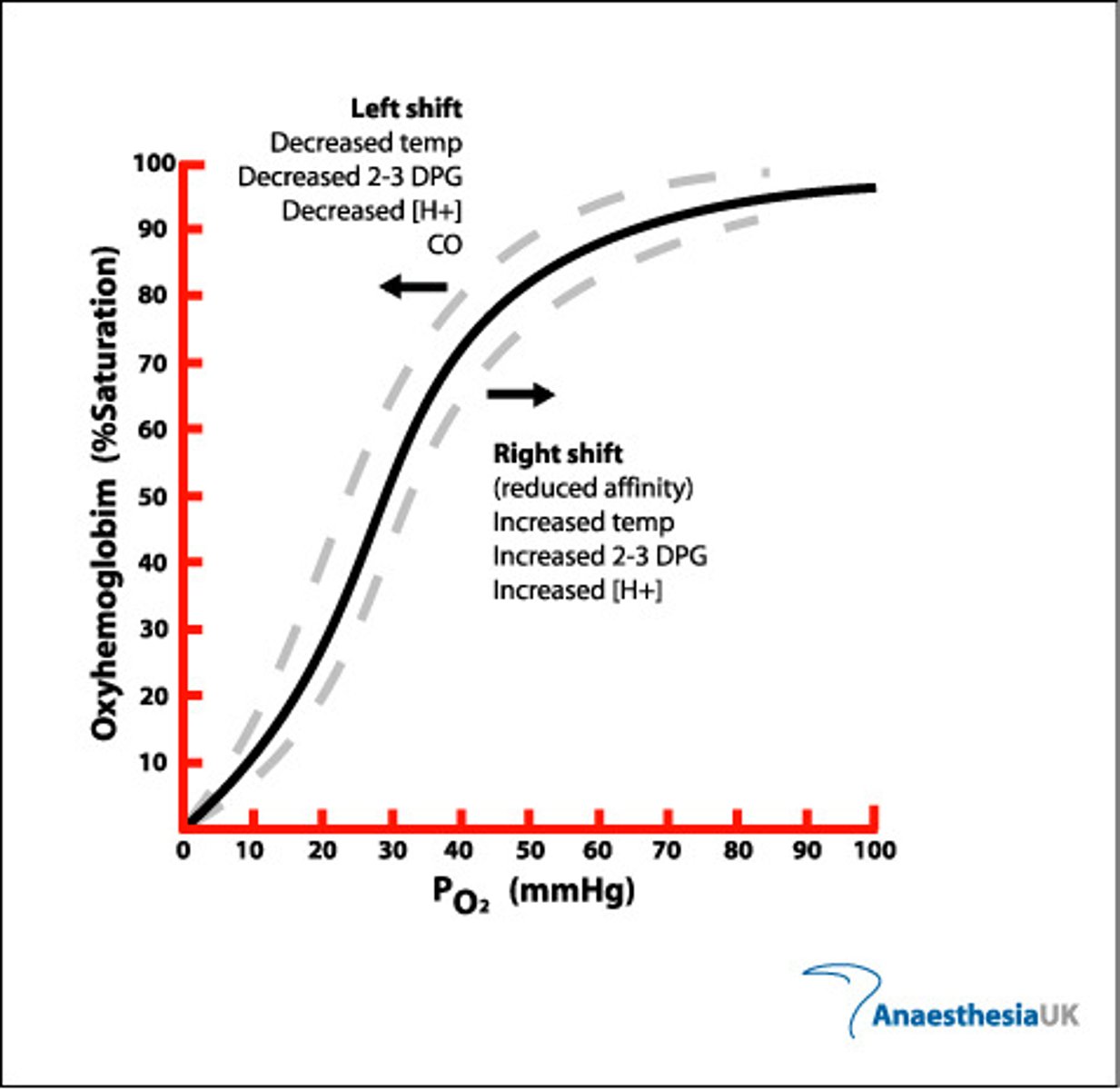
Bohr shift
CO2 lowers blood pH & decreases the affinity of hemoglobin for O2
What determines when hemoglobin grabs or lets go of oxygen?
Partial presure, when the partial pressure ox oxygen is 100 mm Hg, 100% of hemoglobin can bind to oxygen and it is fully staturated and is carrying as much as it can
CO2 is a waste product and forms
- H+ (acid)
- bicarbonate
When pH is low
more acidic (high H+)mand we release more oxygen to the tissues, tissues that are active are using oxygen; more metabolically active are more acidic because they are producing more CO2 and need more O2
pH
concentration of H+
pH=2/(log[H+])
oxygen unloading during exercise (graph)
low pressure (20) and low binding percent (35%)
oxygen unloading during rest (graph)
medium pressure (40) and high binding percent (75%)

oxygen loading (graph)
high pressure (100) and high binding percent (100%)
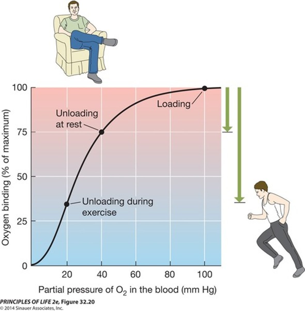
reserve capacity
Store oxygen for emergency issues, never fully deplete hemoglobin or oxygen
Oxygen during exercise
we can unload more oxygen than we can at rest because of reserve capacity; using more oxygen, more blood sent to the tissues, the hemoglobin will let go of more oxygen
Hemoglobin binding
Lungs pick up oxygen because high partial pressure and high hemoglobin binding, when the blood gets to the low oxygen tissue, oxygen has a low binding capacity which tells hemoglobin to let go
CO2 Delivery
just as important as O2, if too much CO2 builds up it is toxic and if we do not have enough, it can also cause problems
CO2 is transported in
blood plasma (10%),
bound to hemoglobin (25%),
As bicarbonate ions (HCO3-, 70%)
Why is so little percentage of CO2 transported in plasma?
Because CO2 cannot be dissolved in water. In hemoglobin it is hidden in the plasma as bicarbonate (CO2 combines with water to form acid and bicarbonate)
Gas Exchange
- Walls of lungs and pulmonary capillaries are simple squamous epithelia
- Two simple squamous right next to each other=thinnest possible layer we can have, gas can move very easily but bicarbonate cannot
how internal respiration works
CO2 enters capillaries & is coverted into HCO3-in the RBC.
HCO3-is carried in plasma to lungs; stick the bicarbonate in the plasma until we get to the lungs at the level of the tissues. Some CO2 will stay in plasma or in the red blood cells but most will interact with water to form bicarbonate in the the red blood cell.
how external respiration works
HCO3-
dissociates into H2O & CO2 in RBC.
CO2 exits capillaries & is removed by lung; we take bicarbonate and throw it back in the red blood cell and it breaks apart, CO2 exits
Exchange System
respiratory surfaces characteristics
Large, moist surfaces, where capillaries are, Gas exchange occurs by diffusion, more surface area=more diffusion=more oxygen
How do you increase surface area
more folding or more branches
examples of respiratory surfaces
outer body surface, skin, gills, tracheae, and lungs
Respiratory mode
oxygen source
Disadvantages of water (oxygen)
There is less oxygen in water and oxygen does not dissolve well in water. Water has 40X oxygen compared to air, water does not move as easy as air so oxygen gets to where it needs to go slower
What affects the dissolve rate of O2 in water?
Warm water and salt water make it harder for salt to dissolve
respiratory organs
- gills
-internal gills
-lungs
-tracheal system
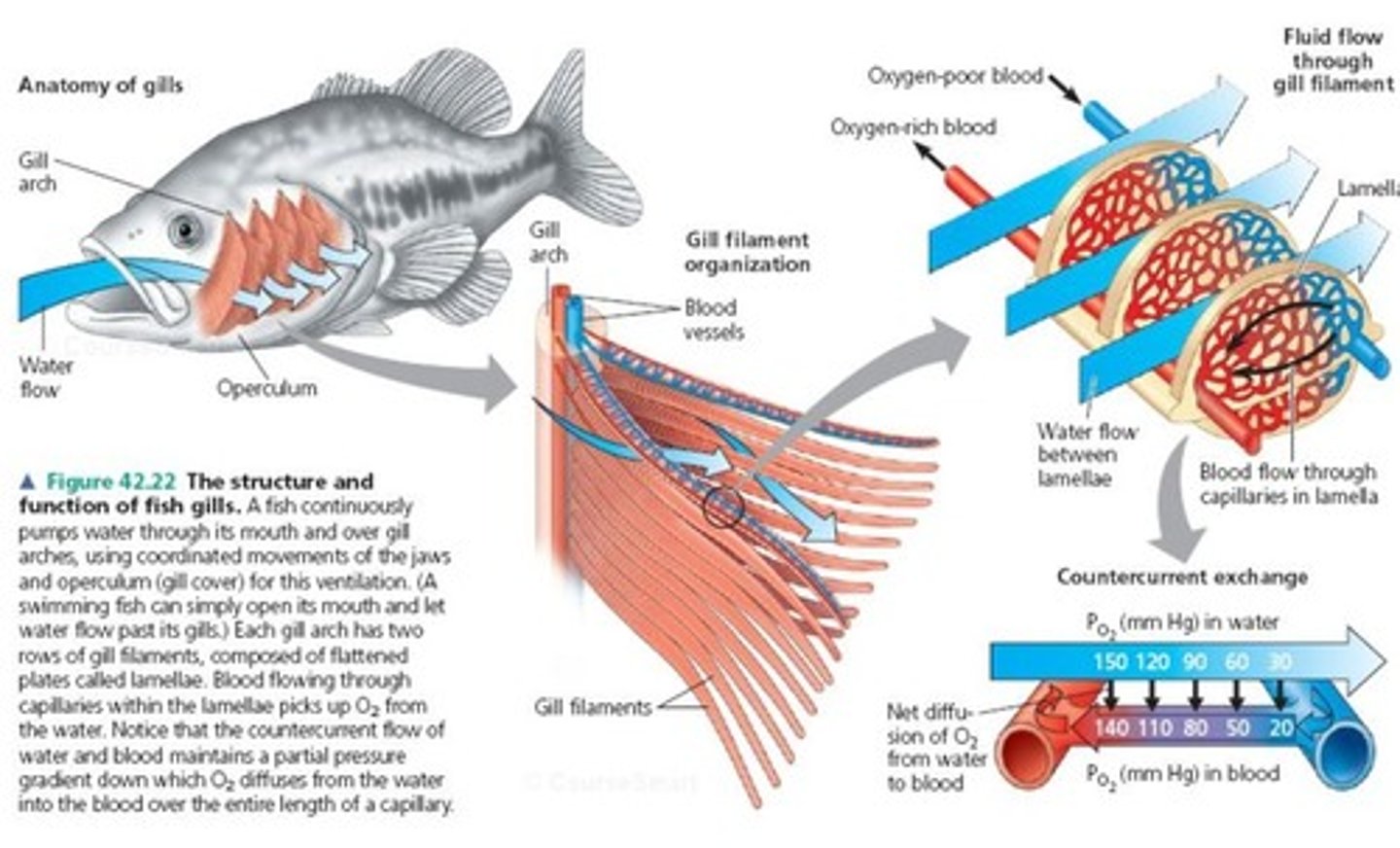
countercurrent exchange
- the opposite flow of adjacent fluids that maximizes transfer rates
(in fish)
- Blood flow in the gill capillary and they move their gills so that the water coming goes over the gills in the opposite friction
-Much more efficient
-Delivers more oxygen from the same amount of water compared to concurrent; gas exchange is enhanced
- Always more oxygen in water than in capillary
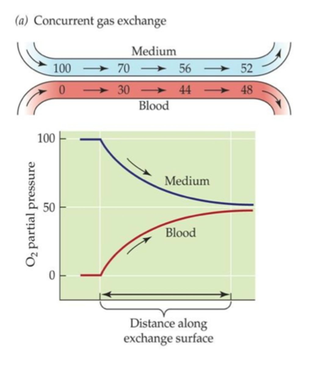
cocurrent gas exchange
Blood moves in the gill capillaries and they move their gills so that the water comes in over the gills and goes in the same direction as the blood
-Capillaries in the gills
-Animal only able to extract 50% of the oxygen from the water
-Gas exchange occurs until they reach equilibrium at the capillary bed
-Disadvantage for animals in water
-If you do not need much oxygen, this one is okay
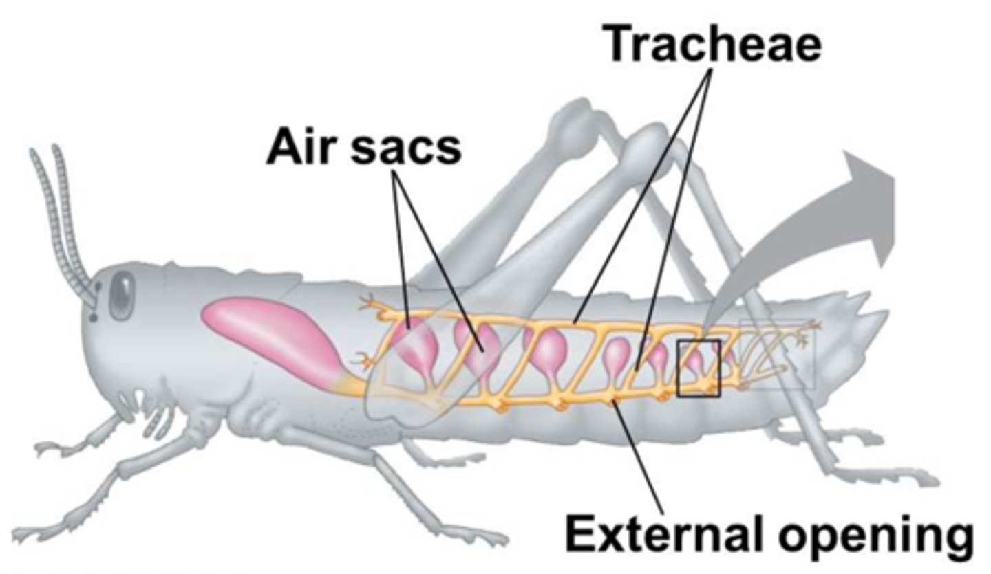
tracheal system
a system of branched, air-filled tubes that extends throughout the body and carries oxygen directly to cells (in insects)
-Open circulatory system
-Lots and lots of opening in the wall of their body, called sphericals, and these go to trachea and these are just tubes
-insects do not have cartilage rings to hold up trachea but instead have rings of cuticles
- Trachea branch into tracheole and they go into air sacs
-Body is filled with air sacs
-Each cell is next to an air sac
Muscles contract and allow air to move in and out through the air sacs and the trachea
nasal conchae (turbinates)
three narrow passages within the nasal cavity, they are moist and warm and lined with mucus
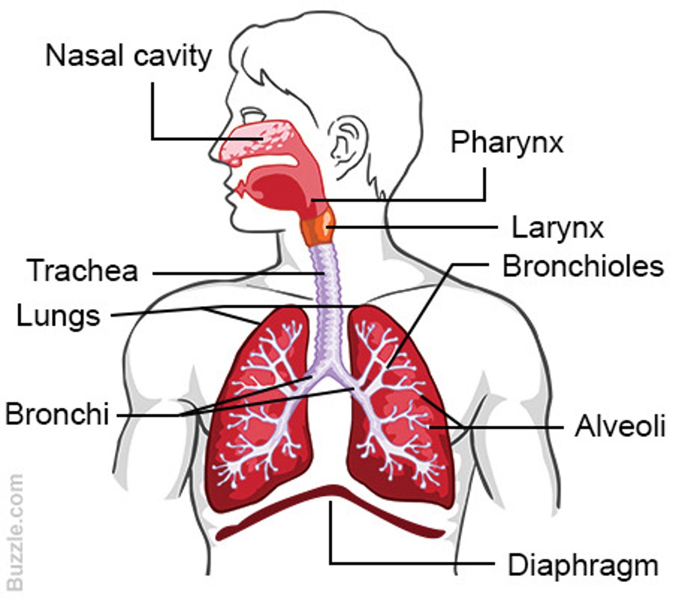
mammalian respiratory system
UPPER: Nasal cavities- filter, warm
and moisten air
Pharynx-
Glottis- opening to larynx
Larynx- sound; Adam's apple, voice box, vocal cords
LOWER: Trachea- air passage,
Bronchi- paired tubes into lungs Bronchioles- branched tubes
Lungs- contain alveoli and blood vessels
Alveoli- The oxygen exchange in the lungs takes place across the membranes of small balloon-like structures called alveoli attached to the branches of the bronchial passages
what does the nasal cavity do to the air?
warms, humidifies, and cleans the air to minimize the damage to the lungs
The tissues of the lungs are
fragile
Nose hairs
coated with mucus and create a filtration system to pick up particles to minimize the damage to the lungs
what part of the respiratory system cleans the air?
nose hairs and mucus
cells in trachea
release mucus and create a thin layer of mucus on the trachea to pick up particles from the air to clean the air before we breathe it in
what pushes mucus away from the lungs?
the cilia in the trachea (up to larynx so it can be swallowed)
mucociliary escalator
in the trachea, combination of cilia and mucus that keep dangerous particles from reaching the lungs
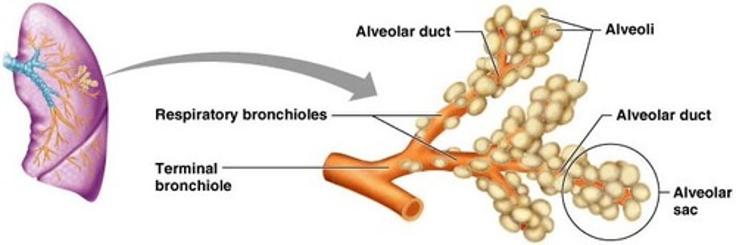
trachea branche into
bronchioles

bronchioles end with
alveoli
lung structure is affected by
metabolic rate
alveoli increases
surface area
Higher metabolic rate=
need more oxygen=highly branches lungs= more surface area= higher diffusion=more oxygen
mechanics of breathing
positive pressure breathing or negative pressure breathing
positive pressure breathing
"push" air into lungs with mouth (frogs)
also seen as swallowing air, contracts muscles to push air into the airway
negative pressure breathing
"pulls" air into lungs (mammals), changes lung volume which creates a pressure gradient and the difference in pressure creates a gradient and the air is moved from an area of high to low
how does negative pressure breathing work?
lower pressure in lungs then air follows gradient
boyle's law
P1V1=P2V2
inhalation (what happens)
diaphram contracts and moves down, rib cage gets larger and the volume increase which create less pressure, pressure decreases, air moving in, we contract muscles that change volume of thorastic cavity
exhalation (what happens)
diaphragm relaxes (moves up), rib cage gets smaller as rib muscles relax, lungs move up, volume gets smaller which creates more pressure, air pressure is larger inside the lungs than outside the lungs, no muscle contraction during exhalation, passive, it is just muscles relaxing and going to their resting state
inhalation volume and pressure
muscles contract, volume increases, pressure decreases
exhalation volume and pressure
volume decreases, pressure increases
What type of breathers are humans?
negative pressure breathers, create internal environment that has a lower atomospheric pressure than outside
What are lungs always being held up by?
The trachea and the trachea has rings of cartilage
what part of the brain controls breathing?
pons and medulla oblongata, neurons from the pons and medulla tell the muscles in the thoracic cavity to contract or relax
Peripheral Chemoreceptors
monitor blood has in the systemic arteries (just the big ones)
What are the peripheral chemoreceptors looking for?
high oxygen levels and low CO2 in branches of the carotid arteries (carrying oxygen to the brain) and in the aorta(carries oxygenated blood from the left ventricle to the systemic system to deliver oxygen throughout the body)
What do chemoreceptors do when CO2 is too high?
They send a signal to pons and medulla which then tells the muscles in the diaphragm to contract, Detection of an increase in pCO2 leads to an increase in ventilation. More CO2 is exhaled, the pCO2 decreases and returns to normal. Detection of a decrease in pCO2 leads to a decrease in ventilation. Less CO2 is retained in the lungs, the pCO2 increases and returns to normal.
What do chemoreceptors do when oxygen levels go down?
feedback increases ventilation to increase oxygen intake; send a signal to the pons and medulla which tell the muscle in the diaphragm to contract
How does pH effect breathing?
If it is too low, or more acidic
CO2 + H2O <->
H+ + HCO3-
peripheral chemoreceptors
in carotid arteries and aorta; monitor blood gases
central chemoreceptors
monitor blood gases, in pons and medulla, do not measure oxygen
Why do we respond more to CO2 than oxygen for a stimulus to breathe?
-CO2 is a major stimulus, we do not need a big change in CO2 for a response to be triggered but we do need a big change in O2 for a response to be triggered do to oxygen reserve capacity
All communication systems release a
chemical
Two cells of the nervous system
neurons and glial cells
neuron structure: soma
cell body, big round area
neuron structure: dendrites
attached to cell body, used to receive incoming information, the more branches, the more incoming information it can receive. Different neurons have a different number of branches, it depends on the neuron and what its job is
What does the cell do with the information received from the dendrites?
-Process information and then decide to send signal out or not (create an action potential or not)
If they do, action potential is sent down the axon
If the cell sends a signal, it starts _______ and then it will travel down the ______ until it gets to the _______ _______
at the axon hillock, axon, axon terminal
neuron structure: axon hillock
attaches cell body to the axon
neuron structure: axon
long branch coming off the cell body
neuron structure: axon terminal/synaptic terminal
the split ending on the axon to send the signal to another place, where the signal reacts with the next cell and send a signal to tell the other cells what to do.
neuron structure: myelin sheath
insulation around axon to make the signal travel faster
Myelin sheath is made up of
individual cells called Schwann cells and they wrap themselves along a small part of the axon and protects it
neuron structure: nodes of ranvier
the "pinched" parts of the axon, the little gaps between each schwann cell
does every neuron have myelin?
no
synapse
the small gap between the axon terminal of one neuron and the next cell
Neurons are part of a pathway
every neuron in a signal pathway create two signals, electrical and chemical