Autonomic Nervous System
1/10
There's no tags or description
Looks like no tags are added yet.
Name | Mastery | Learn | Test | Matching | Spaced |
|---|
No study sessions yet.
11 Terms
Autonomic nervous system
visceral efferent nerve fibers that innervate the heart, smooth muscle, and glands
coordinates cardiovascular, respiratory, digestive, urinary, and reproductive functions
works in conjunction with the endocrine system to maintain homeostasis in the body
quick to respond to a stimulus. nut effects on ANS don’t last as long as the endocrine system
autonomic nervous system influences other systems of the body because of its role in homeostasis
DO NOT NEGLECT THIS CHAPTER FOR THE EXAM
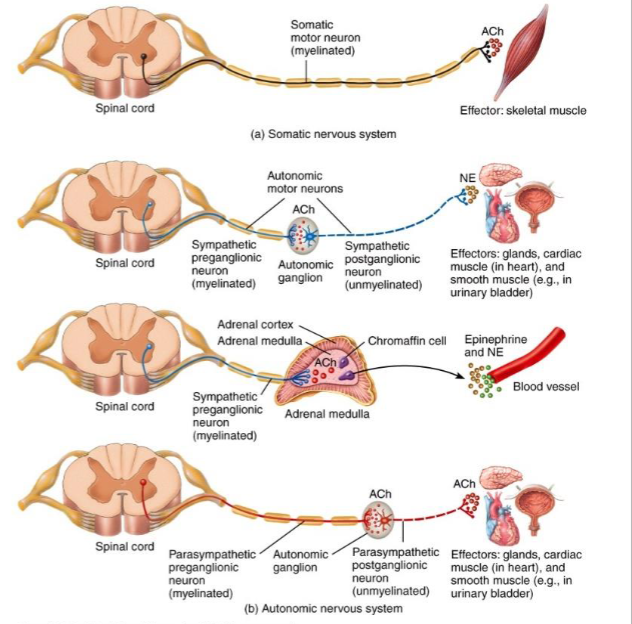
Somatic efferent fibers
somatic efferent fibers innervate skeletal muscles.
one neuron pathway- cell body of a neuron that stimulates a skeletal muscle is found in the CNS
two neuron pathway- In ANS, neuron with a cell body in CNS exits CNS and goes to ganglion where it forms a synapse with a second neuron
preganglionic neuron/first order neuron/central neuron- neuron that exits CNS
myelinated
postganglionic neuron/second order neuron- neuron that foes from the ganglion to the autonomic effector
unmyelinated
Divisions of ANS
sympathetic and parasympathetic division
dual innervation- when an organ has fibers from one division, usually has fibers from the other division
one division stimulates the organ and the other inhibits
allows ANS to maintain homeostasis
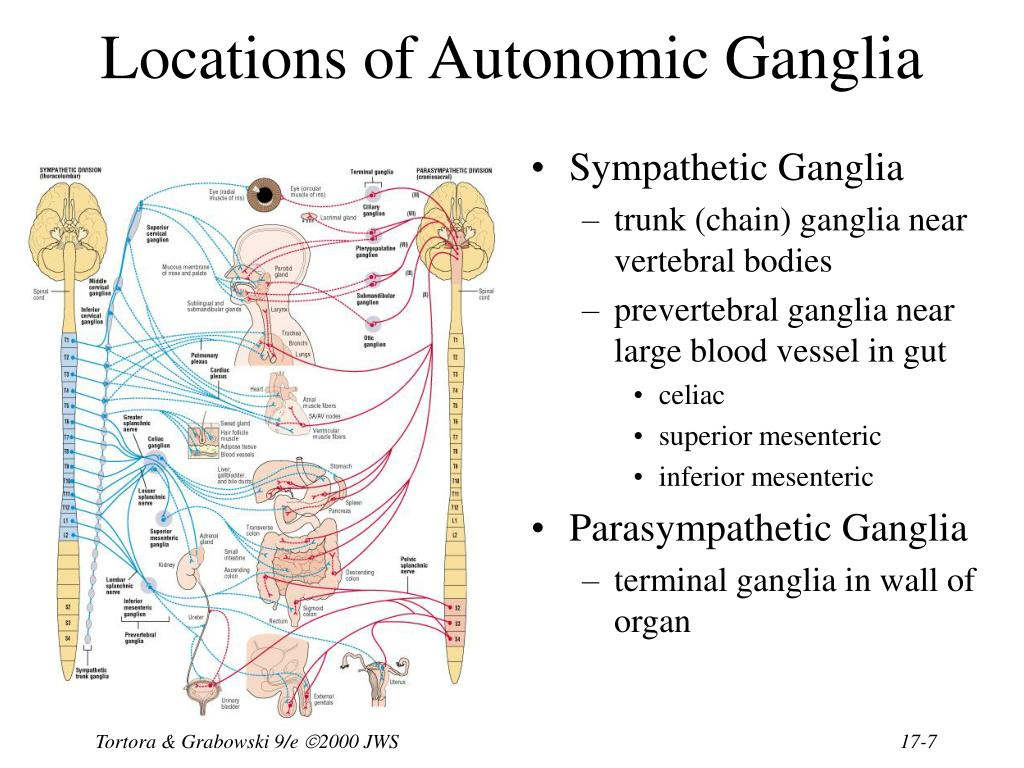
Autonomic Ganglions
sympathetic chain (trunk) ganglion/ paravertebral/ lateral ganglia
series of ganglia that extend from the skull to the coccyx on each side of the vertebral column
part of the sympathetic division of ANS
Collateral/ prevertebral ganglion-
located in front of the spinal column close to large arteries in the abdomen
associated with the sympathetic division of the ANS
Terminal ganglion/intramural ganglion-
located near the effector (“terminal”- at the end of the path) or in the walls (“intramural”) of the effector
associated with the parasympathetic division
since ganglia are closer to target organ in PS division, preganglionic fibers of the PS division tend to be longer than those in the sympathetic division
Parasympathetic division
dominant division during rest
“rest and digest”
usually slows activity of a target organ
MAJOR EXCEPTION- digestive tract
SLUDD- salivation, lacrimation, urination, digestion, defecation
activities that are stimulated
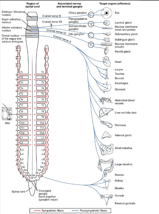
Parasympathetic Division Structures and Pathways
cell bodies of preganglionic neurons of PS division (right side, pink) located in the nuclei of cranial nerves III, VII, IX, and X and the lateral gray horns of sacral nerves 2-4
Parasympathetic division also called the craniosacral division
preganglionic fiber may stimulate several postganglionic fibers, but they all usually go to the same target organ
most of the cranial outflow (80%) of this division is through cranial nerve X, the vagus nerve
note the long preganglionic fiber that synapses with the postganglionic fiber close to, or in the wall of, the target organ
Sympathetic division
dominant division during periods of stress, exertion, or emergencies
increases activity of a target organ
causes activities associated with fear and anger'
Heart rate increases, respiratory rate increases, pupils dilate
“fight or flight”, “acute stress response”, “alarm reaction”
survival mechanism that responds to potentially dangerous situations
MAJOR EXCEPTION: digestion
Sympathetic Division Structures and Pathways
cell bodies of the preganglionic neurons (left side, blue) located in the lateral gray horns of all of the thoracic nerves and the first two lumbar nerves
sympathetic division also called the thoracolumbar division
Preganglionic fiber synapses with several postganglionic fibers
may affect several target organs
some fibers synapse in the chain ganglia that lie beside the spinal cord, but other fibers pass through these ganglia to synapse in collateral ganglia

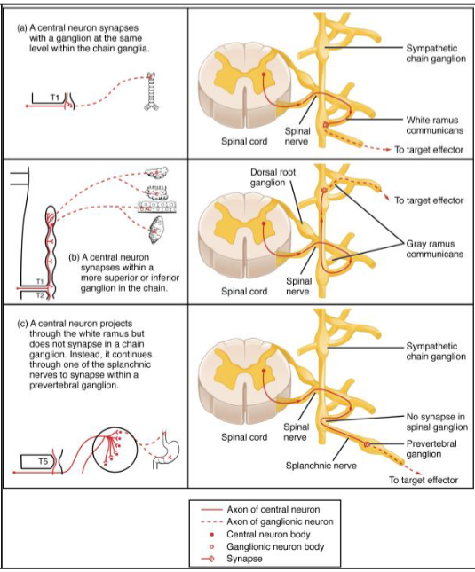
Rami
rami- connections between spinal nerve and a ganglion of the sympathetic trunk
White ramus-
contains myelinated axon of the preganglionic neuron
Gray ramus
contains the unmyelinated postganglionic fiber
note that some preganglionic fibers pass through the trunk and exit the ganglion as the splanchnic nerves
these preganglionic fibers synapse in the collateral ganglia
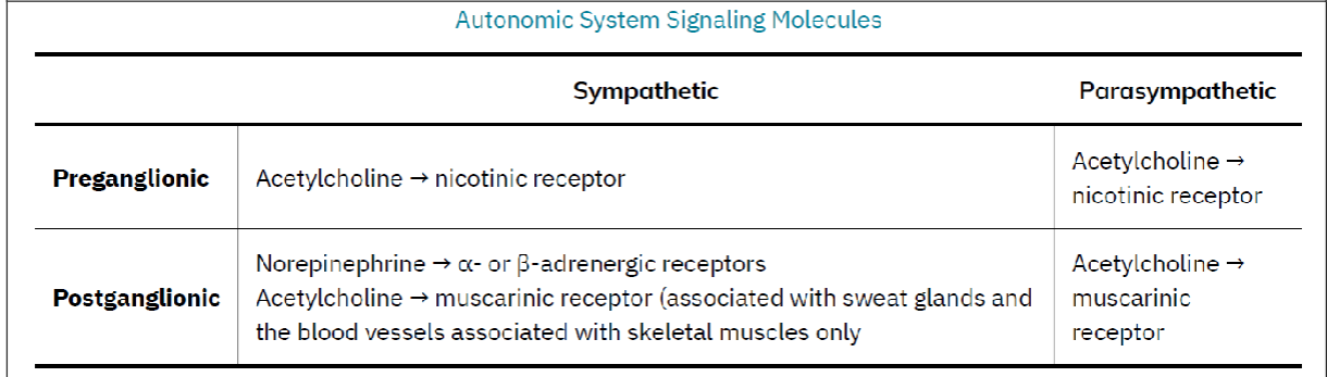
Autonomic System Signaling Molecules
preganglionic fibers of the ANS use Acetyl Choline as the transmitter
postganglionic parasympathetic neurons also use acetyl choline as a transmitter
Called CHOLINERGIC NEURONS
Acetyl choline is broken down very quickly by the enzyme acetyl cholinesterase
Two types of cholinergic receptors
nicotinic receptors- located on the postganglionic neurons
muscarinic receptors are on parasympathetic effectors
adrenergic- almost all sympathetic postganglionic fibers (ignore sweat glands) are this
release noradrenalin/norepinephrine at the neuroeffector junction
alpha- and beta-adrenergic receptors on sympathetic effectors
noradrenalin is broken down slowly by COMT or MAO so the effects of sympathetic stimulation last longer than parasympathetic stimulation
Autonomic nervous stimulation exceptions
supra renal gland/adrenal gland
gland that rests above kidney
divided into a cortex (outer) and medulla (inner)
adrenal medulla is the exception
does not have a two-neuron pathway
preganglionic neuron from CNS directly stimulates the adrenal medulla
Adrenal medulla does not have dual innervation
only has fibers from the sympathetic division
endocrine gland
secretes adrenalin (epinephrine) and noradrenalin into the blood
molecules bind to the alpha- and beta-adrenergic receptors of autonomic effectors
SYMPATHOMIMETIC- cause the same response as direct sympathetic stimulation
hormones important part of the alarm reaction
fact that these molecules are in the blood is another factor in causing the effects of the SNS to last longer than the PSNS
In the past, it was thought that the ANS was automatic, and not subject to control by the conscious mind.
can be affected by higher centers of the brain
biofeedback- people that are connected to a device can alter their heart rate or brain waves
other individuals are able to inhibit SNS without being connected to a device
these techniques are applied through yoga or meditation and require long periods of training