Male and female pelvis and Peroneum Chapter 6
1/17
There's no tags or description
Looks like no tags are added yet.
Name | Mastery | Learn | Test | Matching | Spaced | Call with Kai |
|---|
No analytics yet
Send a link to your students to track their progress
18 Terms
Identify the bones and ligaments forming the pelvis.
Identify the major foramina of the bony pelvis.
Differentiate between the true pelvis and the false pelvis in terms of their structures and locations.
Recognize the blood vessels that pass from the posterior abdominal wall into the pelvis.
Identify the branches of the anterior and posterior divisions of the internal iliac artery.
Relate the muscles of the pelvic wall and floor to one another and to their attachments.
Differentiate between the pelvic diaphragm and the UG (urogenital) diaphragm.
Describe the lymphatic drainage as it relates to the structures in this region.
Visualize and relate structures of the male and female pelvis with respect to adjacent structures.
Describe the etiology of the most common clinical cases pertaining to the male pelvis.
Identify all bones found in this region and discuss the role of any associated tuberosities, grooves, etc.
Describe the boundaries and regions of the perineum.
Identify the muscles of the male perineum and their attachments.
Differentiate between the urogenital (UG) triangle and the anal triangle.
Identify the blood vessels and nerves supplying the structures of the perineum.
Identify other regions into which the superficial fascial layers of the perineum are continuous.
Identify the major arteries supplying the perineum.
Describe the etiology of the most common clinical cases pertaining to the male perineum.
Compare the organs in the female pelvis with those in the male pelvis.
Describe the clinical significance of the peritoneal fossae related to the uterus, the clinical significance of the peritoneal fossae related to the uterus.
Describe the etiology of the most common clinical cases pertaining to the female pelvis.
Identify the muscles of the female perineum and their attachments.
Describe the etiology of the most common clinical cases pertaining to the female perineum.
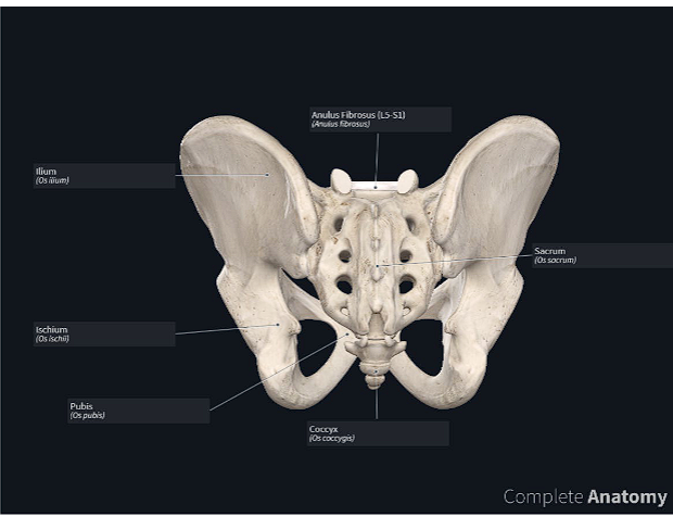
picture of the pelvis
pelvis= basin because the pelvis looks like a basin.
the pelvis is inferiorposterior to the abdomen
the pelvis is the area of transition between the trunk and lower limbs.
The pelvic cavity is the inferiormost part of the abdominopelvic cavity.
pelvis: basin
cavity: hollow, concave
pelvic cavity" is "the hollow basin" or "the hollow space of the basin."
The name is highly descriptive of its anatomy. The pelvic cavity is the funnel-shaped space enclosed by the bony pelvis (the "basin").
Anatomically, the pelvis is the part of the body surrounded by the pelvic girdle (bony pelvis), part of the appendicular skeleton of the lower limb
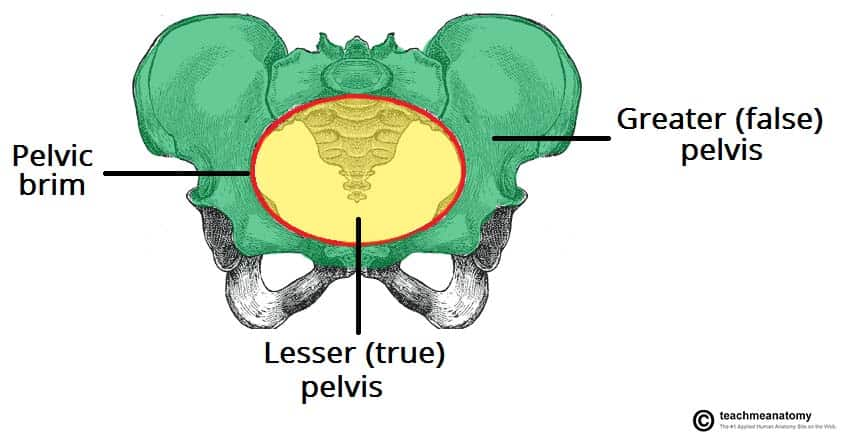
pelvis is subdivided into greater and lesser pelves
The greater pelvis is surrounded by the superior pelvic girdle.
The greater pelvis is occupied by inferior abdominal viscera
The lesser pelvis is surrounded by the inferior pelvic girdle.
Externally, the pelvis is covered or overlapped by the inferior anterolateral abdominal wall anteriorly, the gluteal region of the lower limb posterolaterally, and the perineum inferiorly.
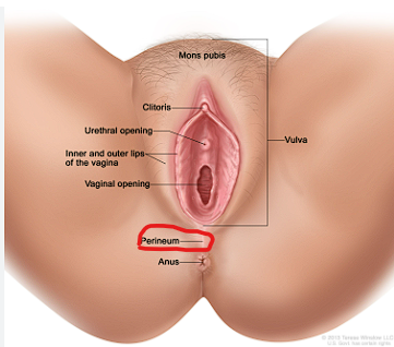
perineum
the area between the anus and the scrotum or vulva.
perineum: (perí): A preposition meaning "around," "about," or "near."
(inéō): An verb meaning "to empty out," "to excrete," or "to discharge." This verb is related to the Greek word for feces.
Literal Interpretation:
Therefore, the most accurate literal meaning of the Greek perineon is "the region around (the points of) excretion." or "that which is around the discharge (areas)."
perineum refers both to the area of the surface of the trunk between the thighs and the buttocks, extending from the coccyx to the pubis, and to the shallow compartment lying deep (superior) to this area but inferior to the pelvic diaphragm. The perineum includes the anus and external genitalia: the penis and scrotum of the male and the vulva of the female
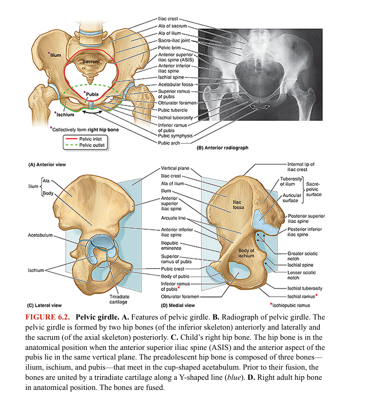
PELVIC GIRDLE
PELVIC GIRDLE
pelvic girdle
etymology
relationship to the femur
primary functions
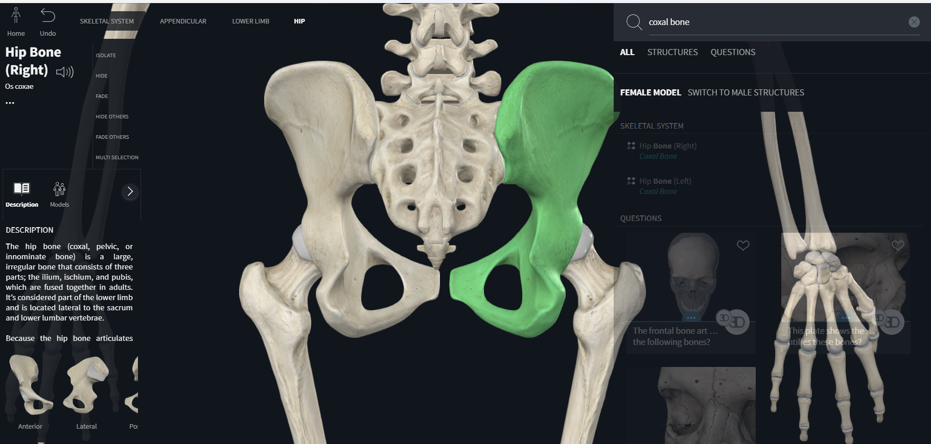
pelvic girdle= coxal bone
The pelvic girdle is a basin-shaped ring of bones (pelvis) that connects the vertebral column to the two femurs.
femur: “thigh”
The primary functions of the pelvic girdle are:
• bear the weight of the upper body when sitting and standing
• transfer that weight from the axial to the lower appendicular skeleton for standing and walking
• provide attachment for the powerful muscles of locomotion and posture and those of the abdominal wall, withstanding the forces generated by their actions
-contain and protect the pelvic viscera (inferior parts of the urinary tracts and the internal reproductive organs) and the inferior abdominal viscera (e.g., intestines) while permitting passage of their terminal parts (and, in females, a full-term fetus) via the perineum
-provide support for the abdominopelvic viscera and gravid (pregnant) uterus
-provide attachment for the erectile bodies of the external genitalia
-provide attachment for the muscles and membranes that assist the functions listed above by forming the pelvic floor and filling gaps that exist in or around it
bones and features of the pelvic girdle
the pelvic girdle is formed by three bones:
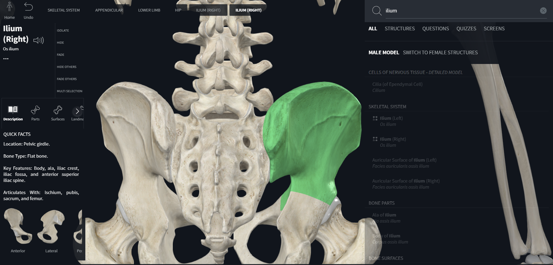
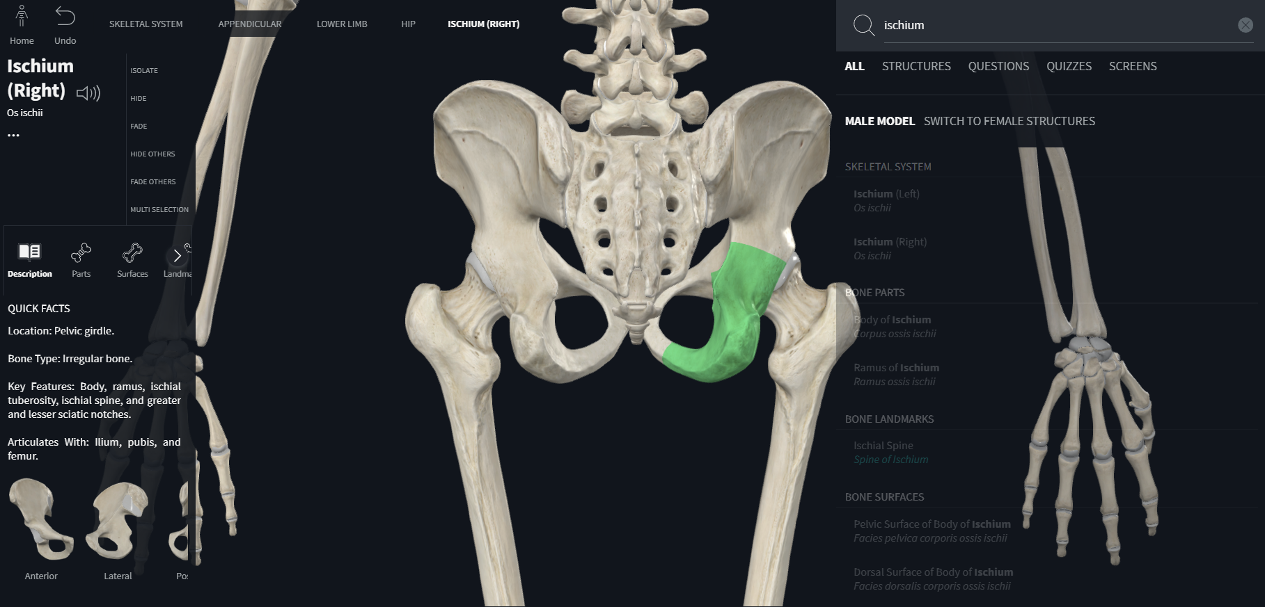
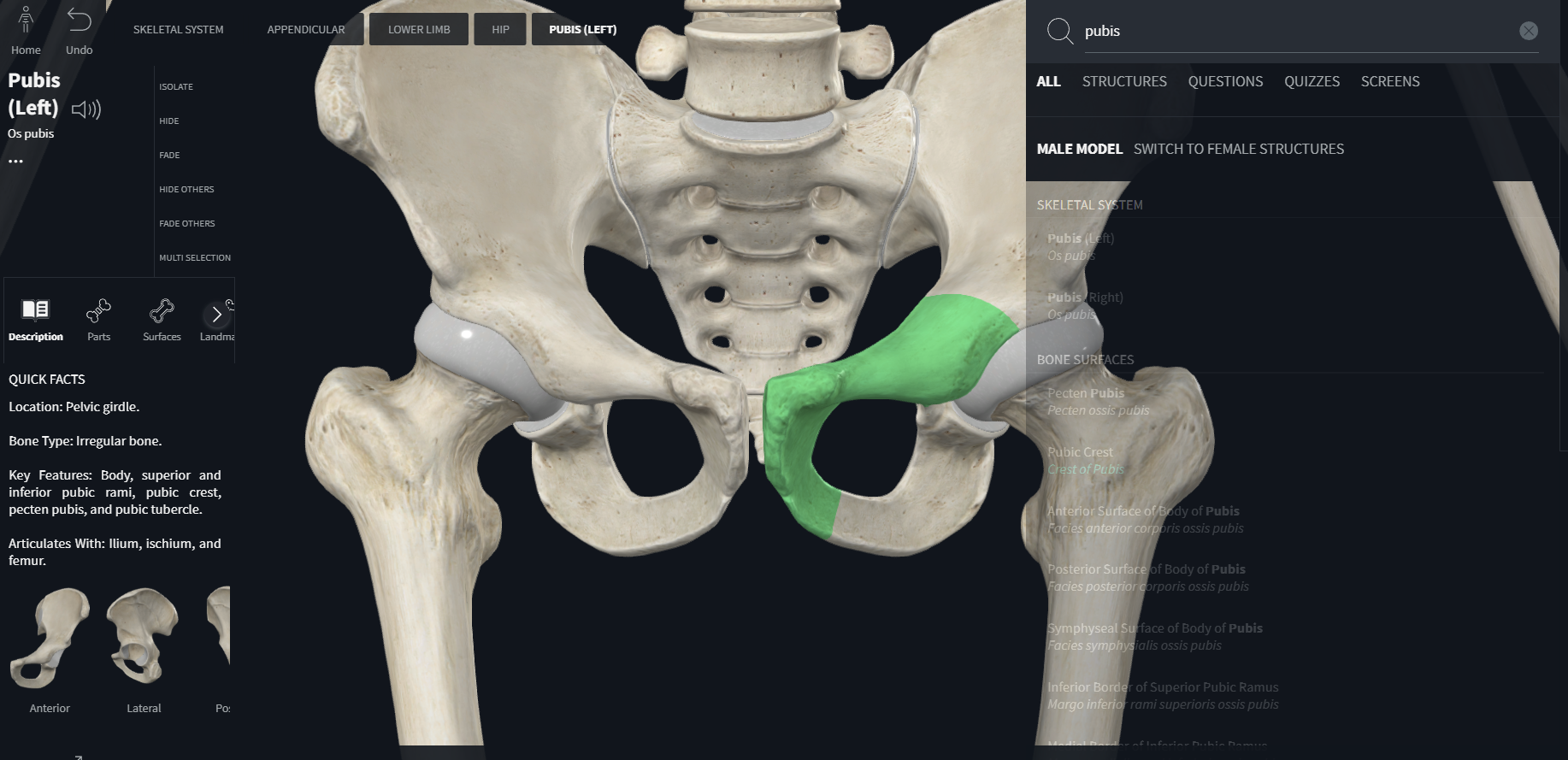
Right and left hip bones (coxal or pelvic bones): large, irregularly shaped bones, each of which develops from the fusion of three bones (ilium, ischium, and pubis)
Sacrum: formed by the fusion of five, originally separate, sacral vertebrae