A&P I Unit 2 Bones
1/178
There's no tags or description
Looks like no tags are added yet.
Name | Mastery | Learn | Test | Matching | Spaced | Call with Kai |
|---|
No analytics yet
Send a link to your students to track their progress
179 Terms
Foramen (plural: foramina)
hole in a bone
head
expanded end of a long bone
tuberosity
Large rounded projection; may be roughened
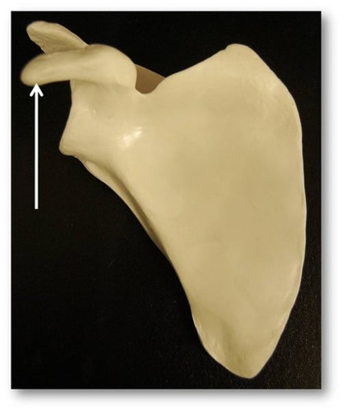
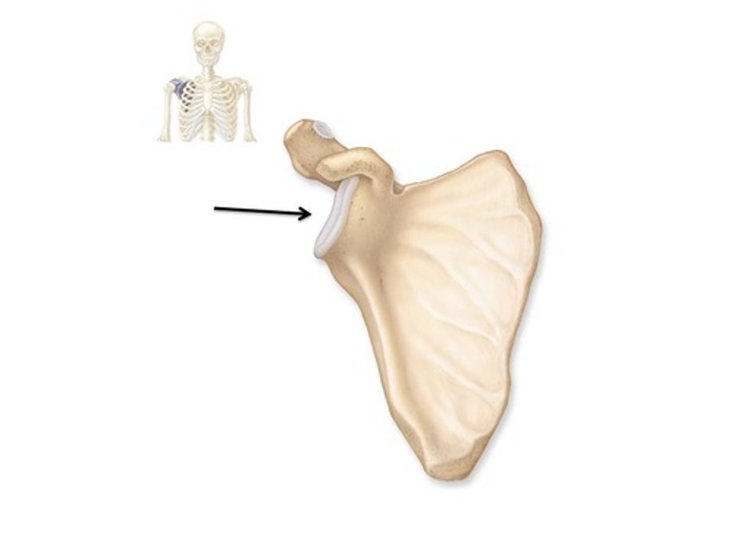
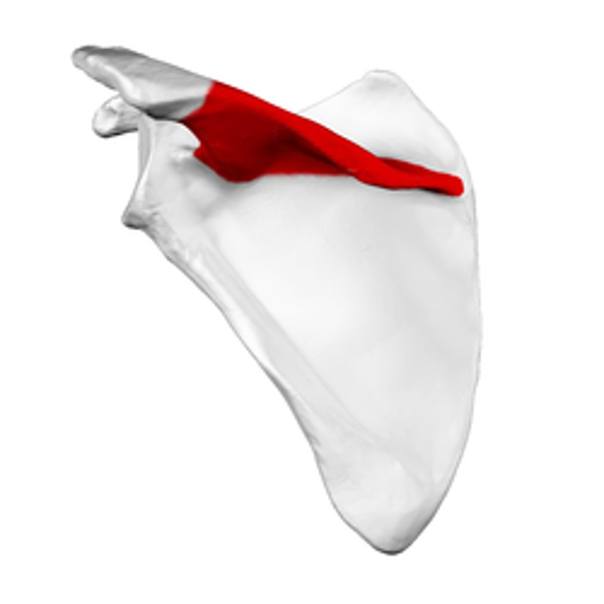
scapula acromion
detach part
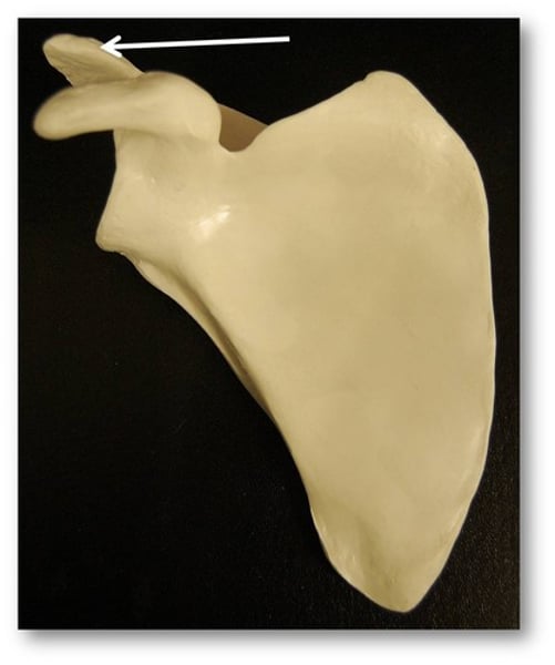
scapula coracoid process
"crow head"
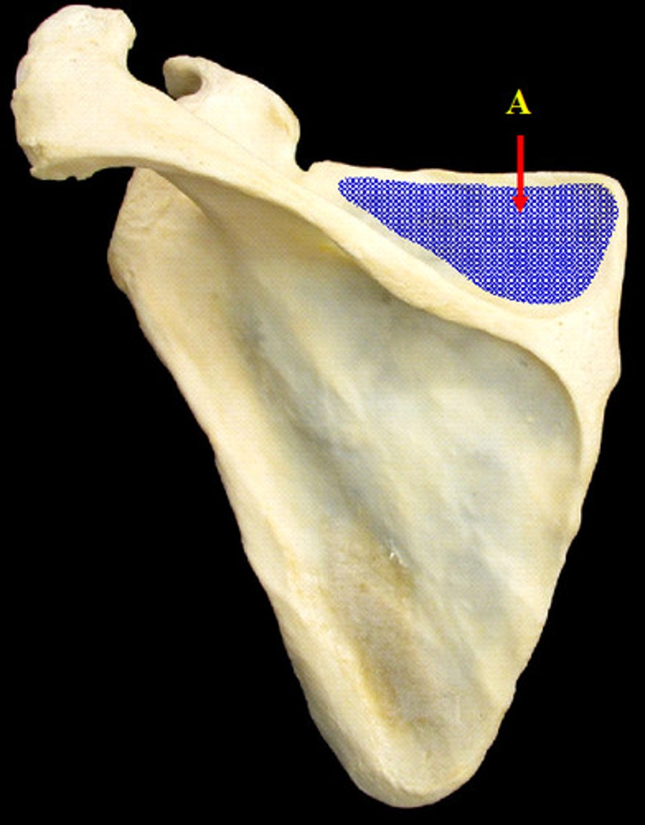
scapula supraspinous fossa
a depression located superior to the spine of the scapula
scapula infraspinous fossa
a broad depression located inferior to the spine of the scapula
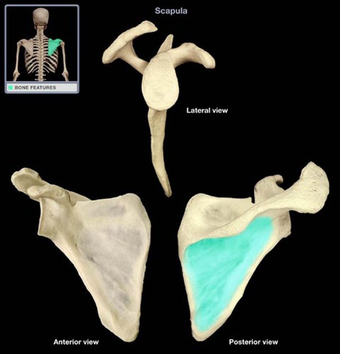
scapula glenoid process
lateral flat surface of the bone - shows when turnt sideways; connects to humerous
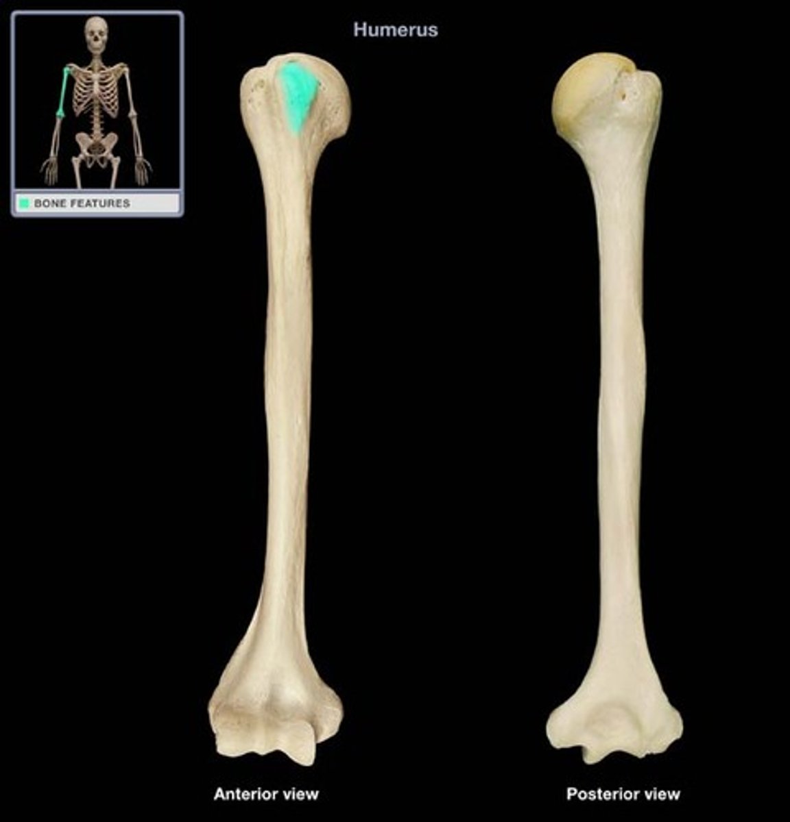
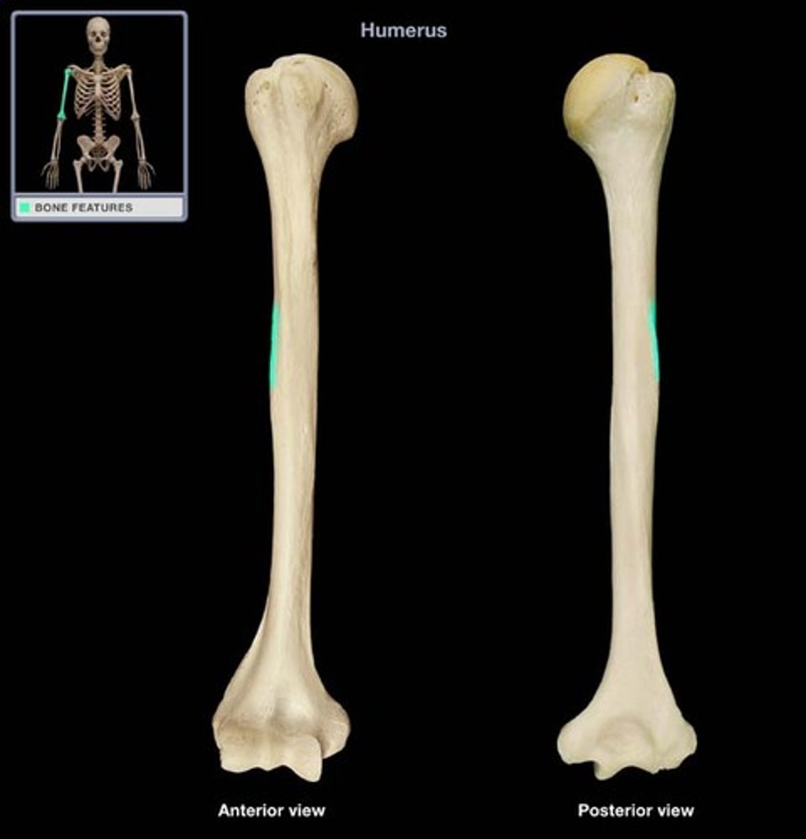
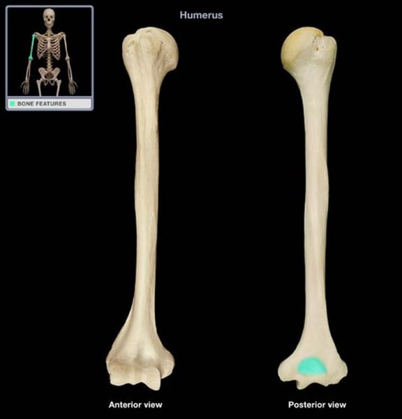
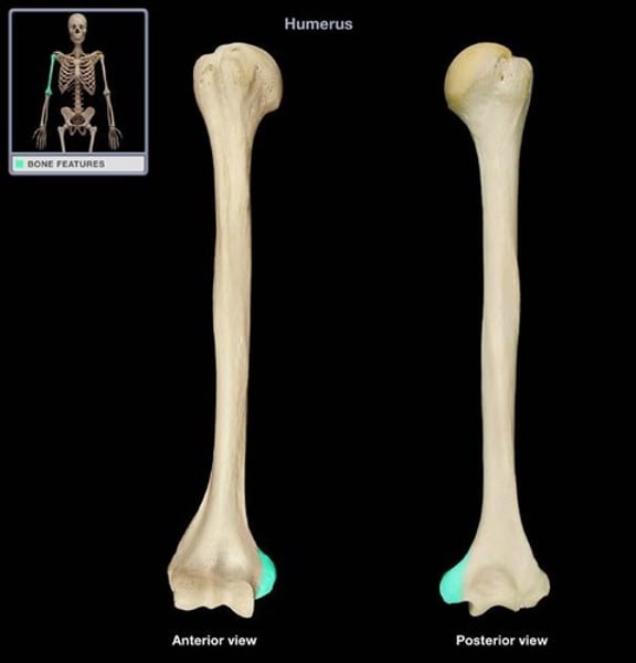
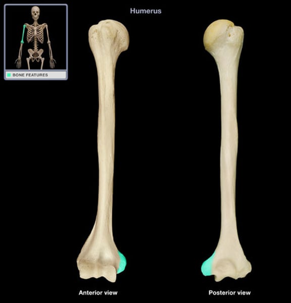
humerus
upper arm bone
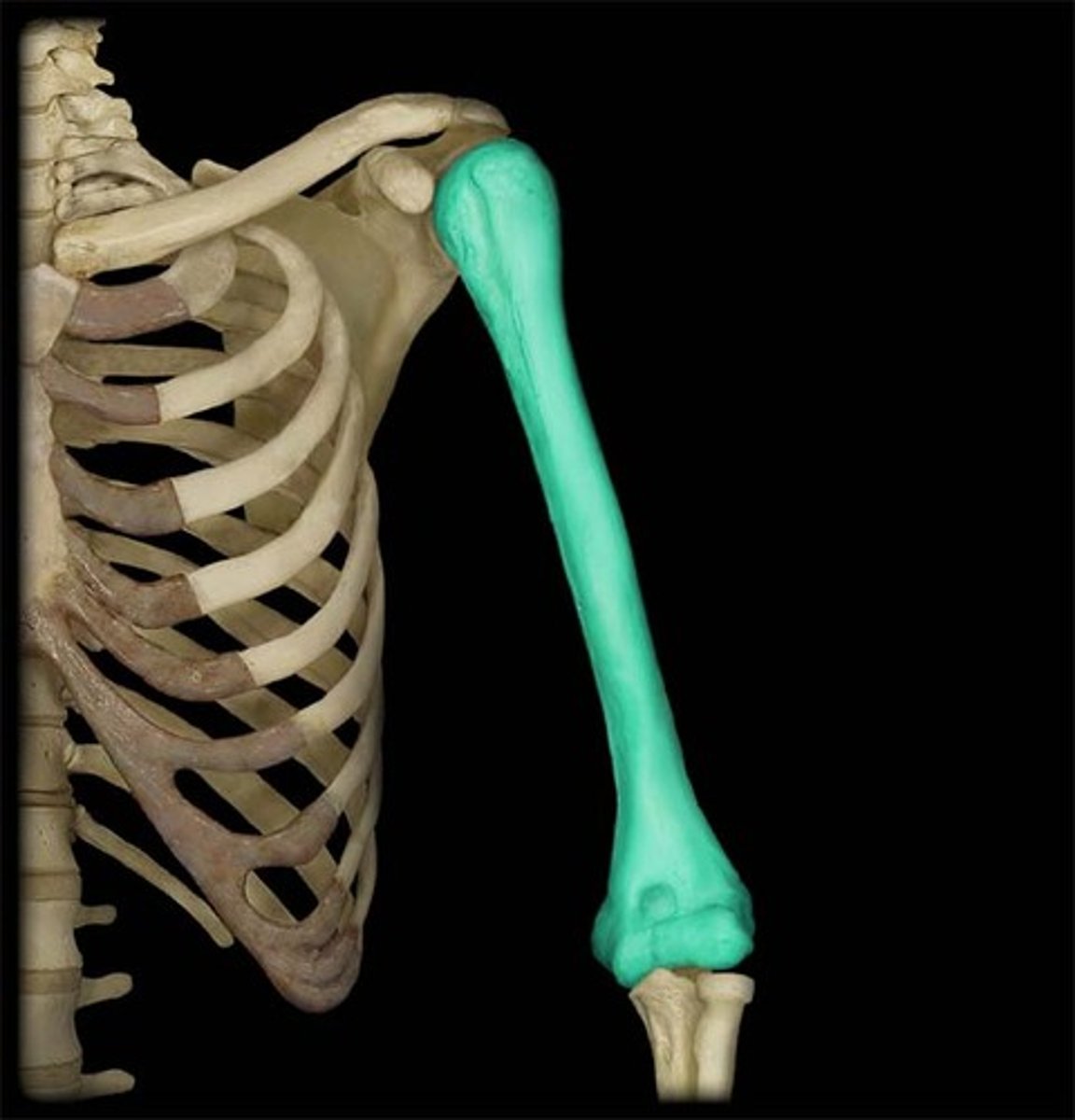
humerus head
rounded section of the humerus that articulates with the glenoid cavity of the scapula
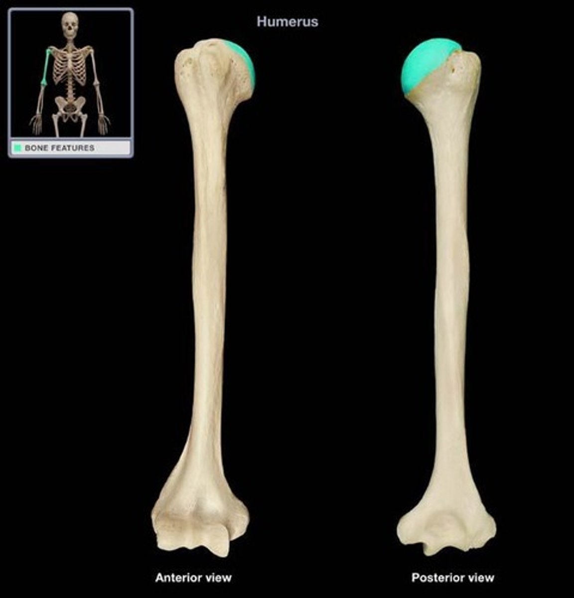
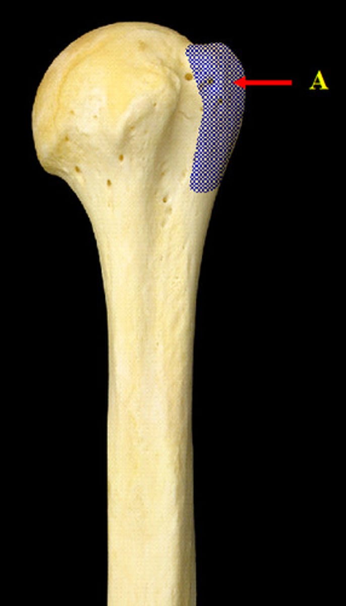
humerus greater tubercle
Large lateral prominence; site of the attachment of rotator cuff muscles
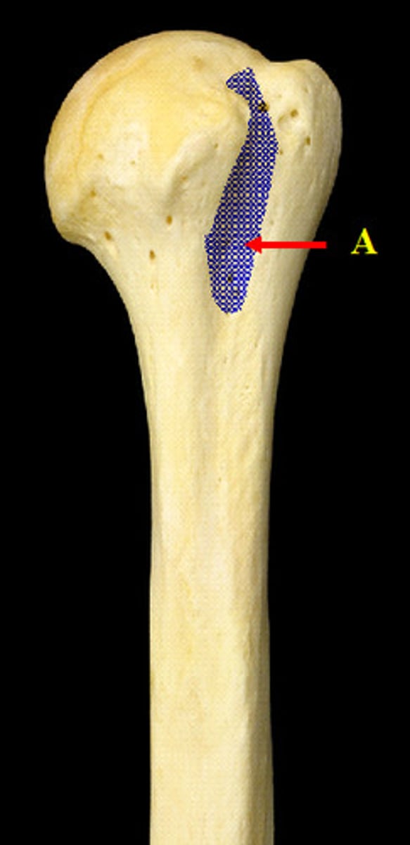
humerus lesser tubercle
a smaller projection on the anterior side between the head and greater tubercle
humerus intertubercular groove
space between greater and lesser tuberacle
humerus deltoid tuberosity
insertion of deltoid
humerus trochlea
lopsided bowtie (pulley system)
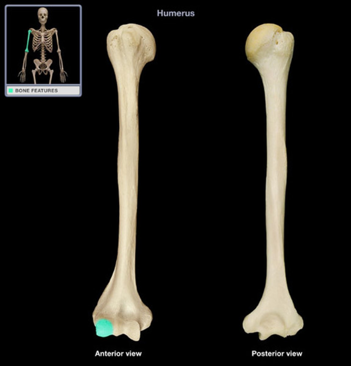
humerus capitulum
rounded bump on the bottom (little head)
fossa
shallow dent
tubercle
button shaped; rough
fissure
large, irregular crack
meatus
opening or canal
facet
Flat surface that forms a joint with another facet or flat bone
spine
pointy projection off body of bone
process
extension of a bone
trochanter
Very large, blunt, irregularly shaped process (the only examples are on the femur)
condyle
knuckle-like process at the end of a bone near the joint
epicondyle
Raised area on or above a condyle
fovea
tiny pit or depression
notch
indentation at the edge of a bone
sinus
Air-filled cavity within a bone
Ramus (pl. rami)
branch
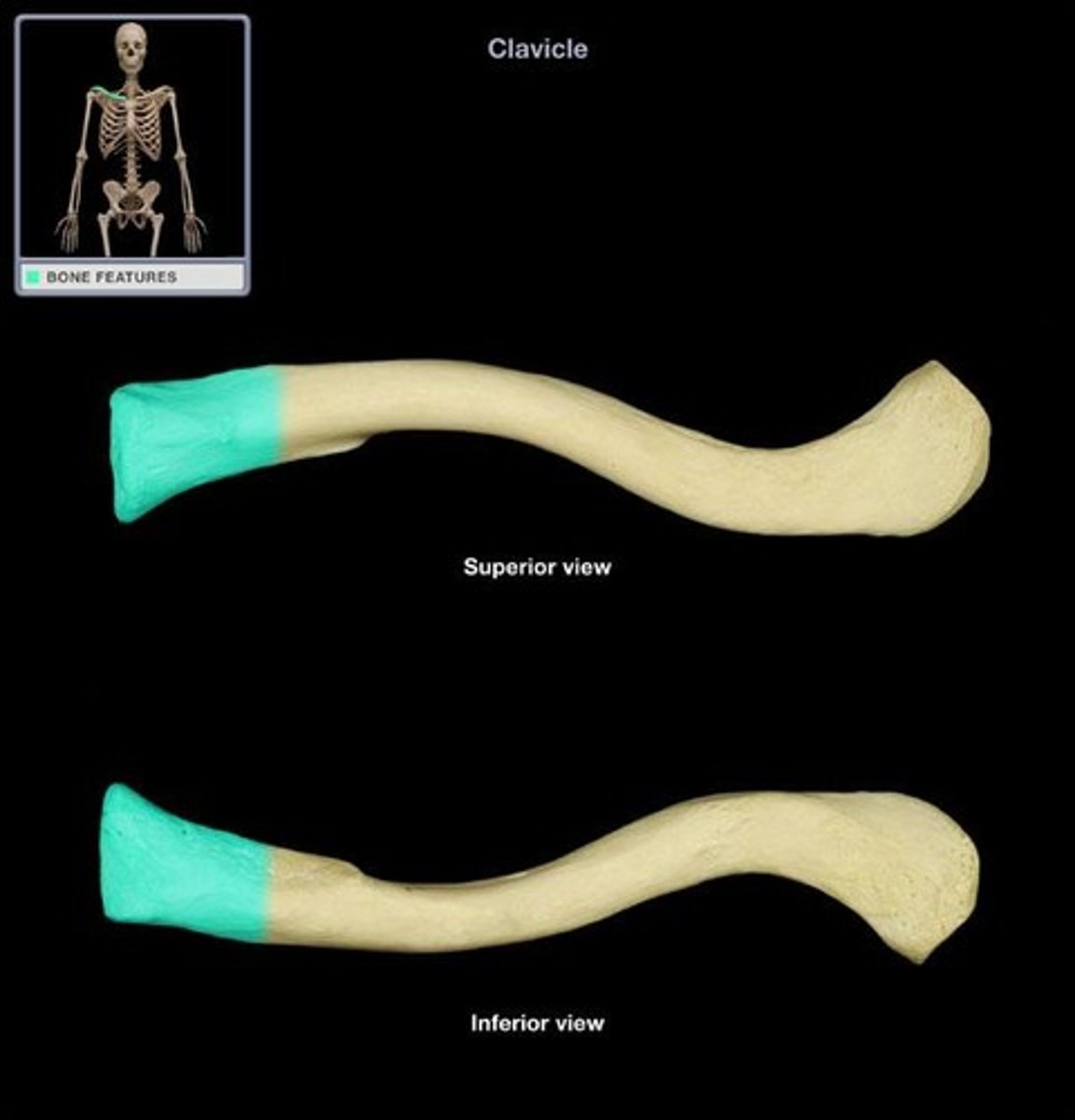
clavical
collar bone
scapula
shoulder blade
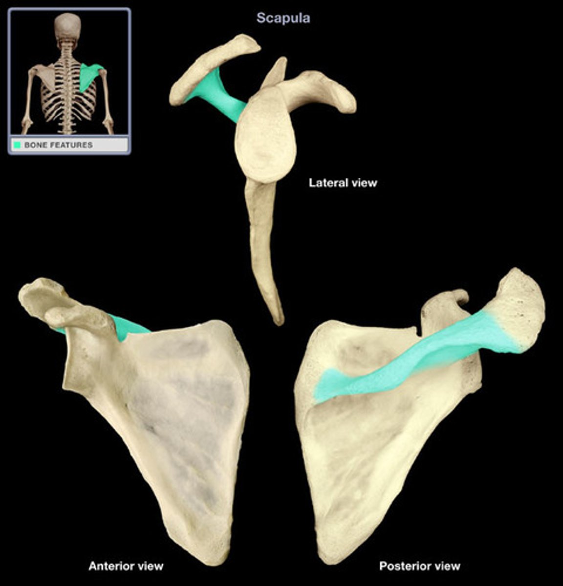
scapula spine
bony ridge off back
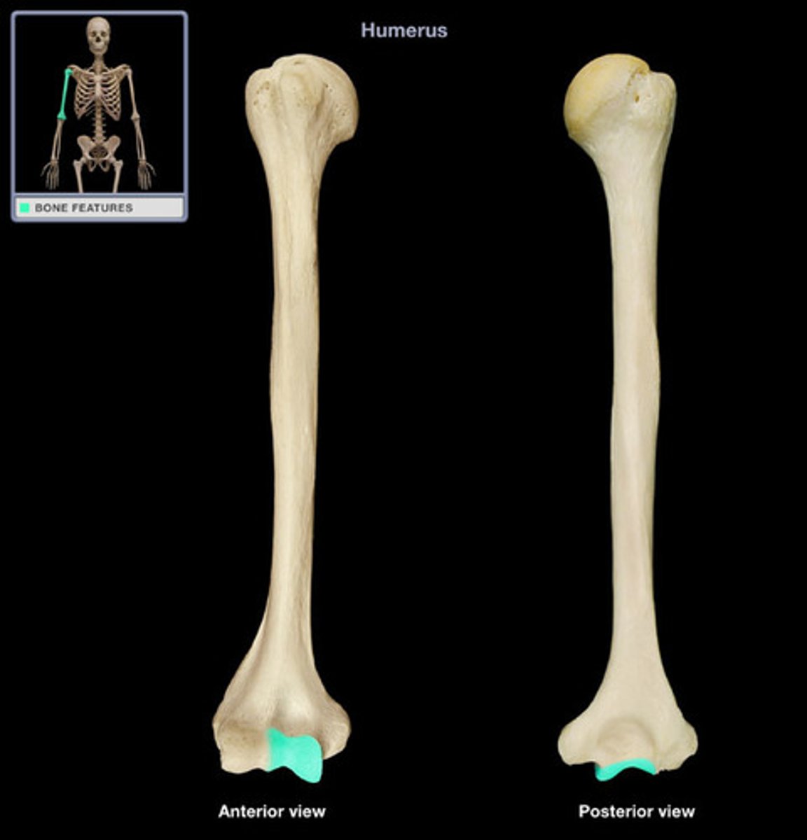
humerus olecranon fossa
large depression on posterior humerus that receives the olecranon of the ulna
humerus medial epicondyle
big jutty on same side of humerus head
humerus lateral epicondyle
small condyle proximal to the capitulum
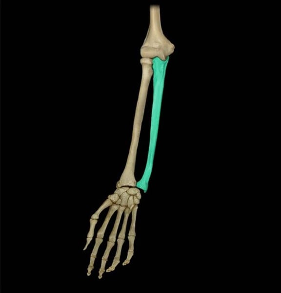
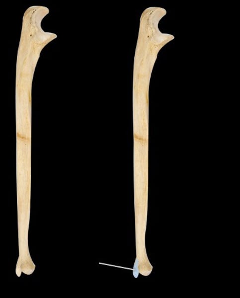
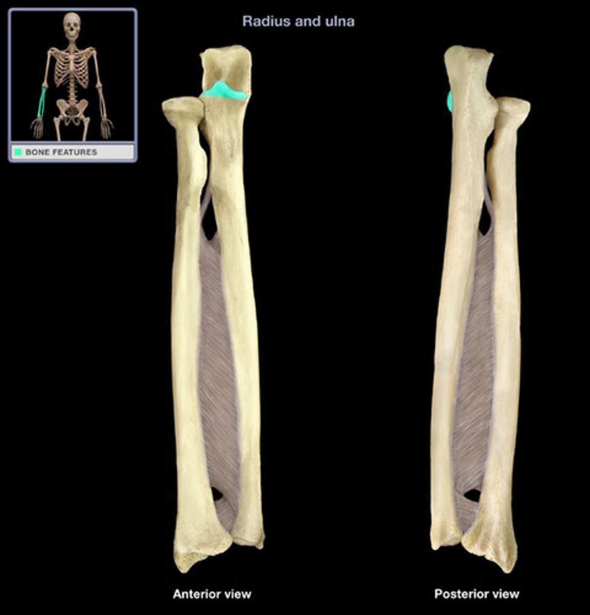
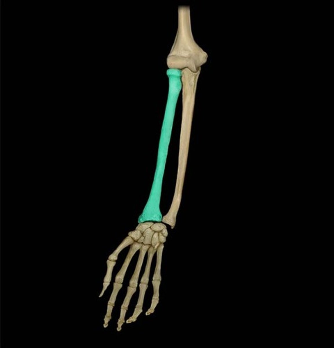
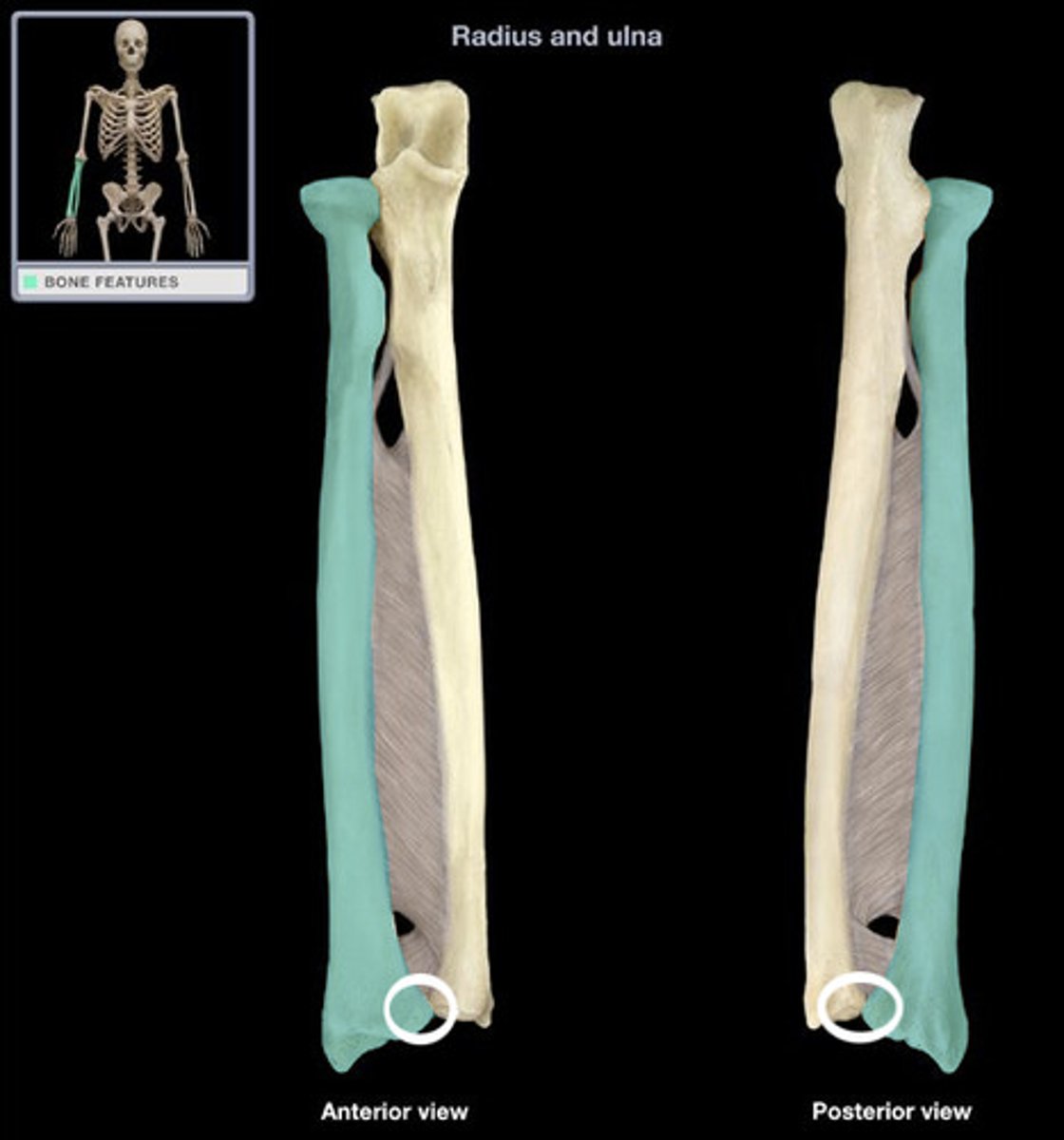
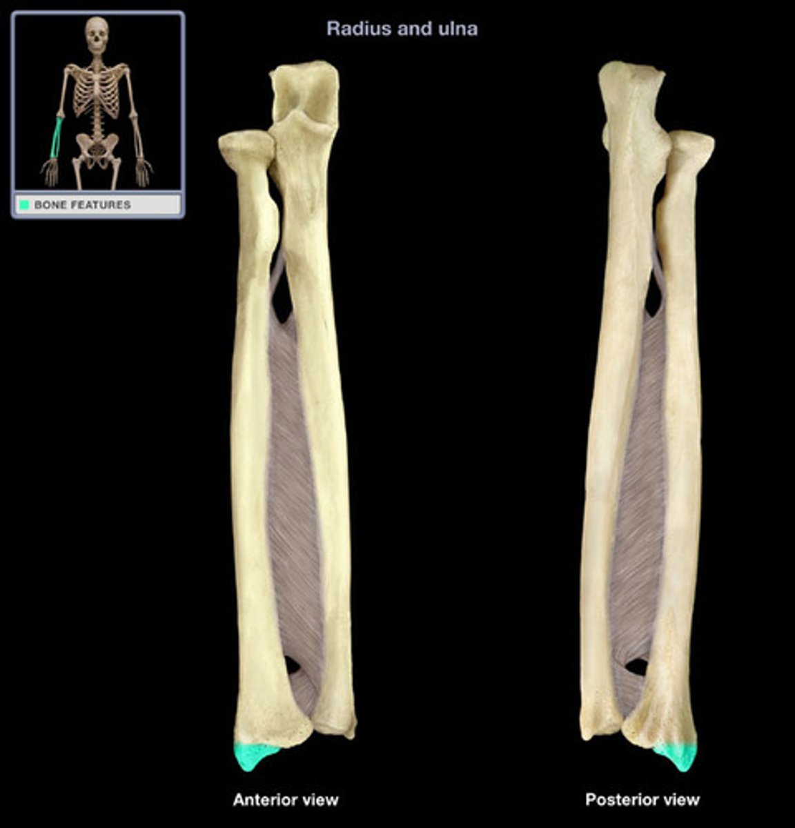
ulna
Inner and larger bone of the forearm, attached to the wrist and located on the side of the little finger (pinky)
ulna styloid process
pointy projection on the distal end
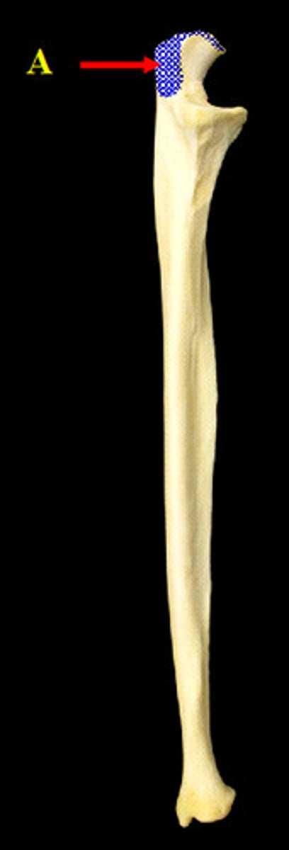
ulna olecranon
prominent process on the posterior proximal ulna; articulates with the olecranon fossa of the humerus when the forearm is extended
ulna coronoid process
shaped like a point on a crown; articulates with the trochlea of the humerus
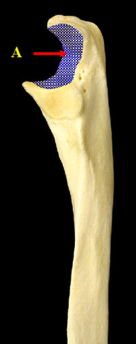
ulna semilunar notch
smooth notch (ice cream scoop)
radius
lateral bone of the forearm
radius head
disk-shaped; articulates with the humerus and the radial notch of the ulna
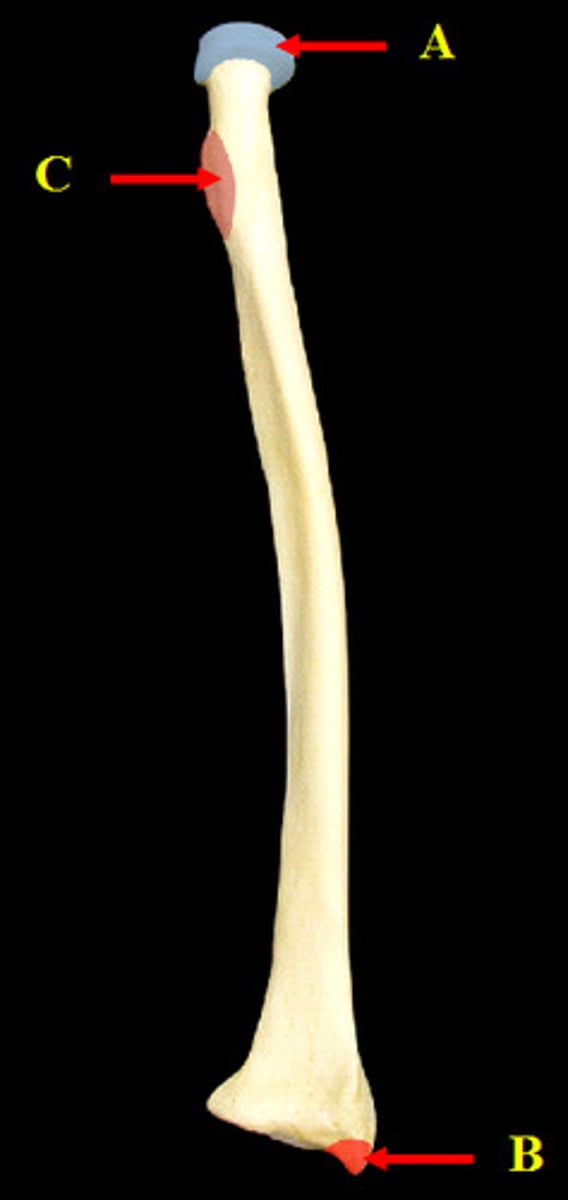
radius radial tuberosity
little bump below neck

radius ulnar notch
articulates with the head of the ulna
radius styloid process
projection of bone on the lateral surface of the distal radius bone
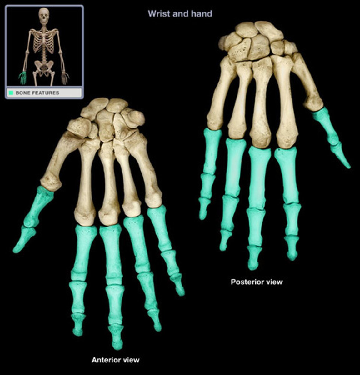
carpals
bones of the wrist
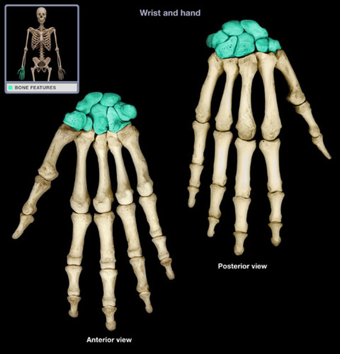
metacarpals
the five bones that form the palms of the hand
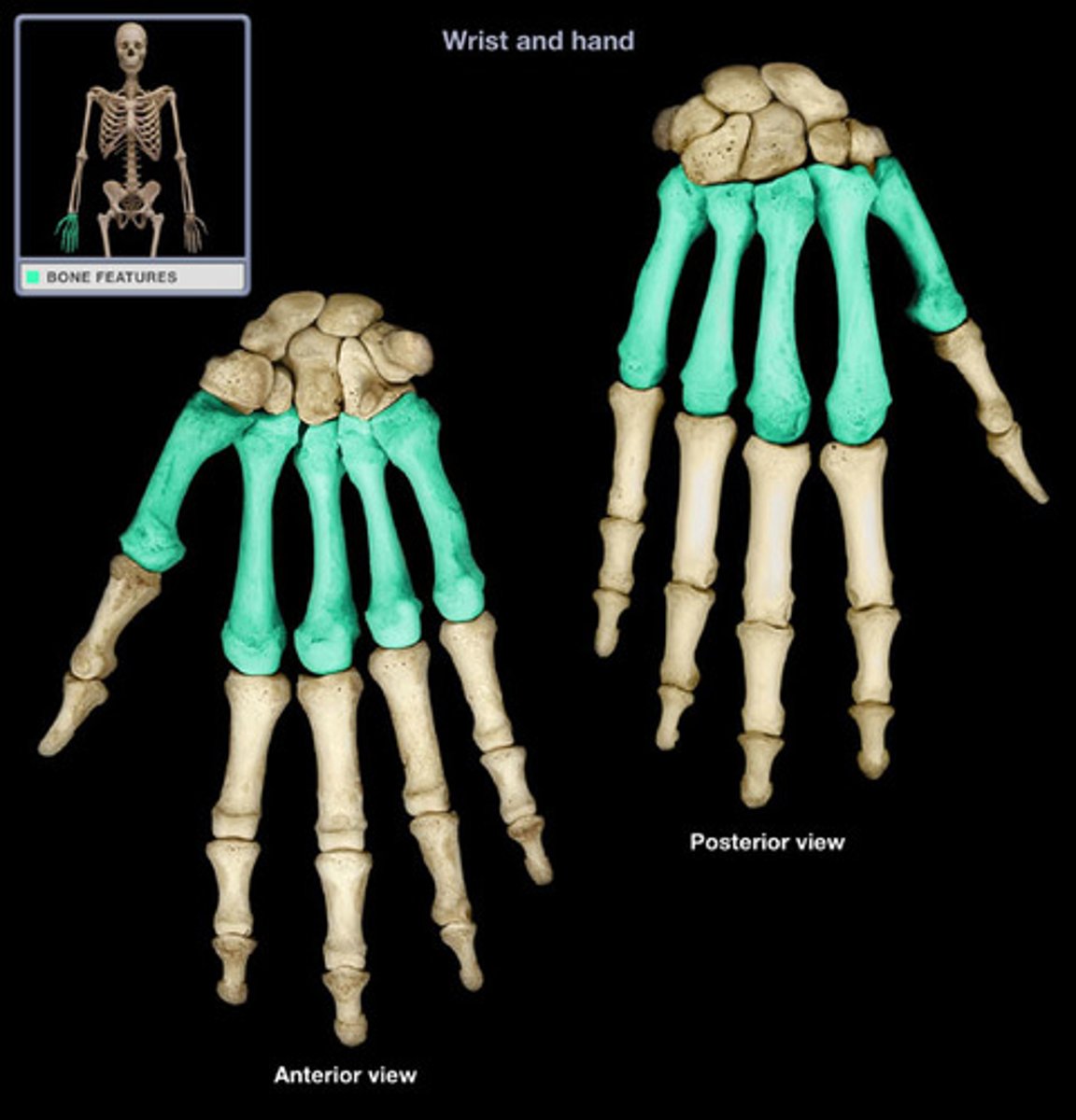
Phalanx (proximal, middle, distal); pl. phalanges
finger bones (thumb has no middle phalanx)
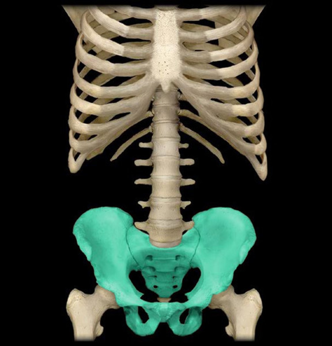
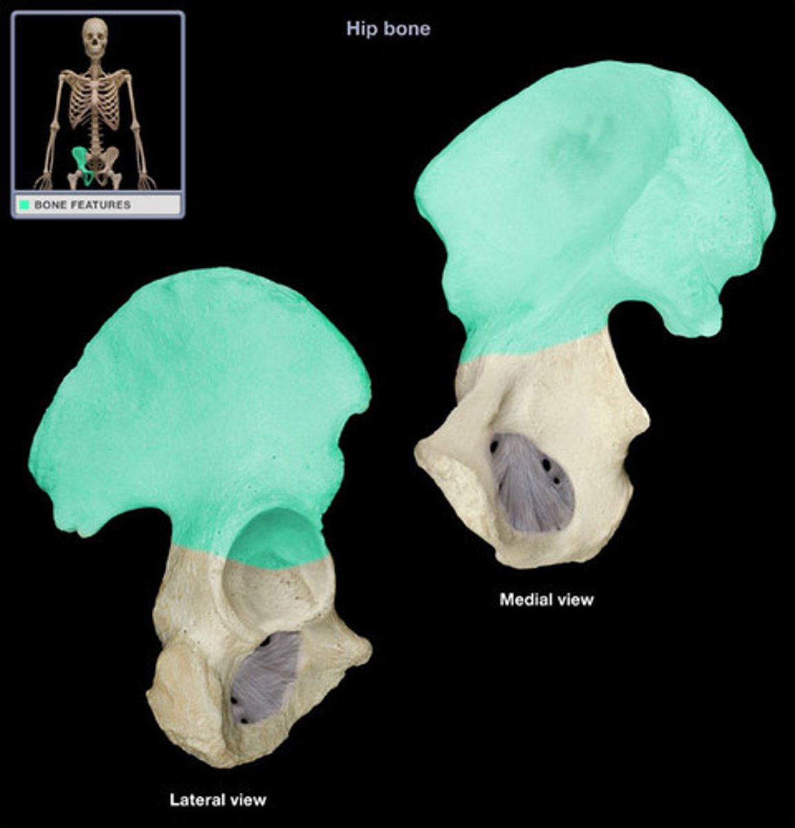
Pelvis
hip bone
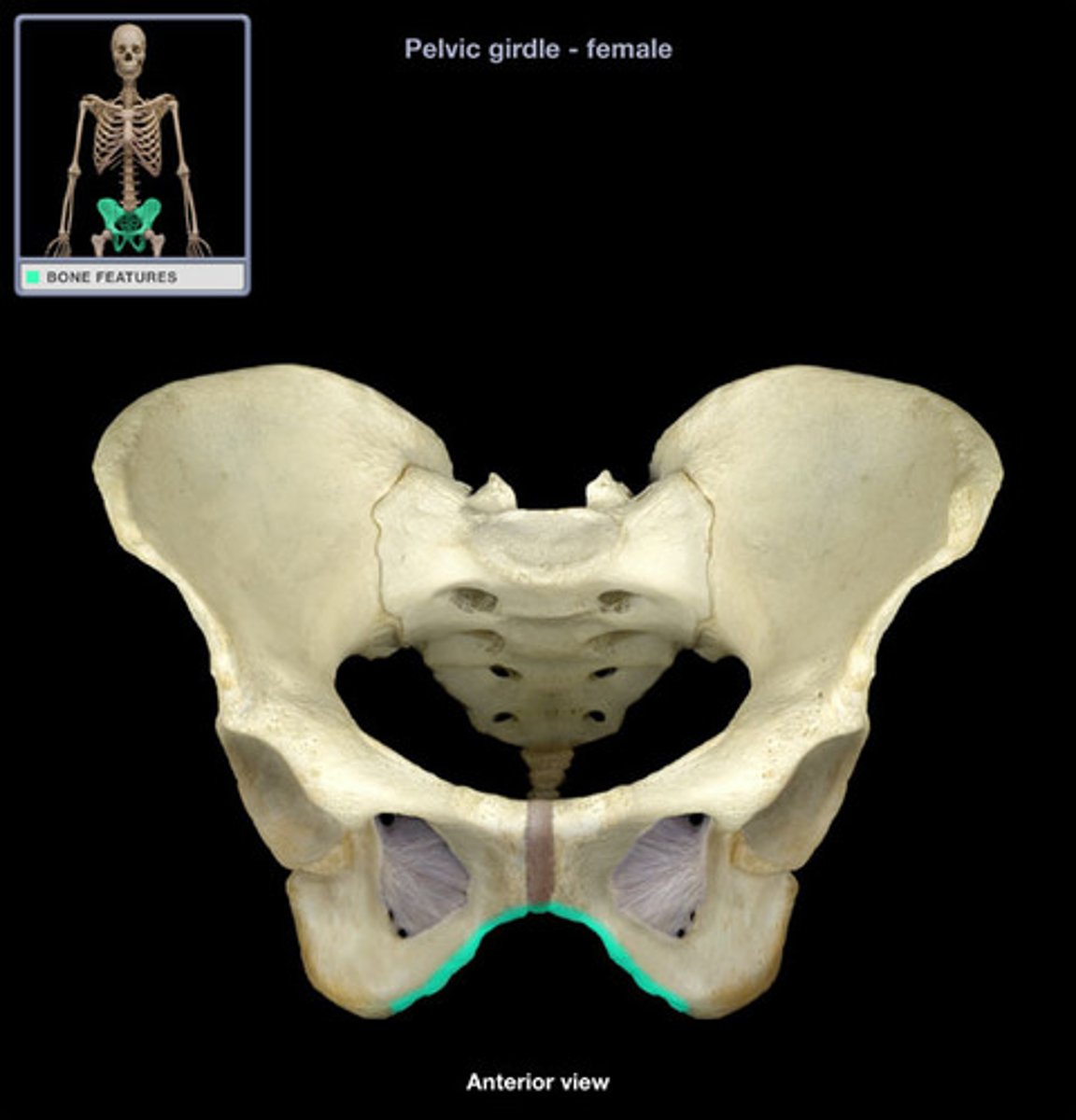
Pelvis: Pubic Symphysis
joint in front that connects pubic bones in the middle
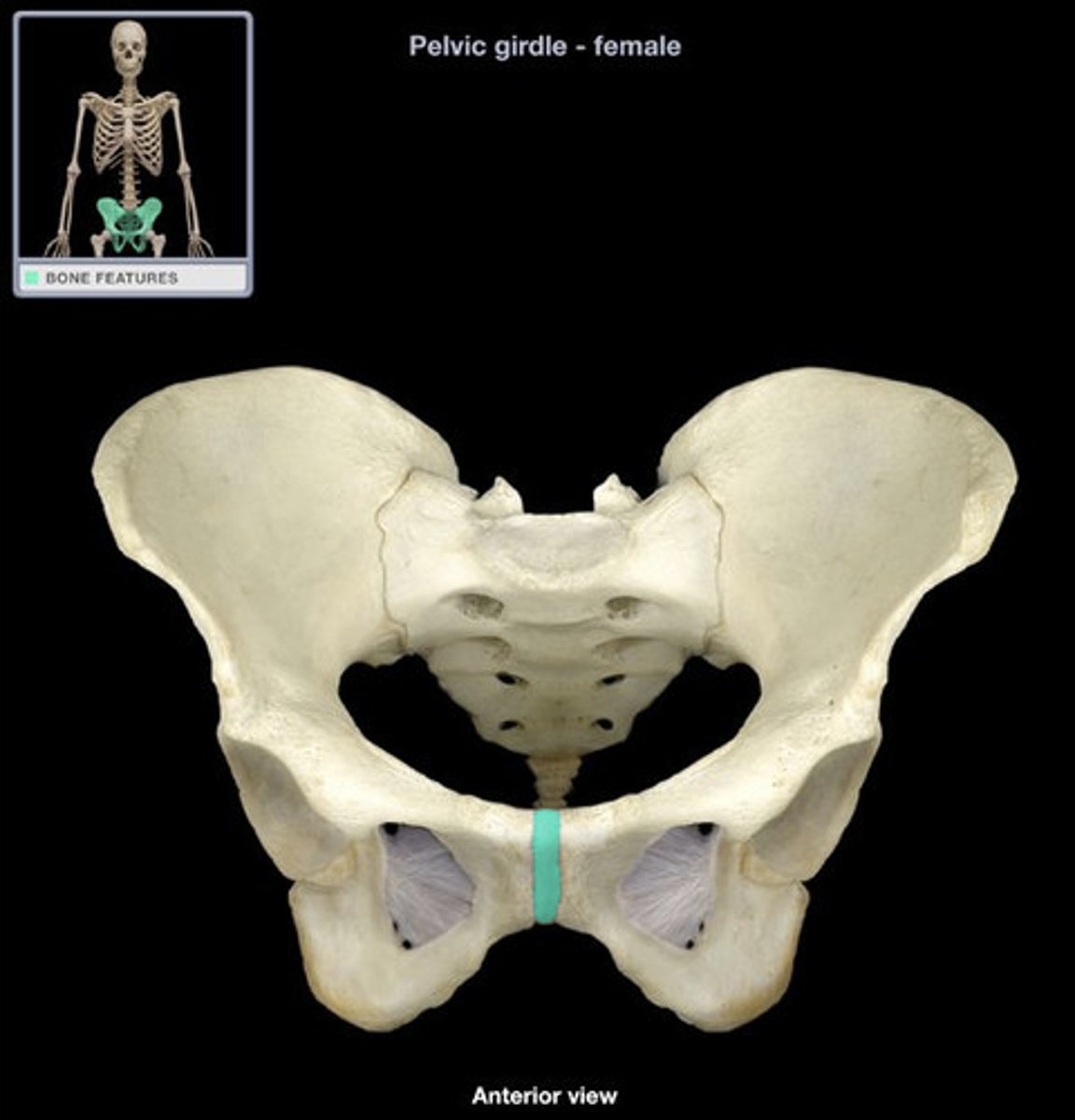
pelvis: pubic arch
U or V shaped
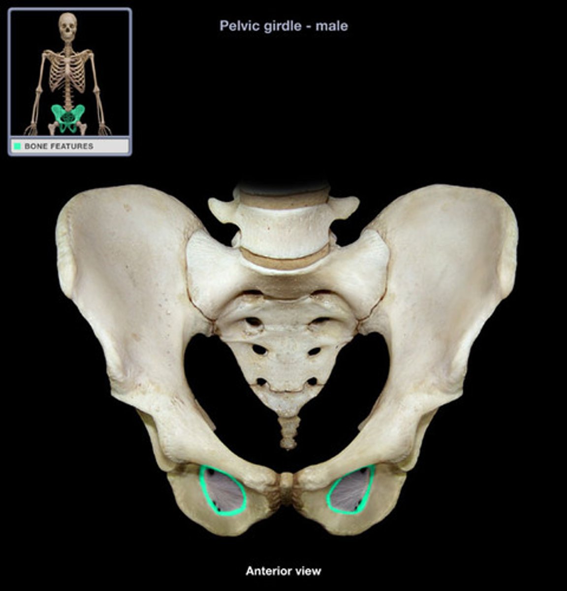
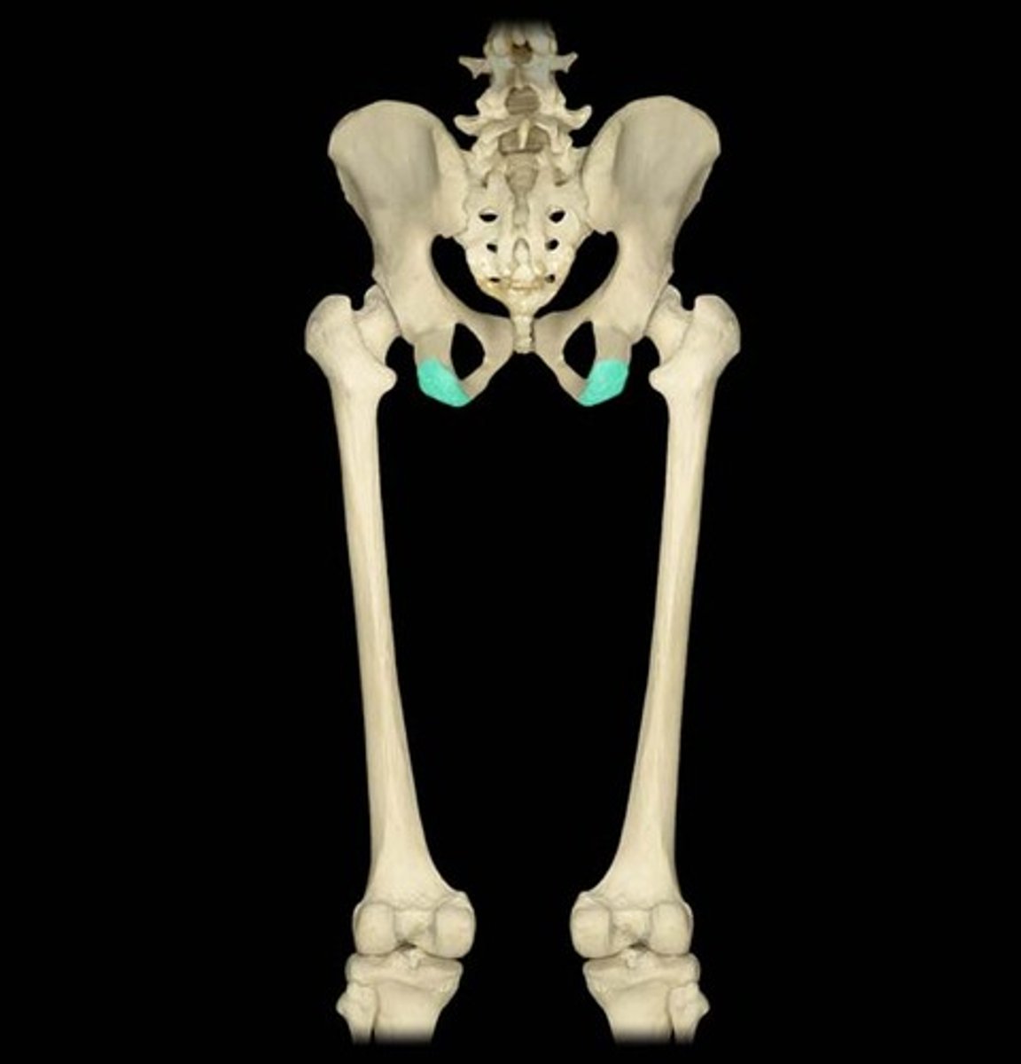
Acetebulum
hip socket

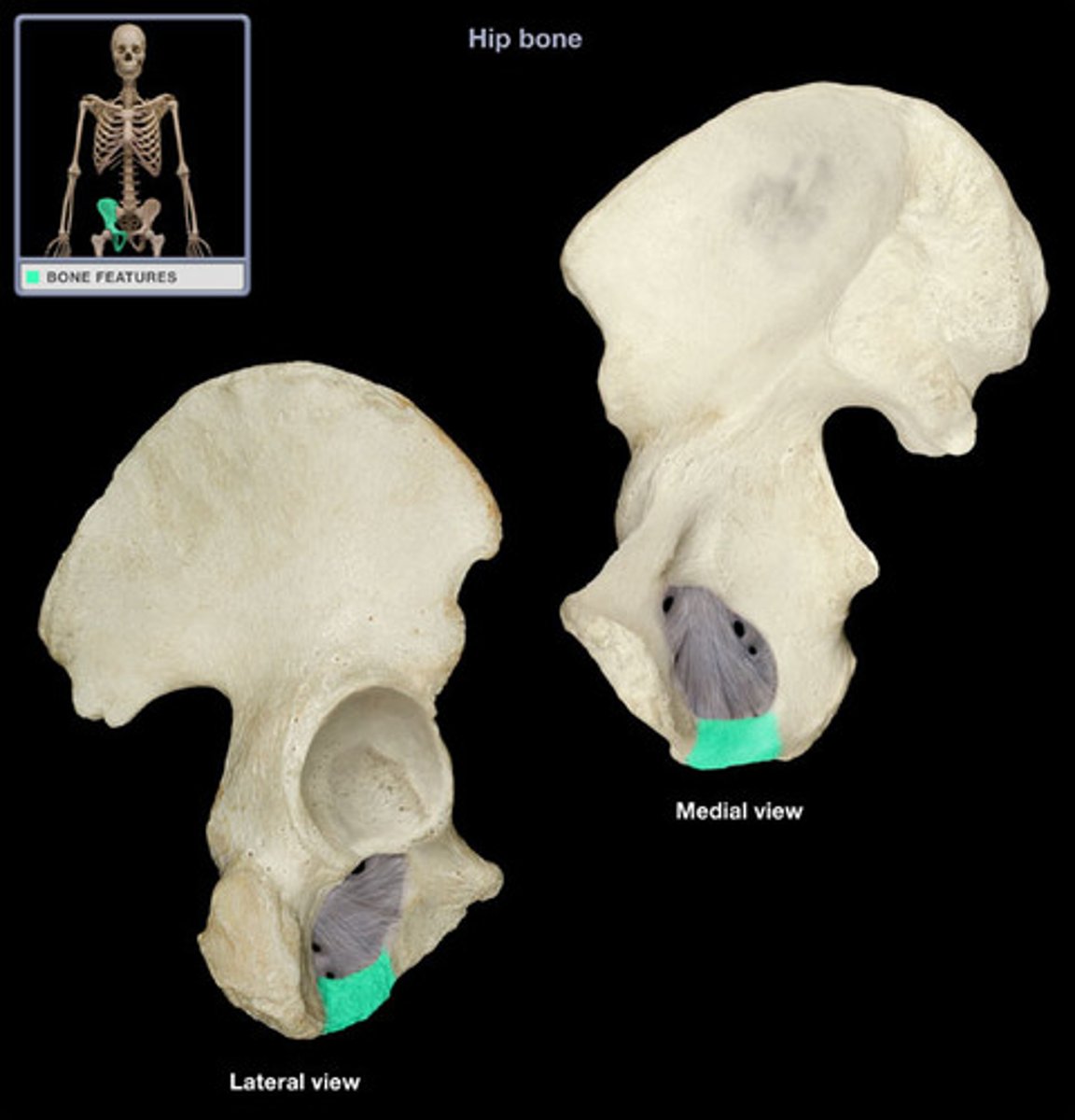
hip bone: obturator foramen
Large opening by pubis and ischium Lower part of hip bone
ilium
part of the hip bone
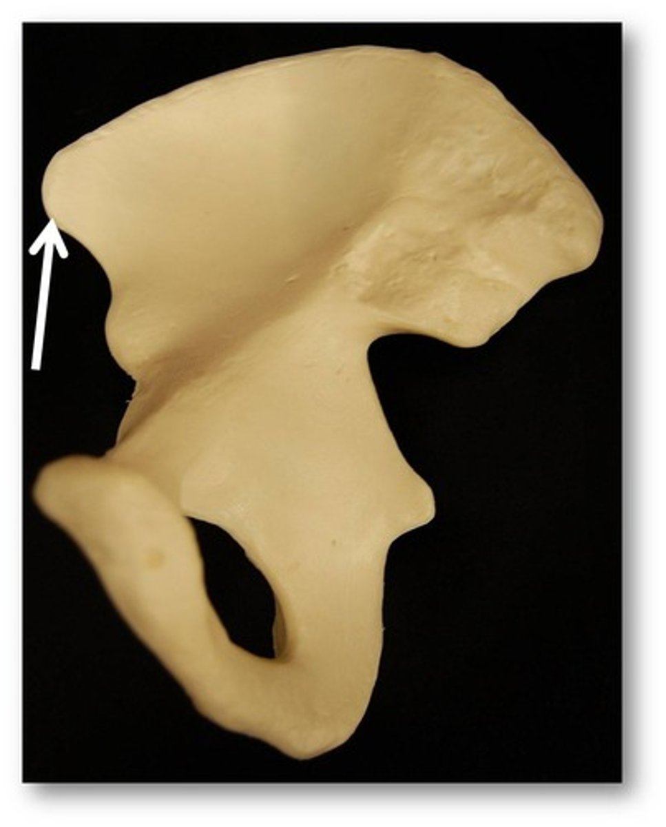
anterior superior iliac spine
Name this specific part of the pelvic bone.
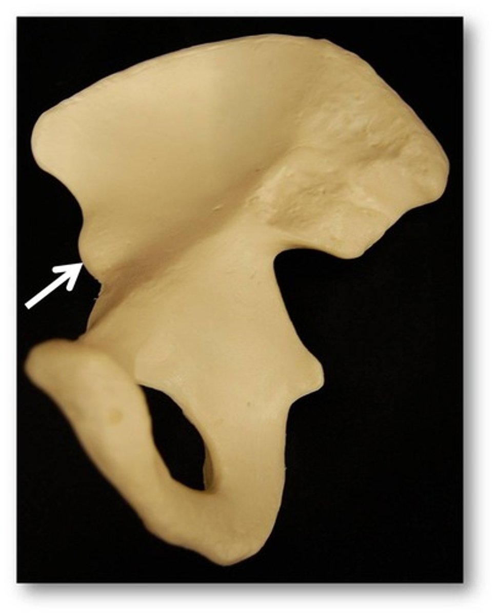
anterior inferior iliac spine (AIIS)
Name this specific part of the pelvic bone.
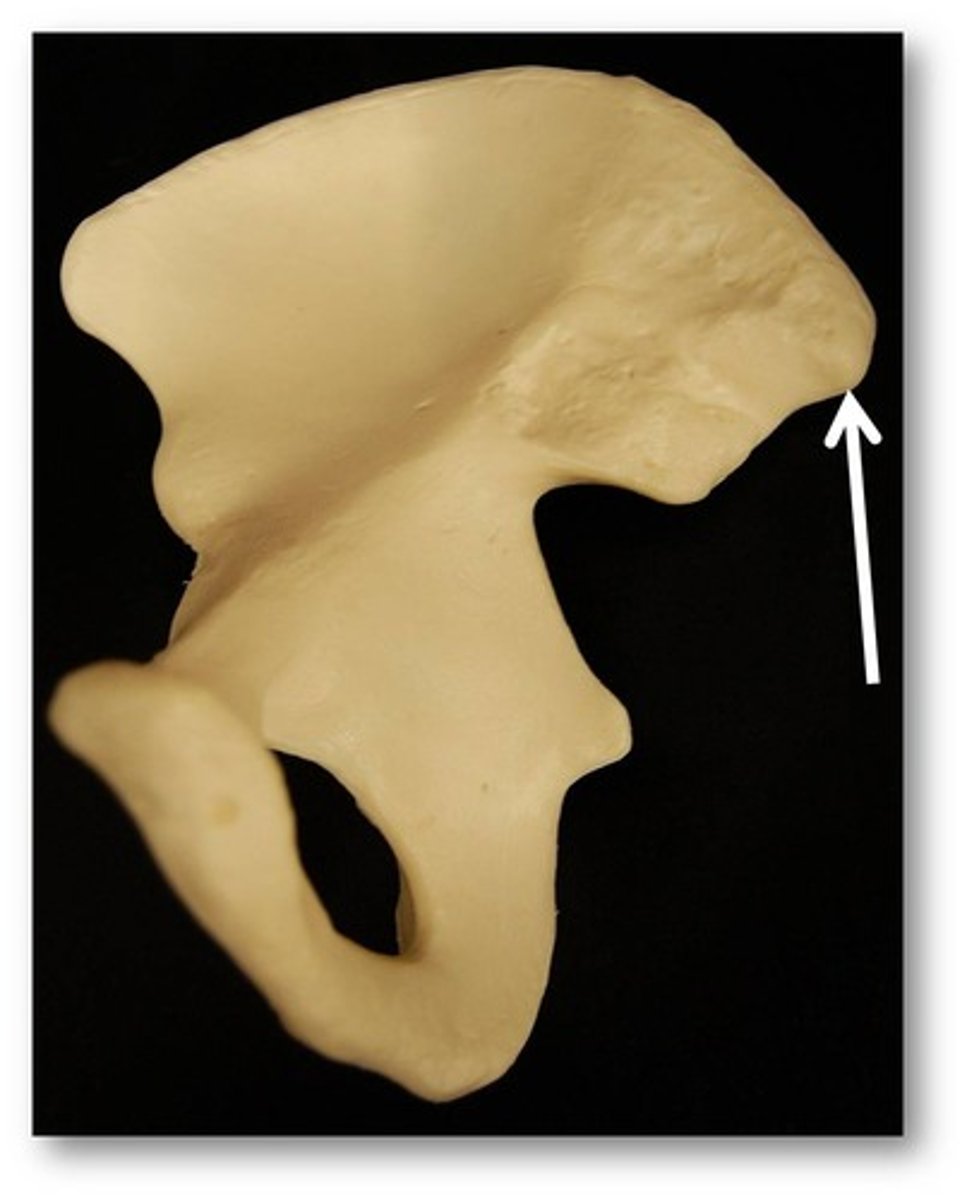
posterior superior iliac spine
Name this specific part of the pelvic bone.
posterior inferior iliac spine (PIIS)
Name this specific part of the pelvic bone.

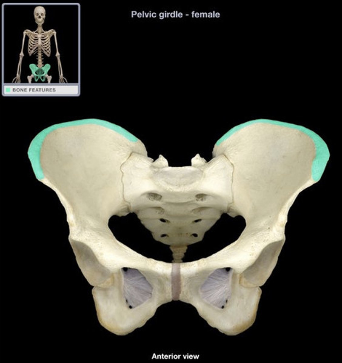
iliac crest
found on the top of the hip bone.
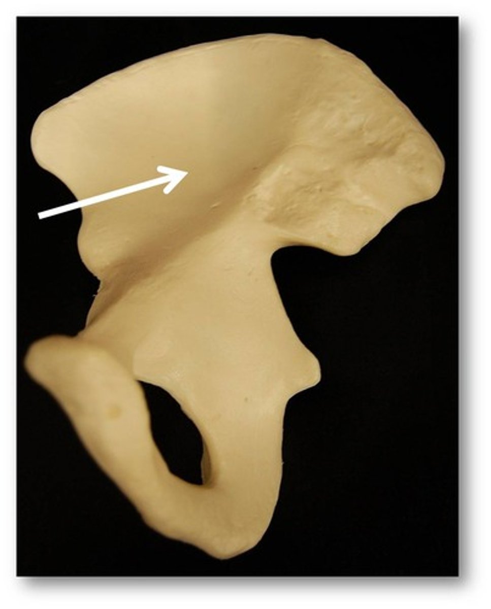
iliac fossa
broad, smooth dent
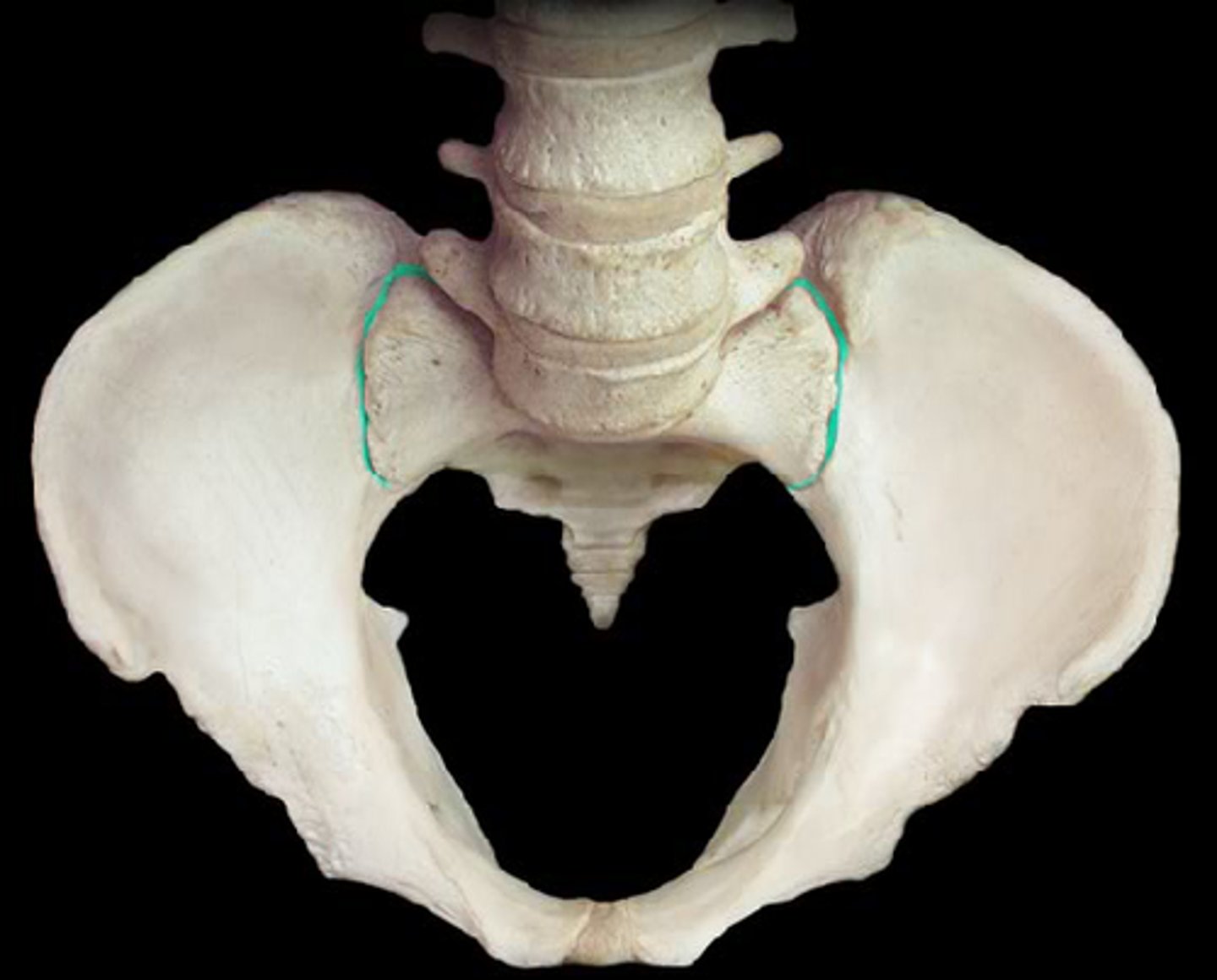
iliac articular surface
rough spot, sacrum forms here
greater sciatic notch
Name this specific part of the pelvic bone.

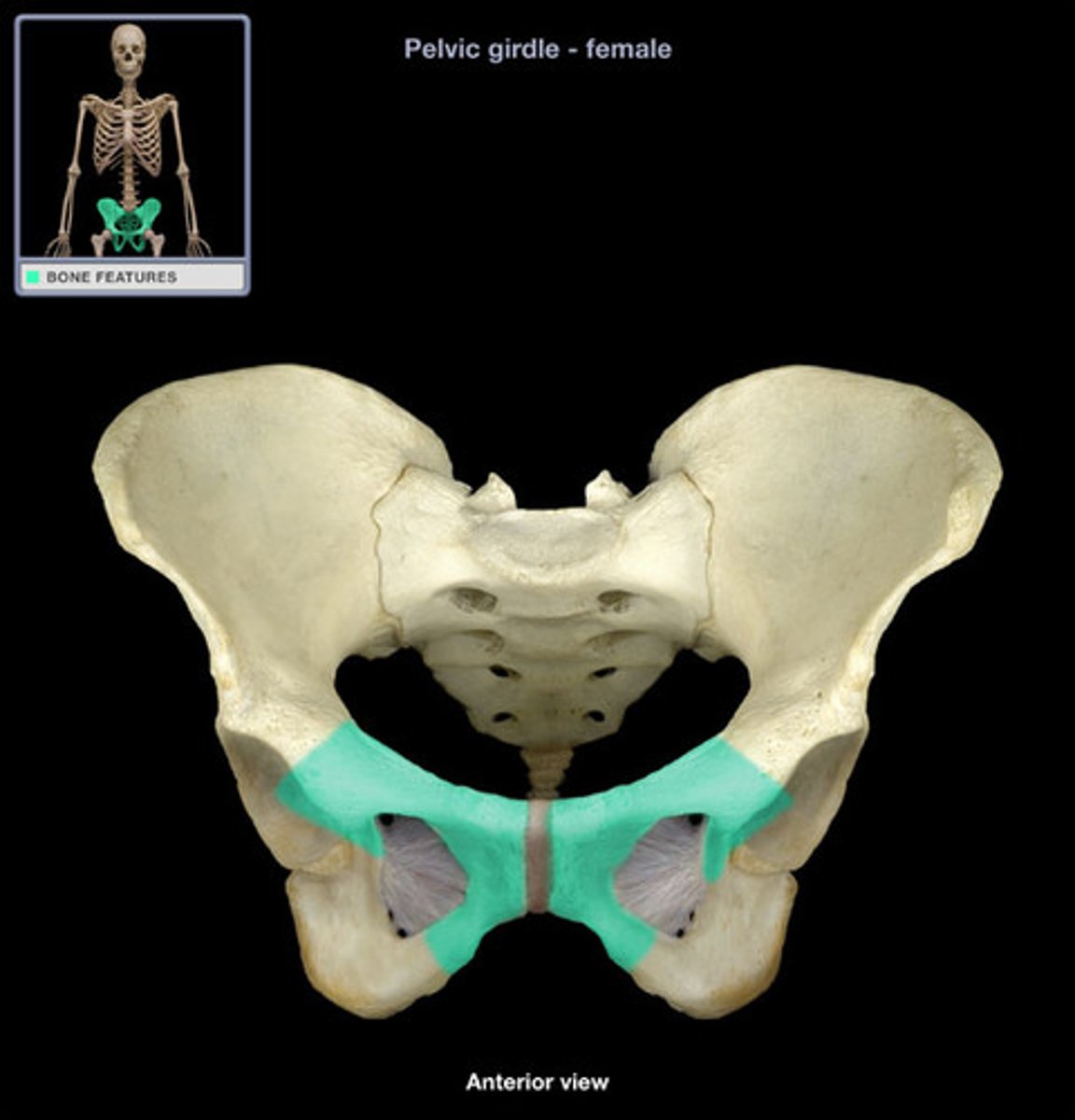
ischium
the lower, posterior portions of the pelvis
ischial tuberosity
receives the weight of the body when sitting
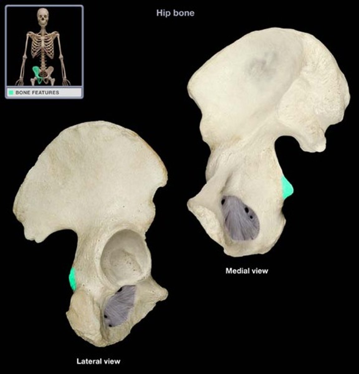
ischial spine
located superior to the ischial tuberosity and projects medially into the pelvic cavity
lesser sciatic notch
inferior to ischial spine

pubis
The medial anterior portion of the pelvis
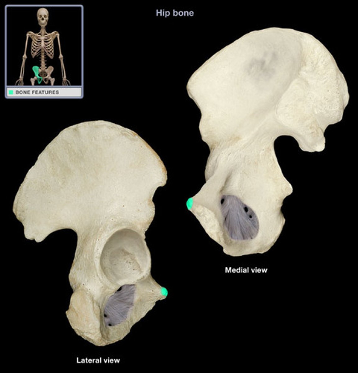
pubic tubercle
anterior prominence superior to the pubic symphysis, lateral end of pubic crest
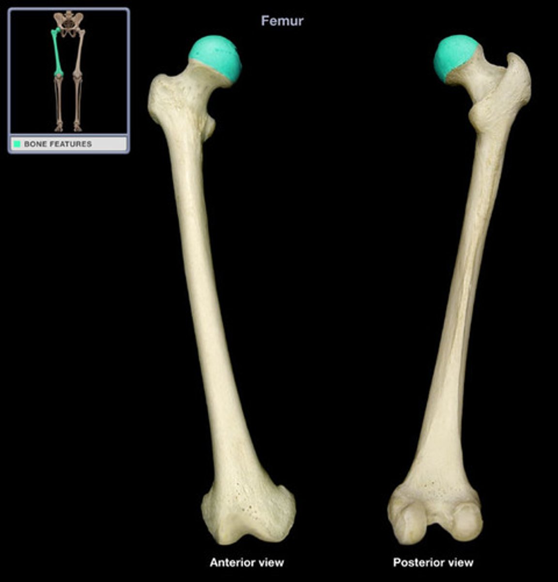
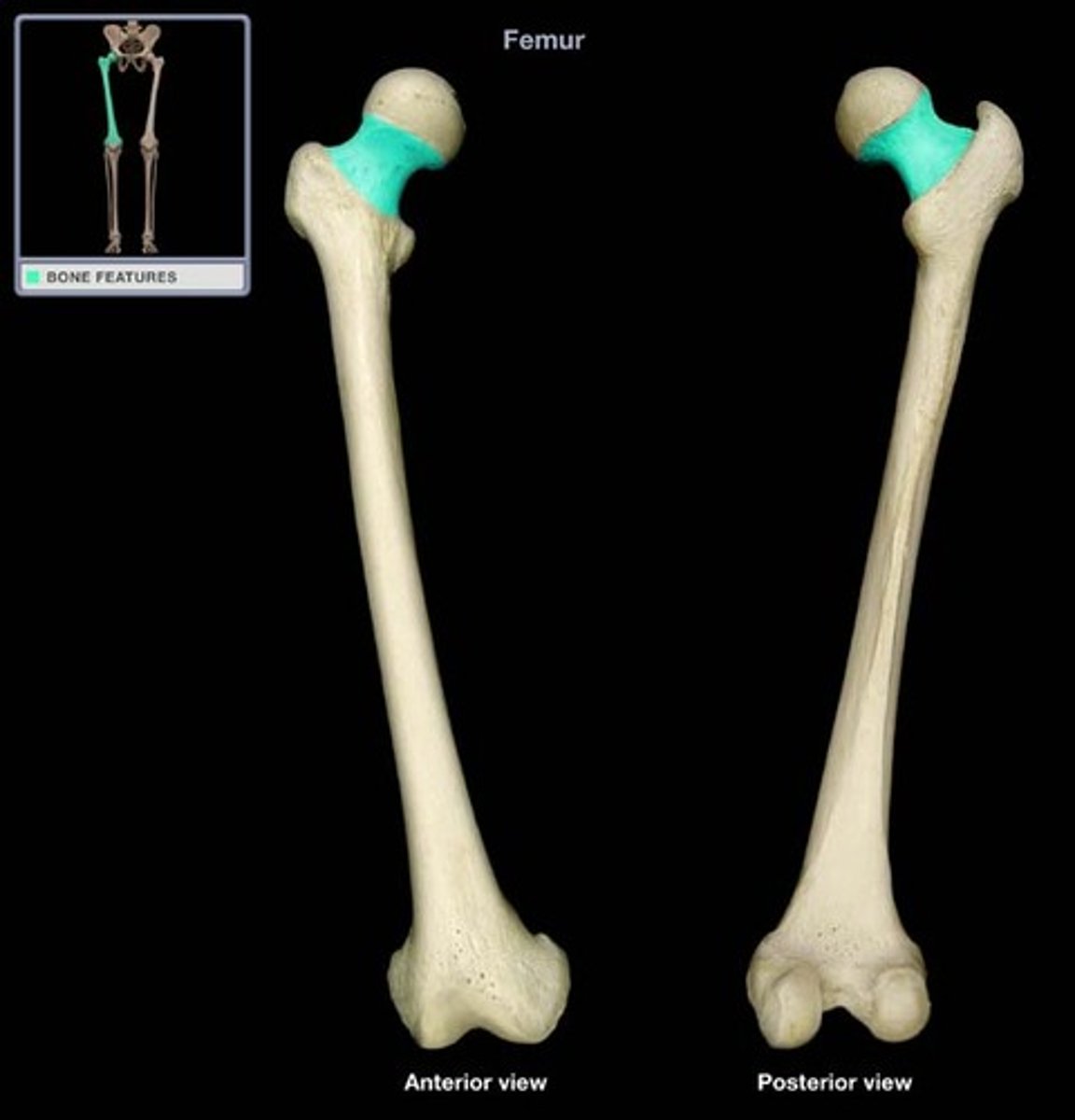
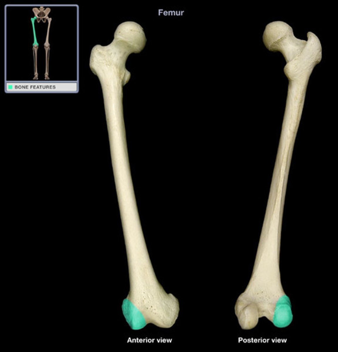
femur
thigh bone
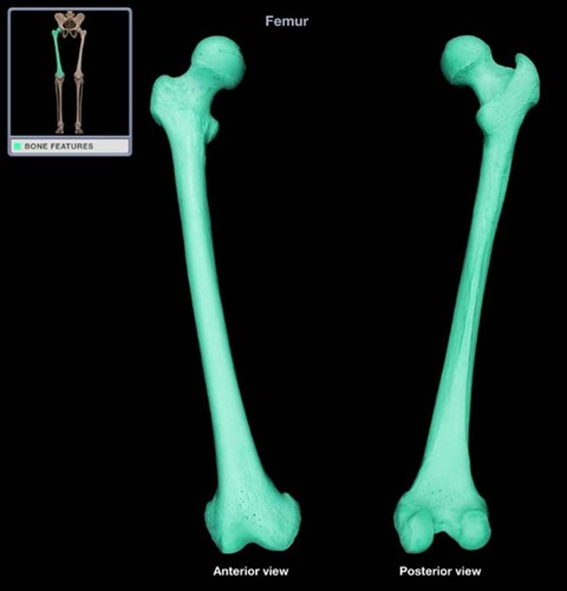
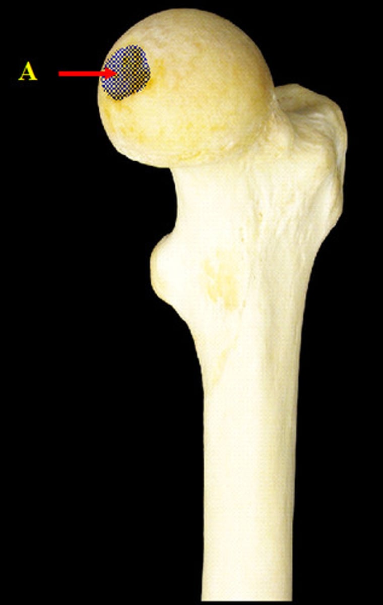
femur head
rounded portion of femur covered with articular cartilage - fits into acetabalum of pelvic girdle
femur fovea capitis
pit in the femoral head
femur neck
Between the head of the femur and trochanter
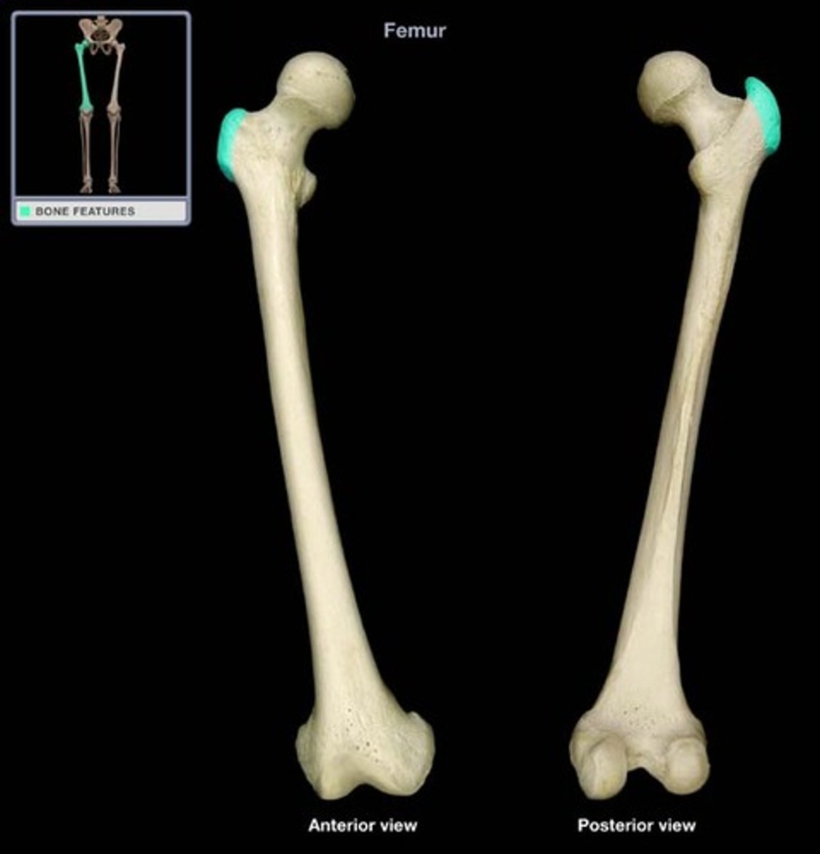
femur greater trochanter
Identify the structure.
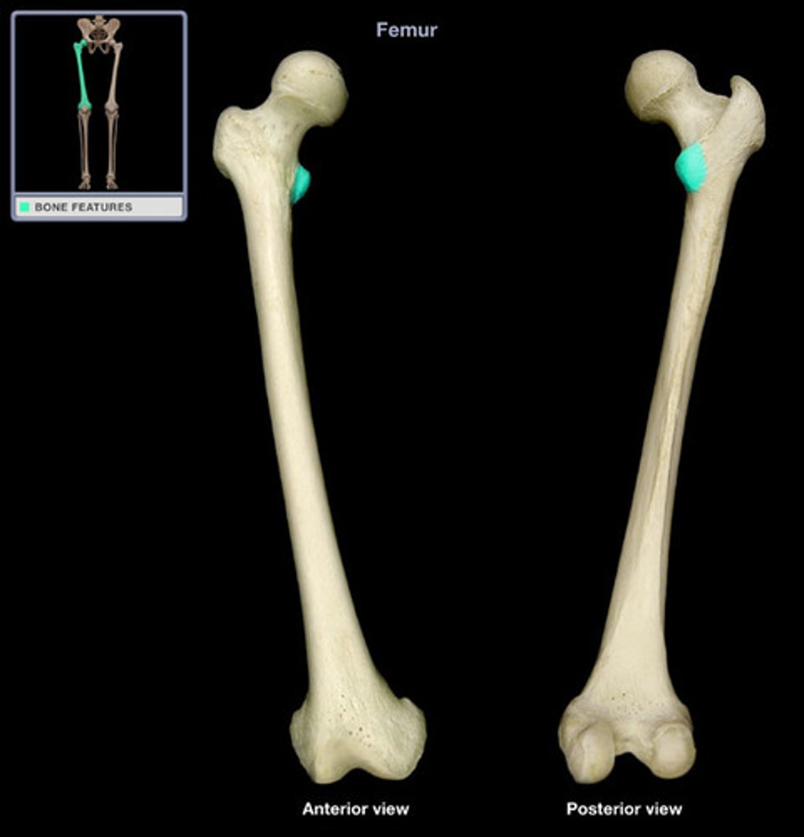
femur lesser trochanter
smaller prominence inferior to the head on the medial side of the femur
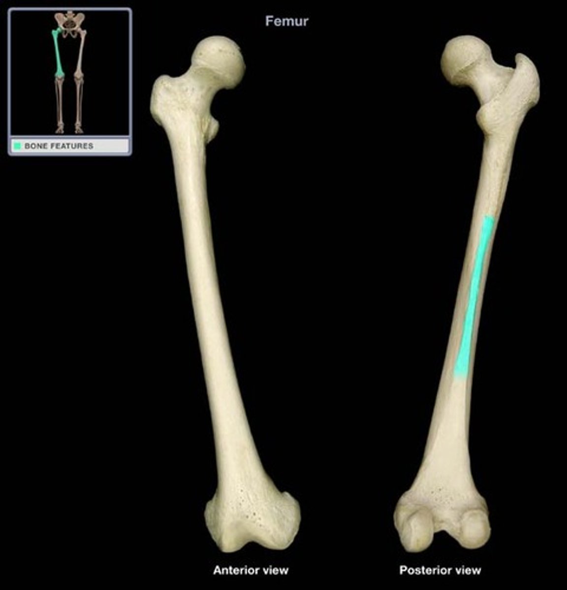
femur linea aspera
Name this structure
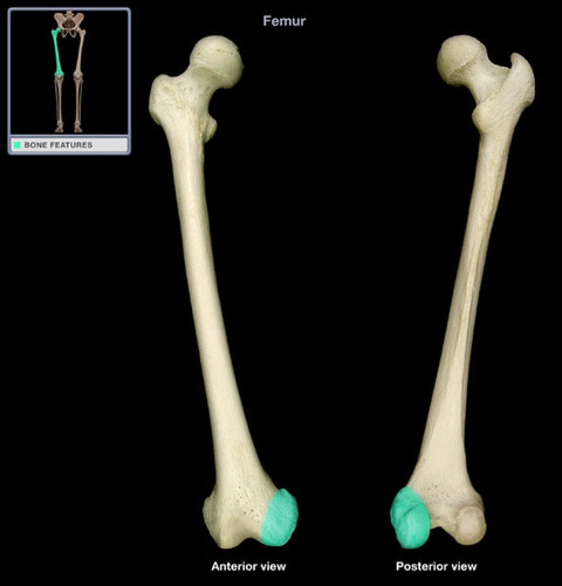
femur medial condyle
Posterior, inferior of femur. "Rounded knob that articulates with another bone."
femur lateral condyle
articulates with the lateral condyle of the tibia
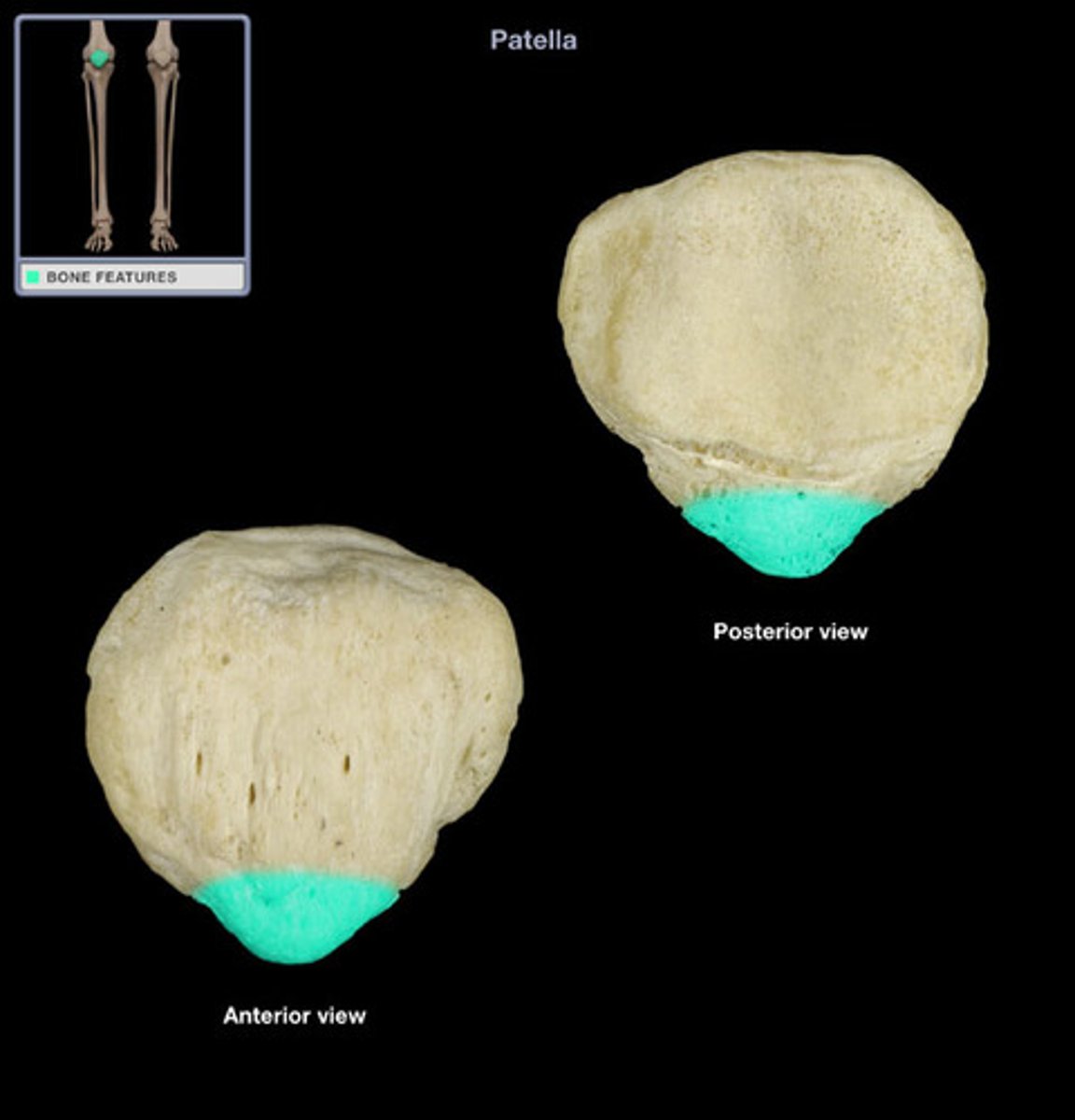
patella
kneecap
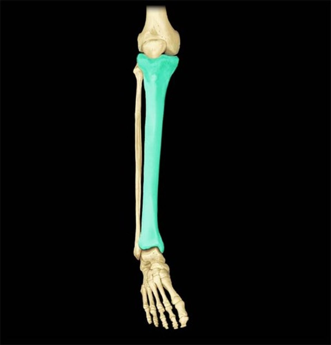
tibia
shin bone
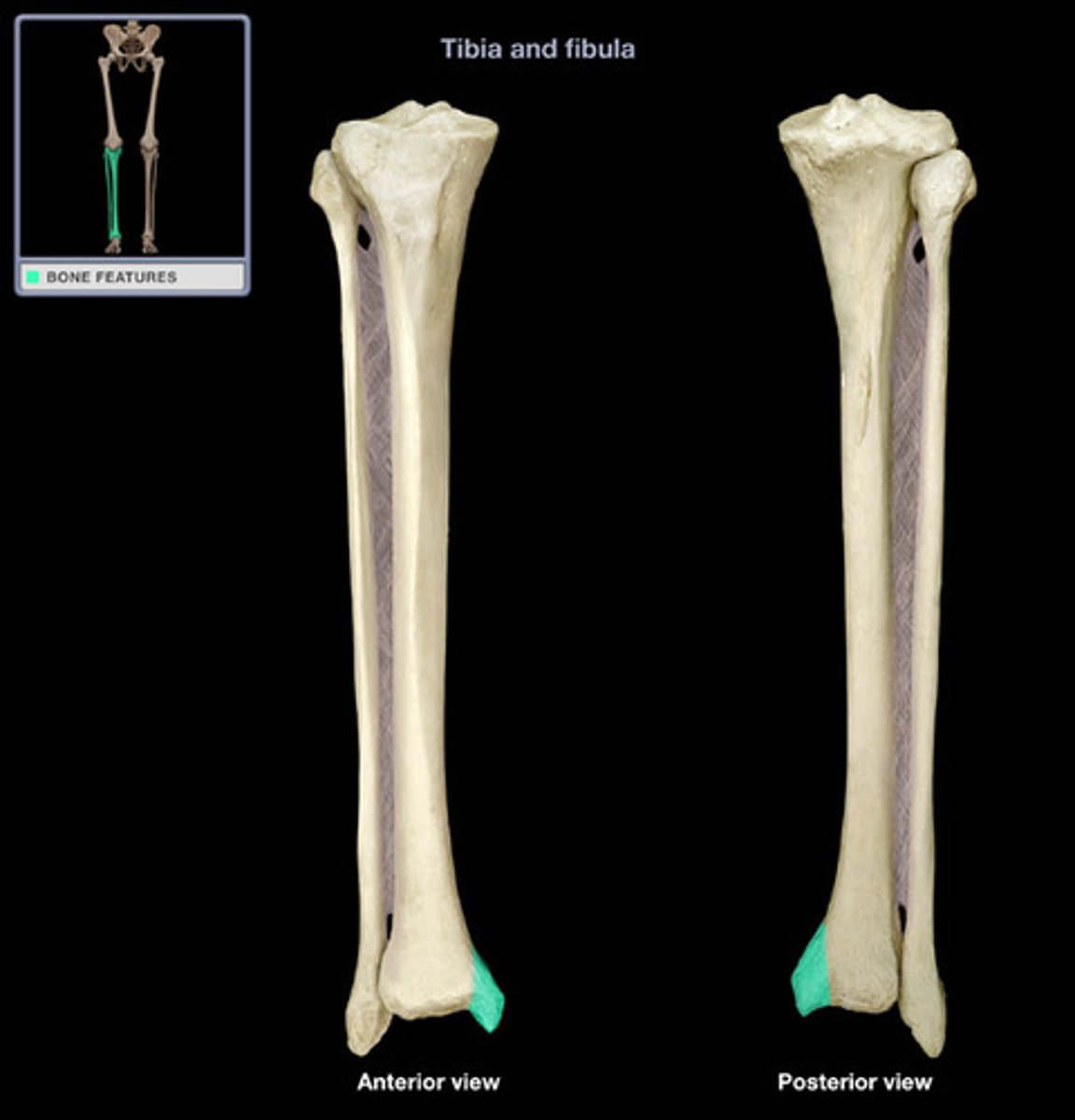
tibial tuberosity
point where the patellar ligament attaches
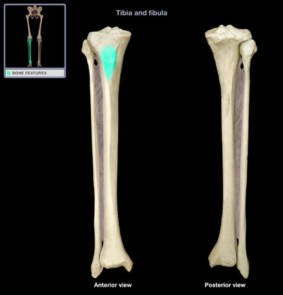
tibia medial malleolus
forms the medial bulge of the ankle
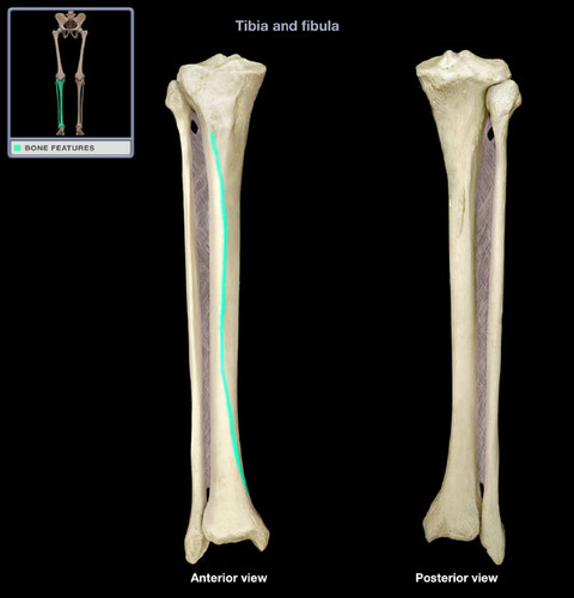
tibia anterior border
sharply angular on shaft
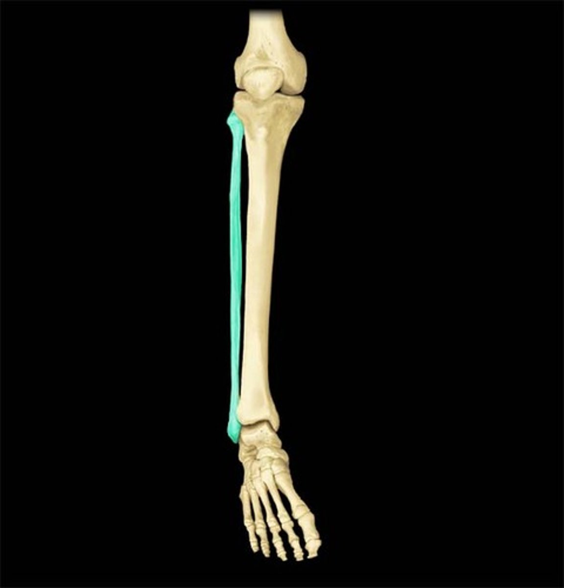
fibula
calf bone
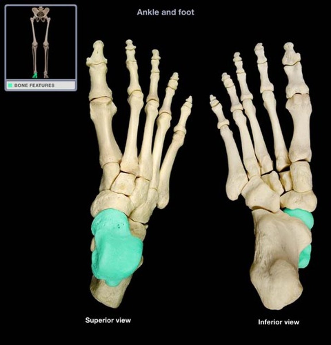
talus
ankle bone that articulates with bones of the leg
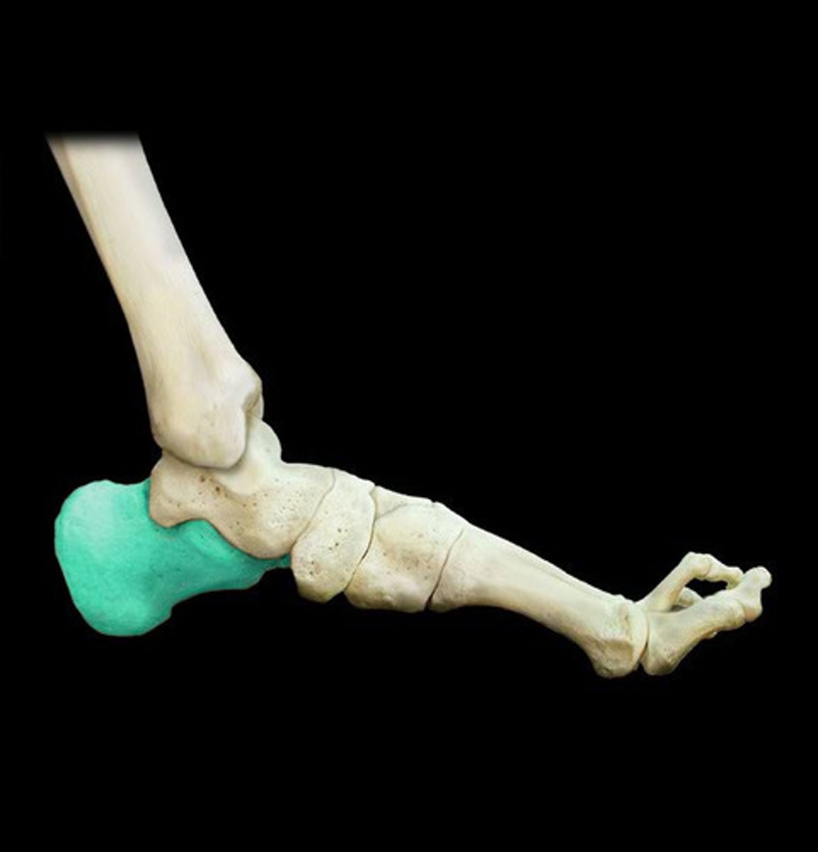
calcaneus
heel bone
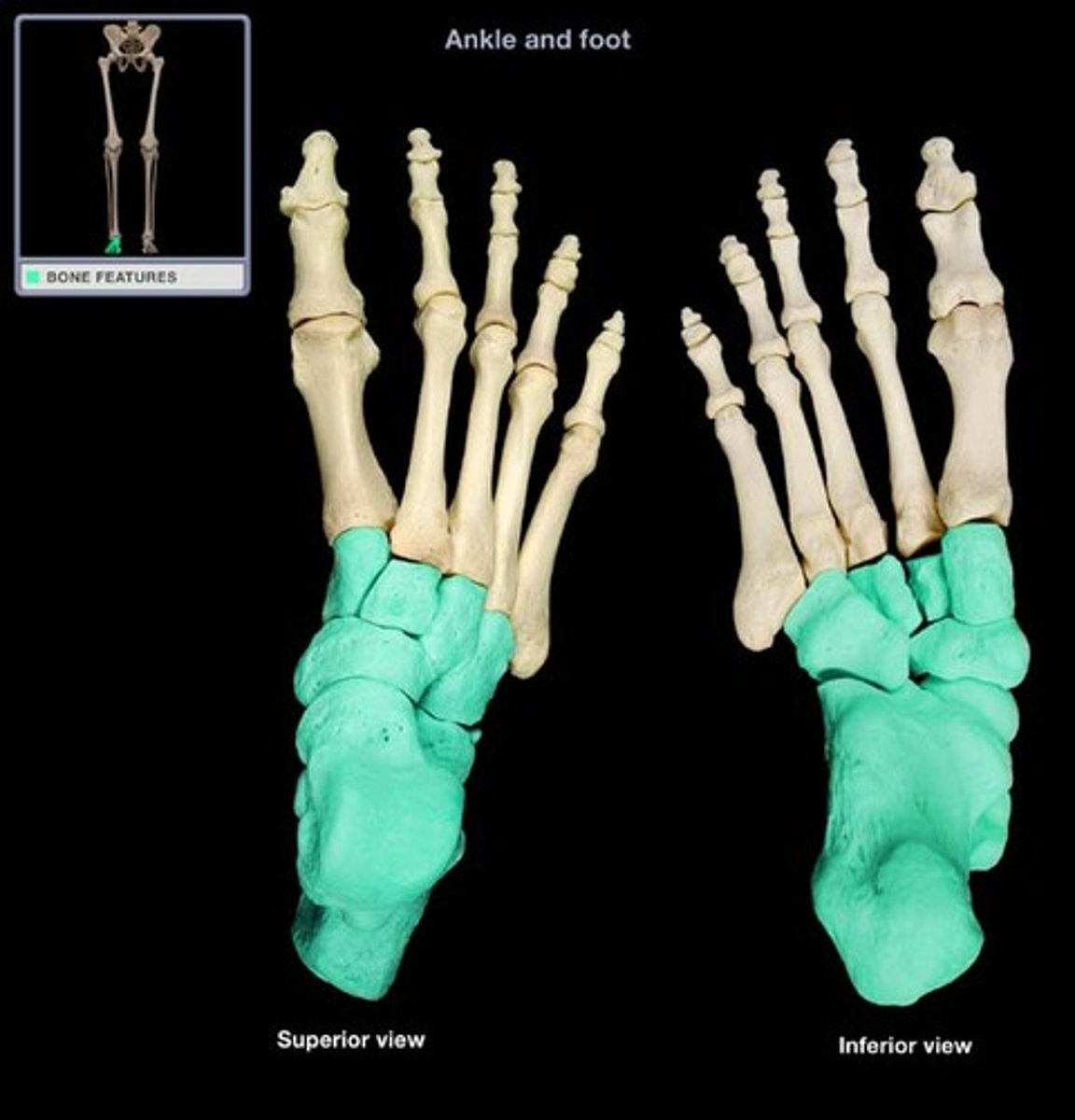
tarsals
ankle bones
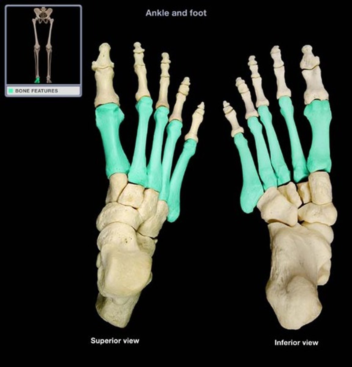
metatarsals
bones of the foot between ankle and toes
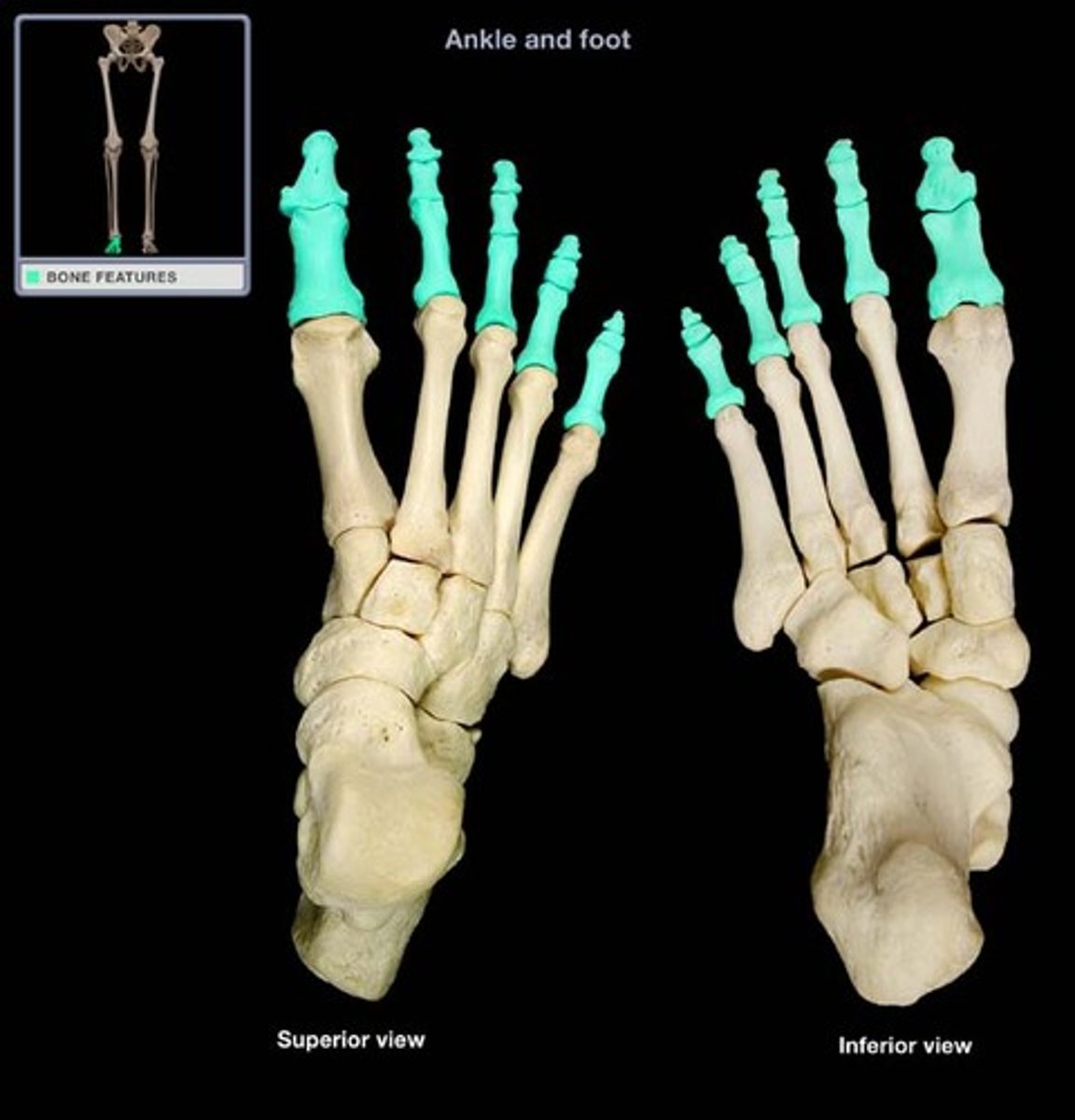
phalanx (phalanges)
toe bones (big toe has no middle phalanx)
skull sutures
coronal, saggital, lambdoid, squamous
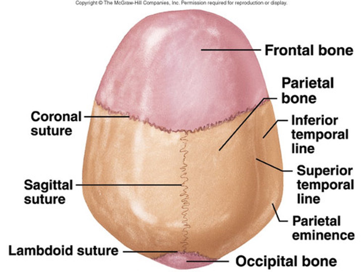
coronal suture
the suture between the parietal and frontal bones of the skull
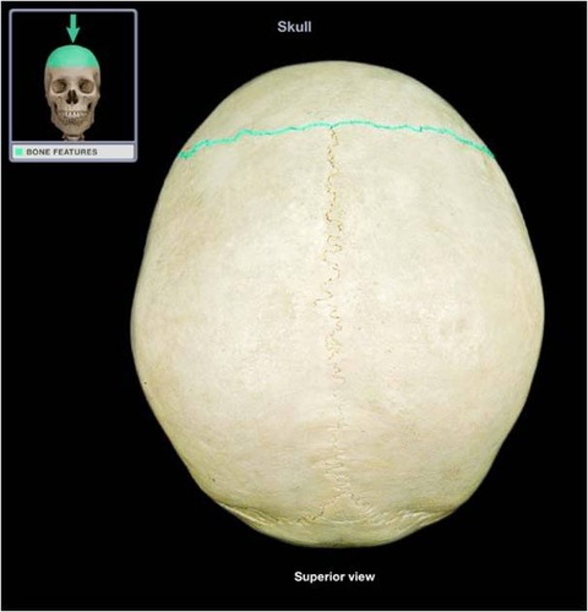
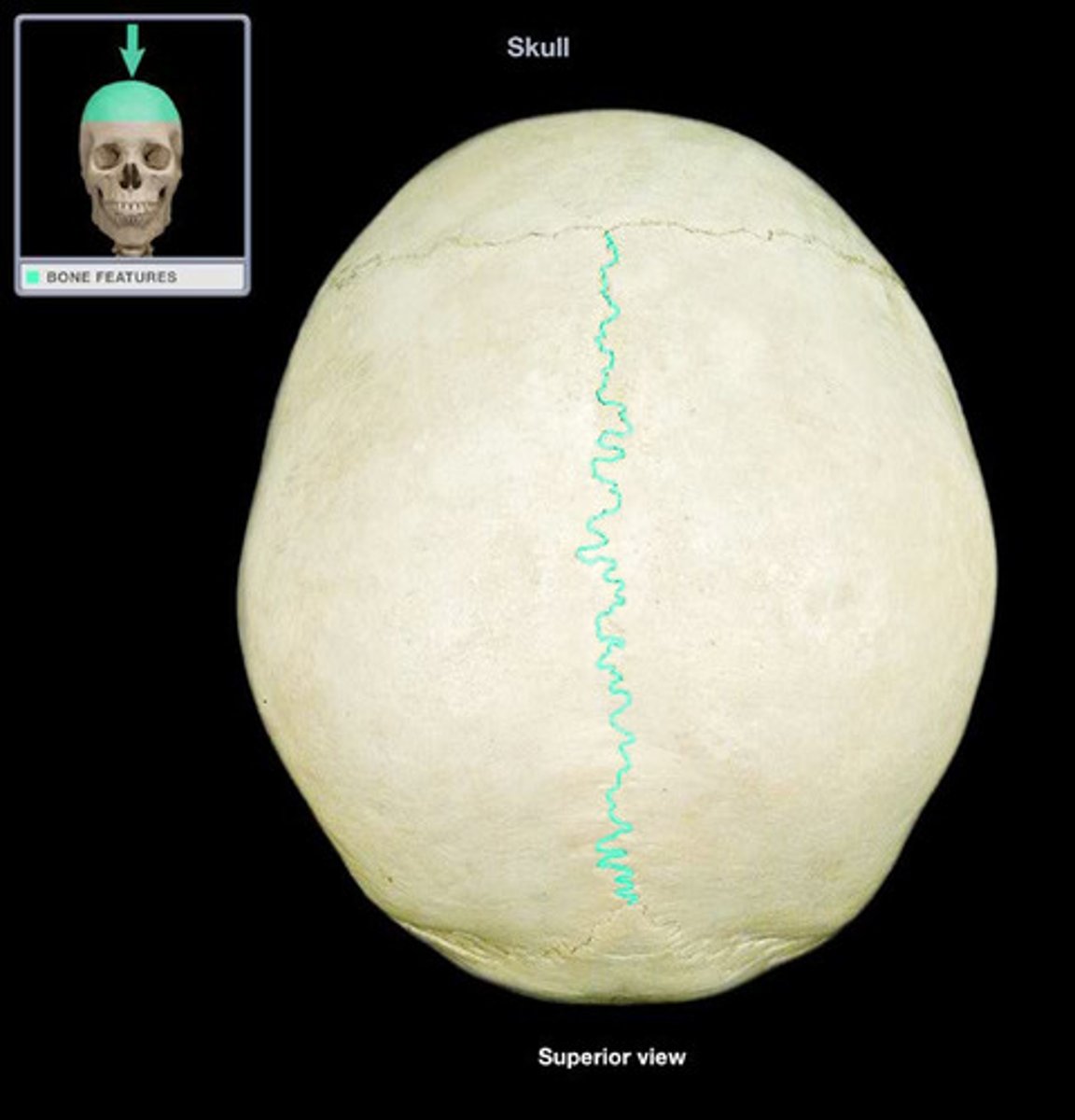
saggital suture
separates the left and right parietal bone
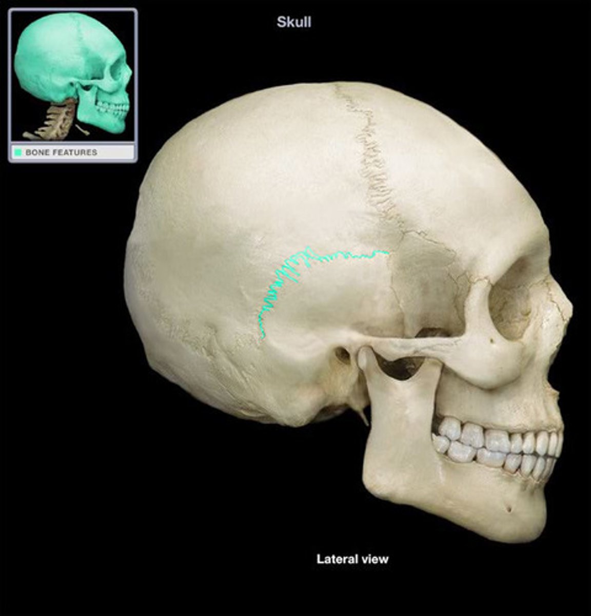
squamous (temporal) suture
thin and flat suture behind ear
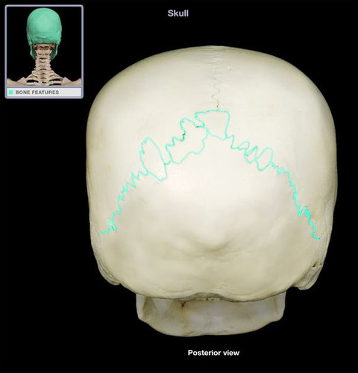
lambdoid suture
between parietal bones and occipital bone
orbit
eye socket
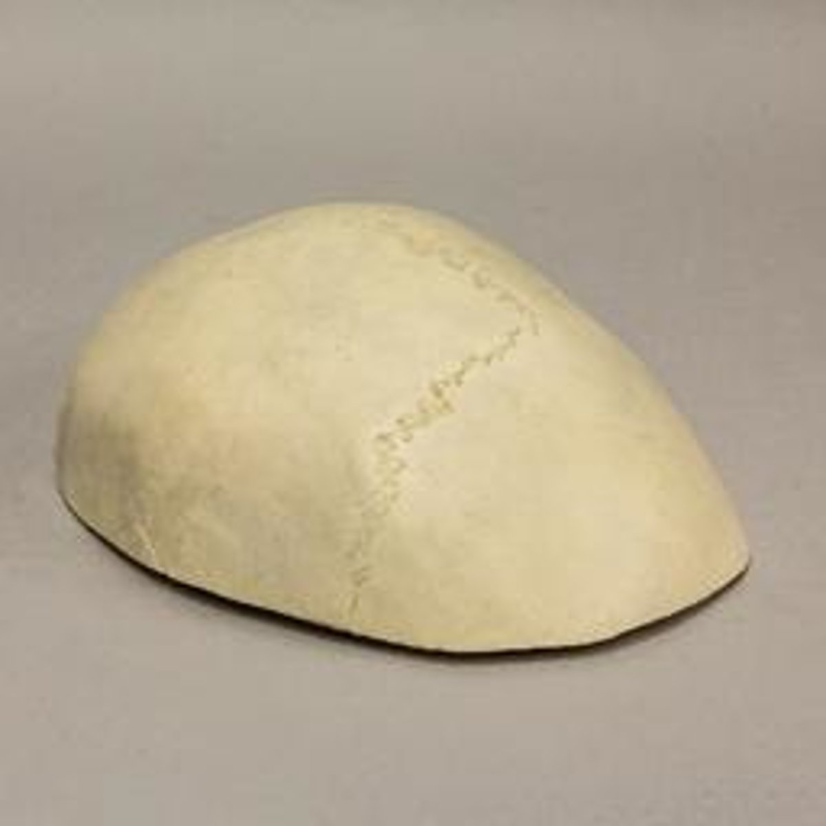
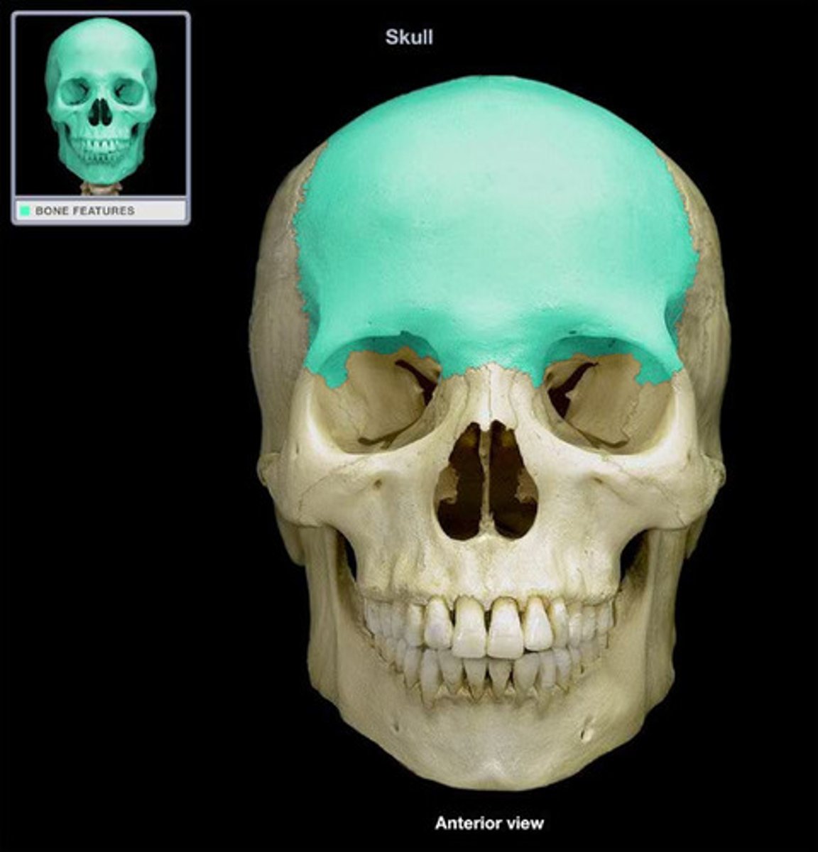
Calvarium
skull cap
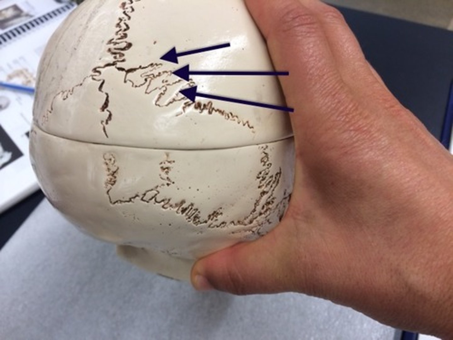
Suture Bones (Wormian Bones)
Little bones in the skull that were separated by sutures
frontal bone
forehead
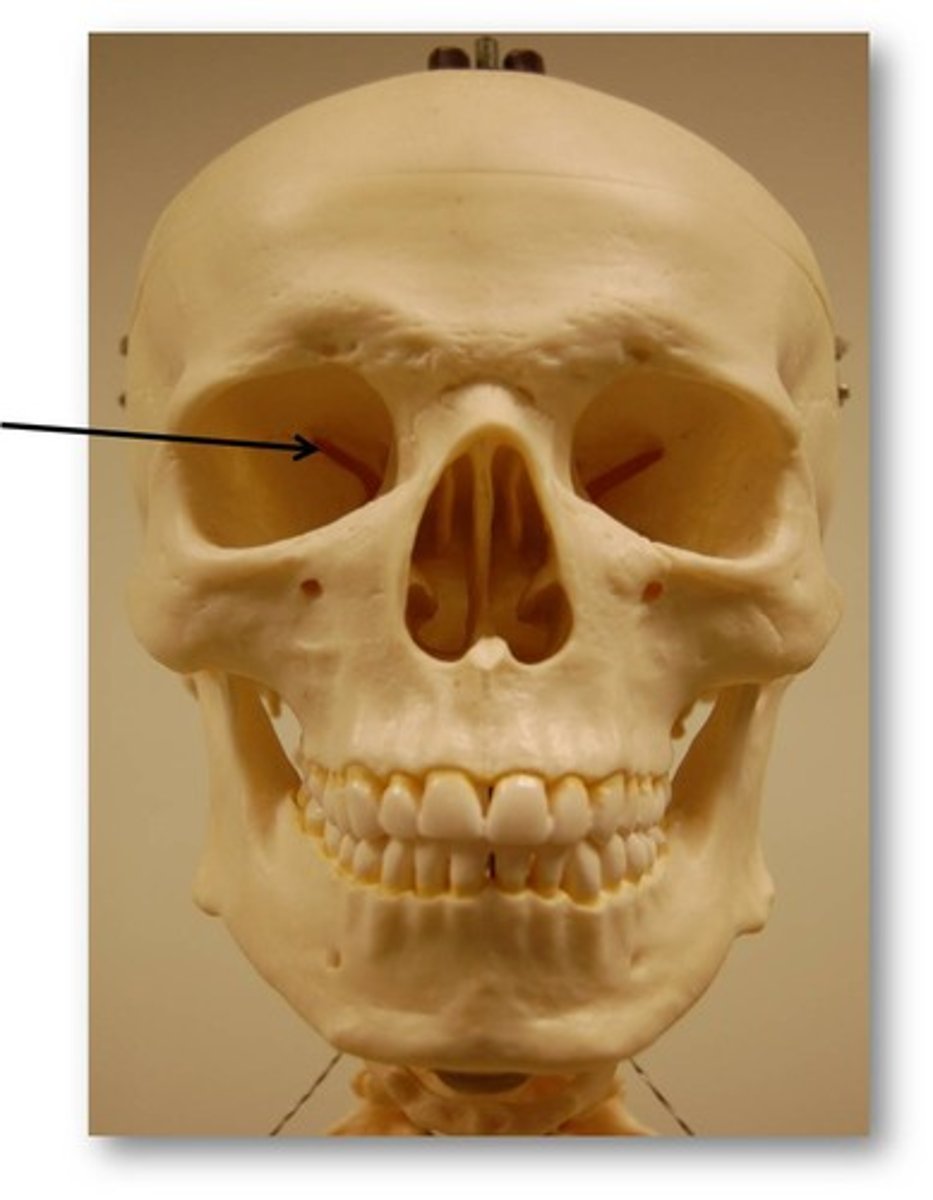
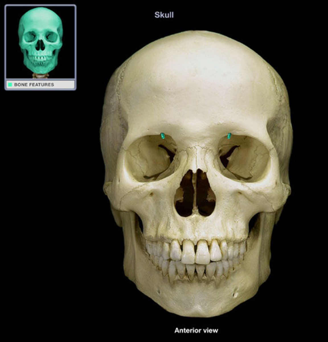
frontal bone: supraorbital foramen
opening above each orbit allowing blood vessels and nerves to pass
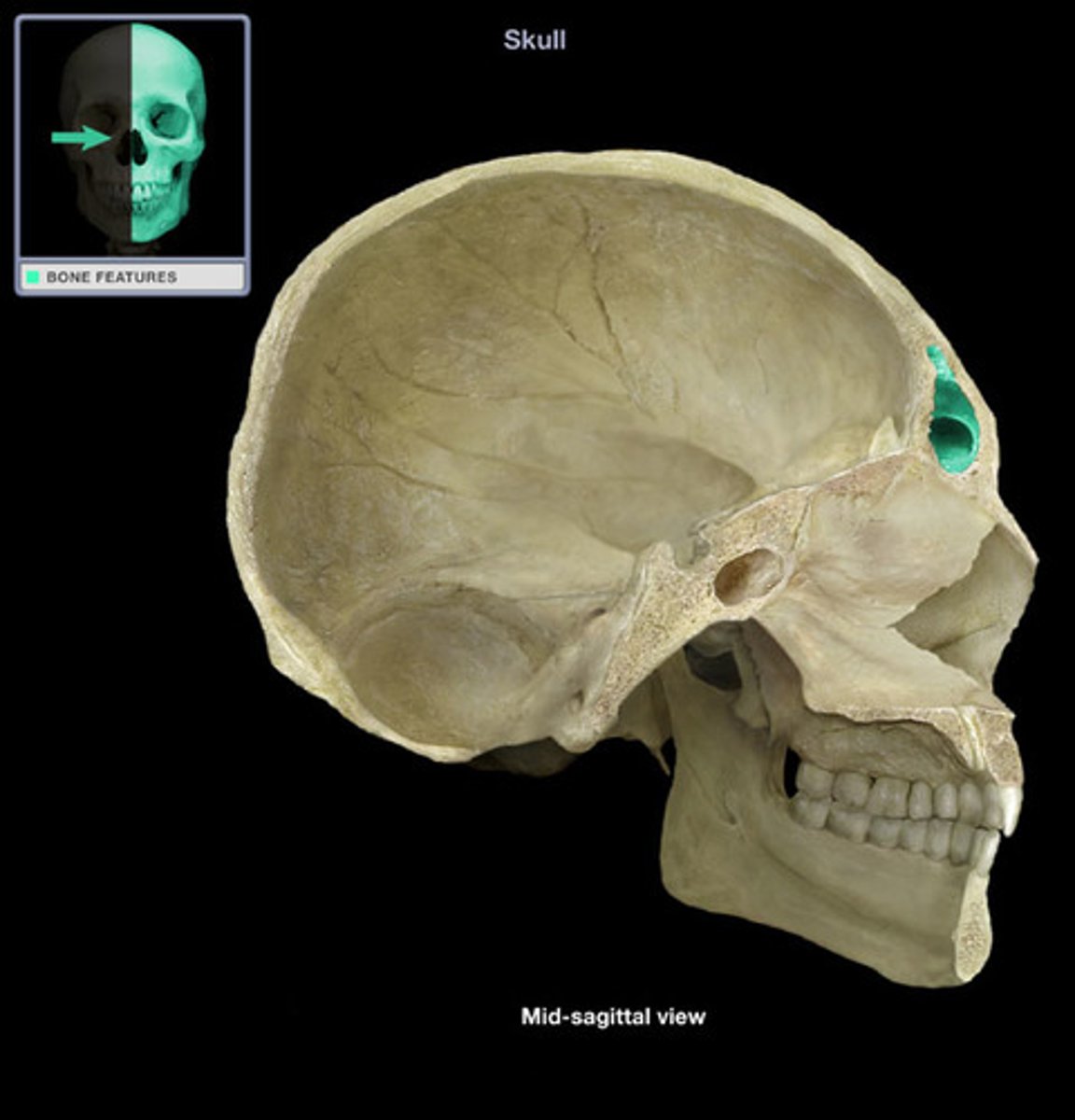
Frontal Bone: Frontal Sinus
cavity within the frontal bone