Bones
1/95
There's no tags or description
Looks like no tags are added yet.
Name | Mastery | Learn | Test | Matching | Spaced |
|---|
No study sessions yet.
96 Terms
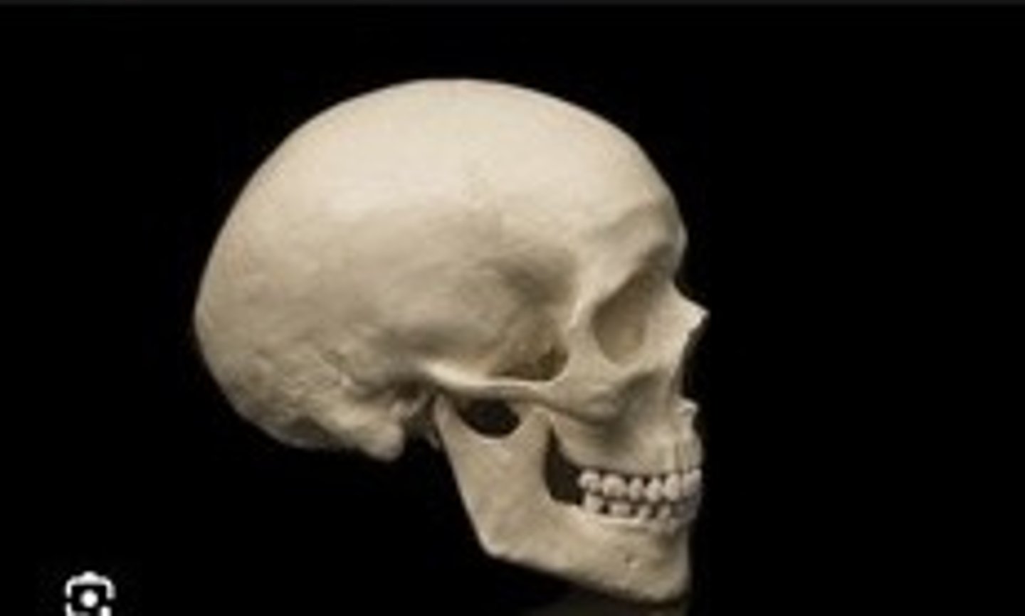
Cranium
The skull that encases the brain.
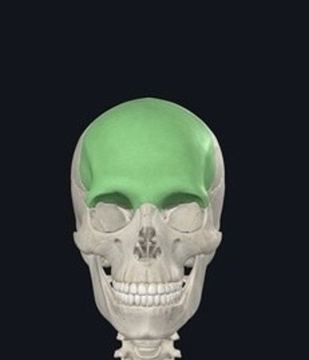
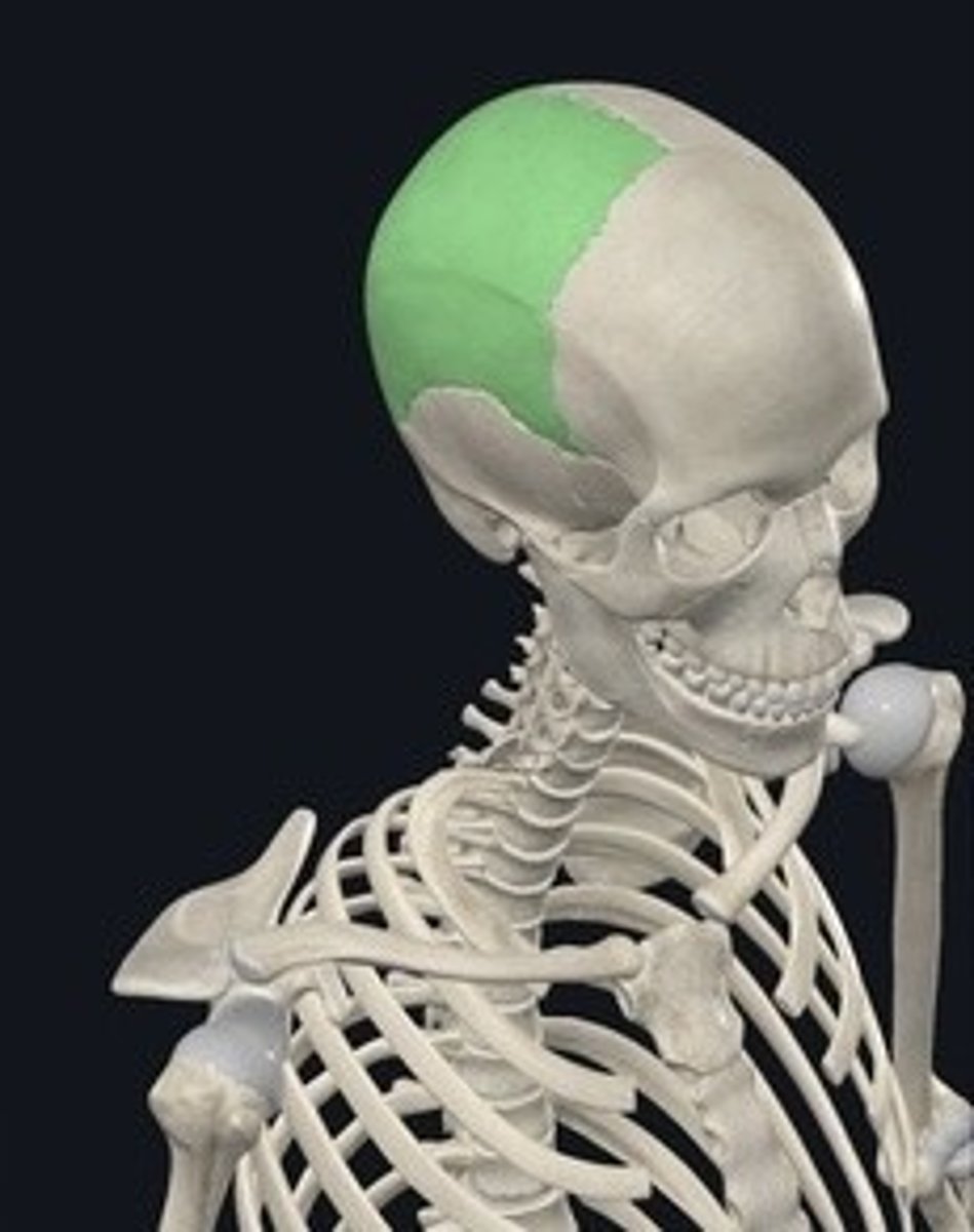
Frontal
Forehead bone; forms the anterior skull.
Parietal
Two bones forming the top and sides of the skull.
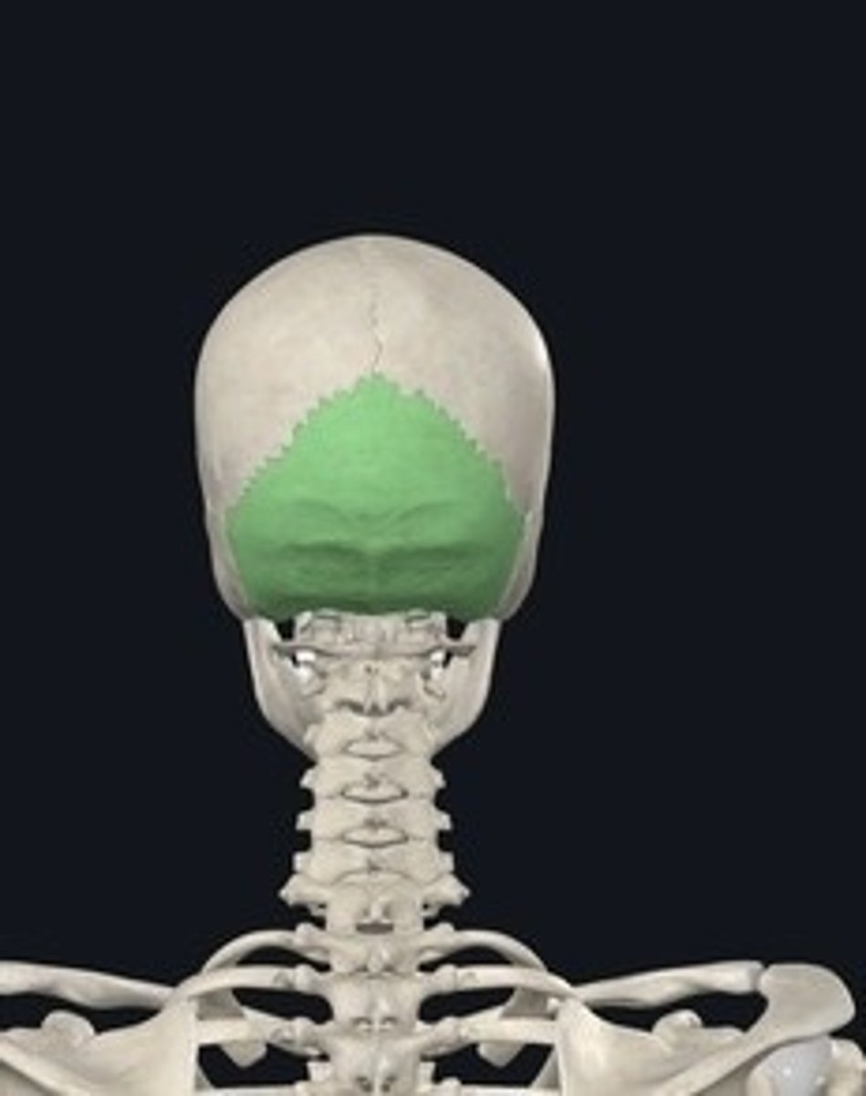
Occipital
Bone at the back of the skull.
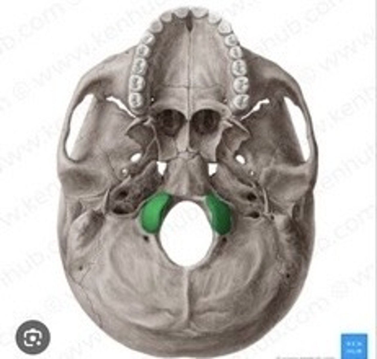
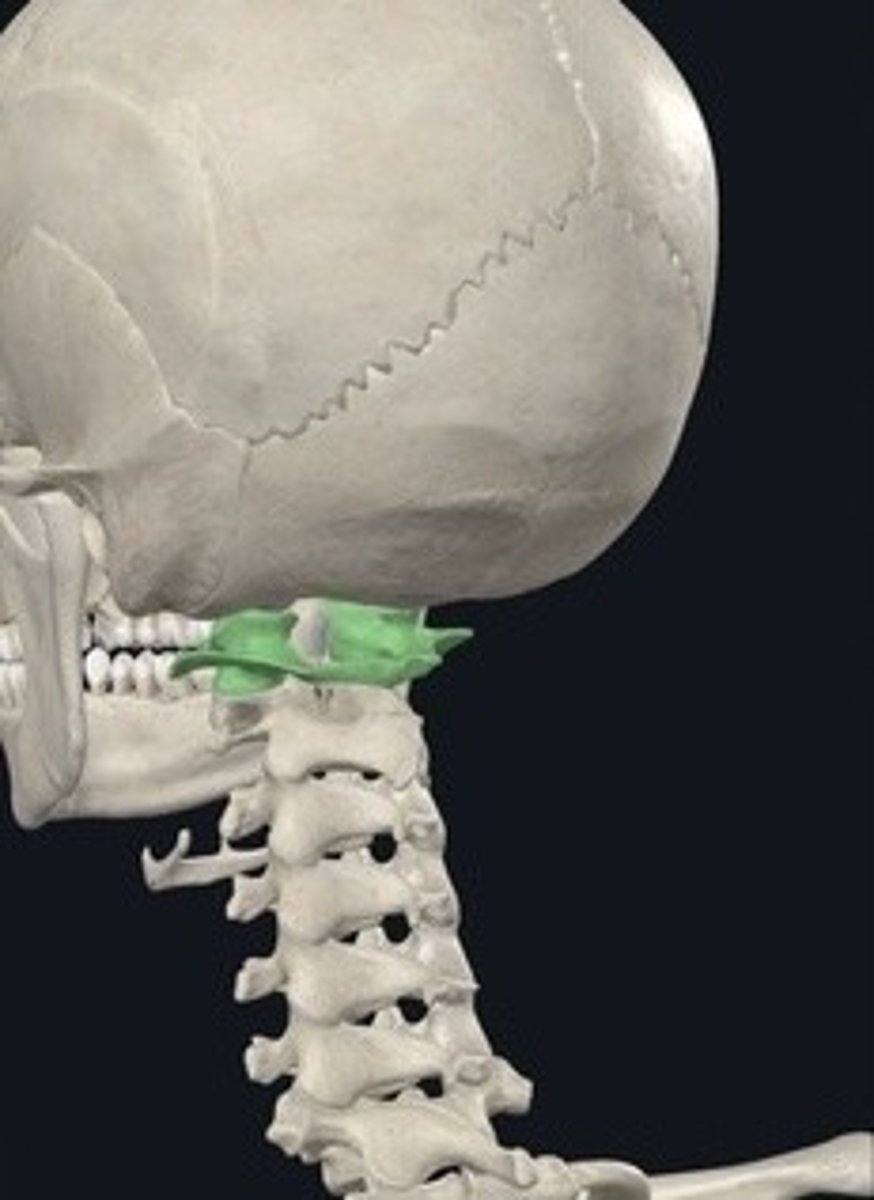
Occipital Condyle
Articulates with the first cervical vertebra.
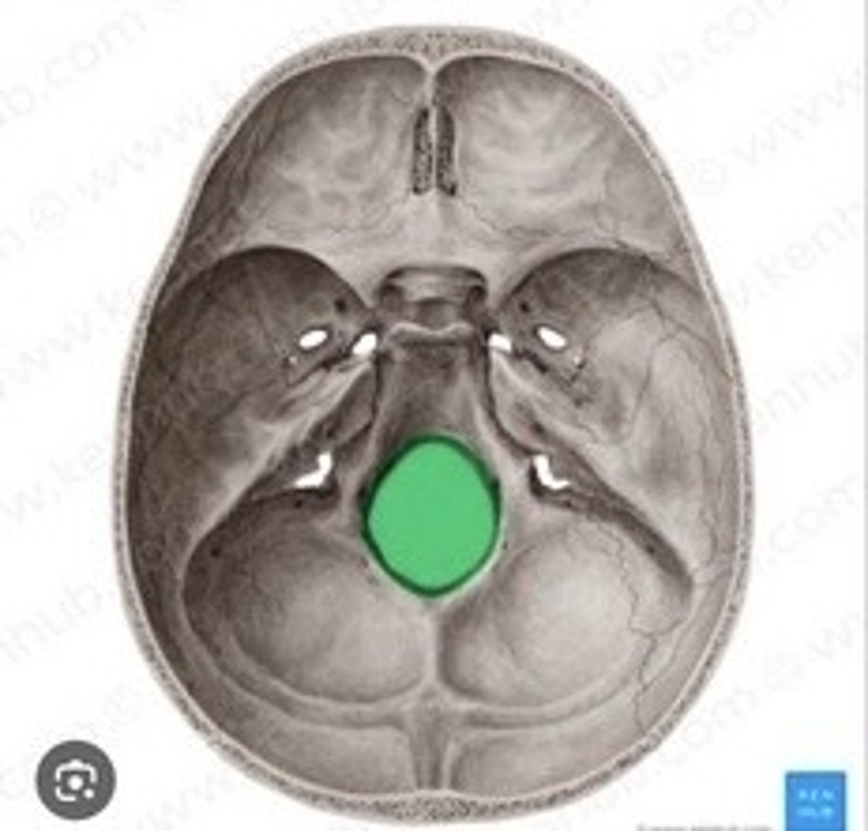
Foramen Magnum
Large opening for spinal cord passage.
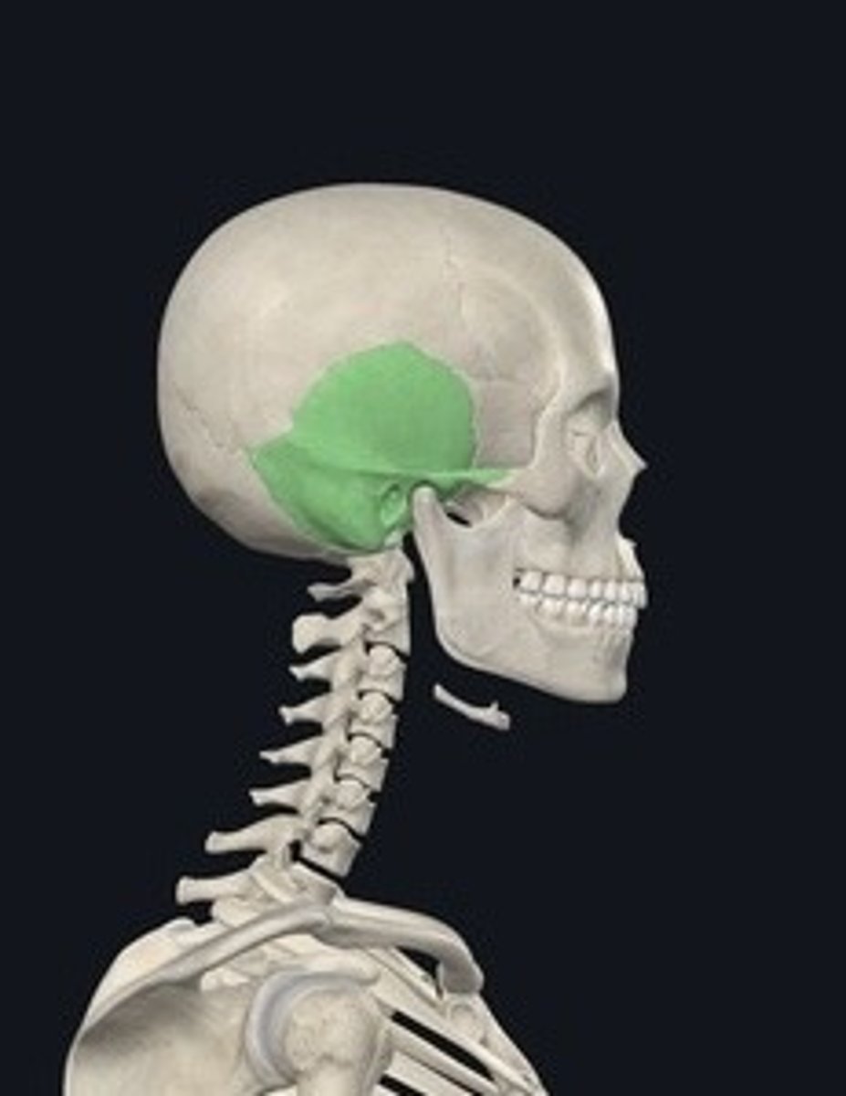
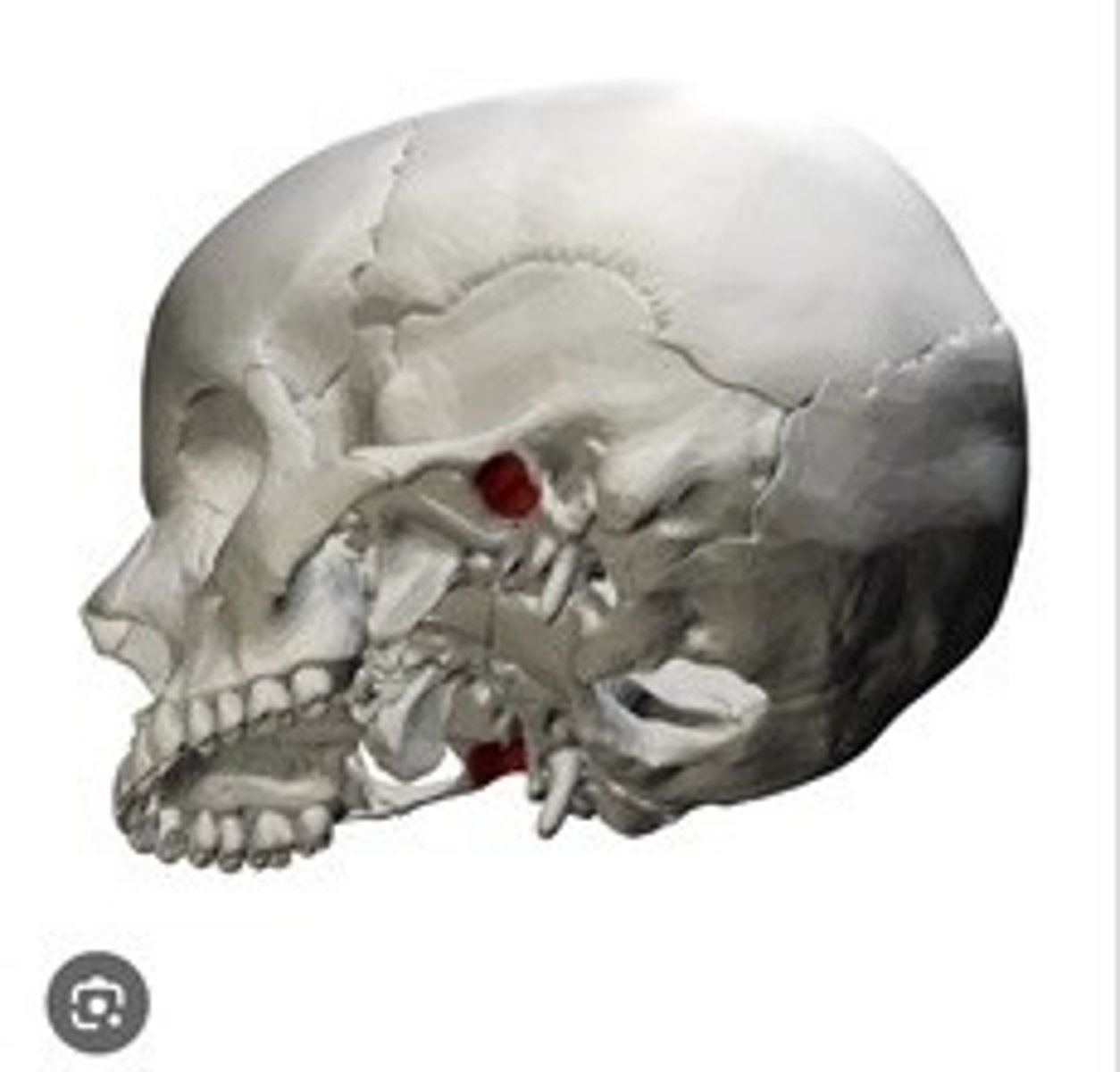
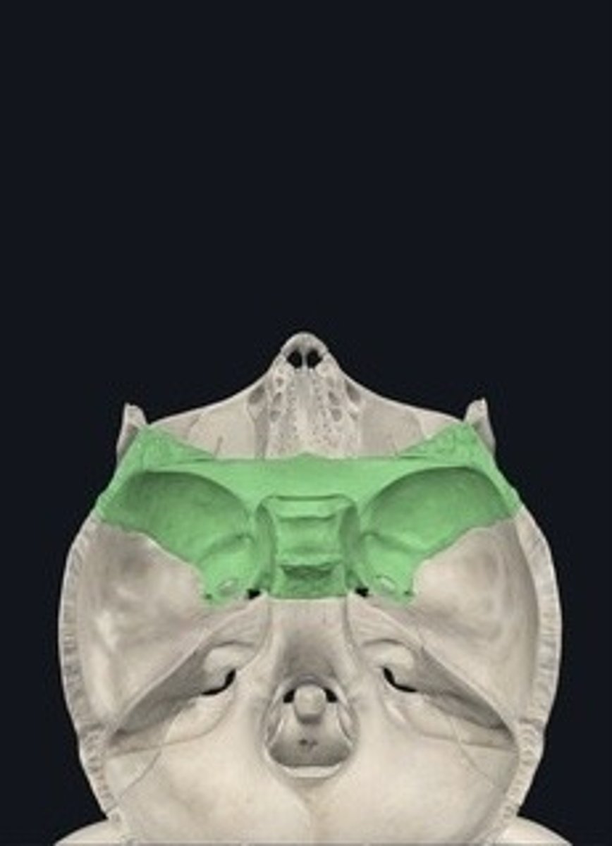
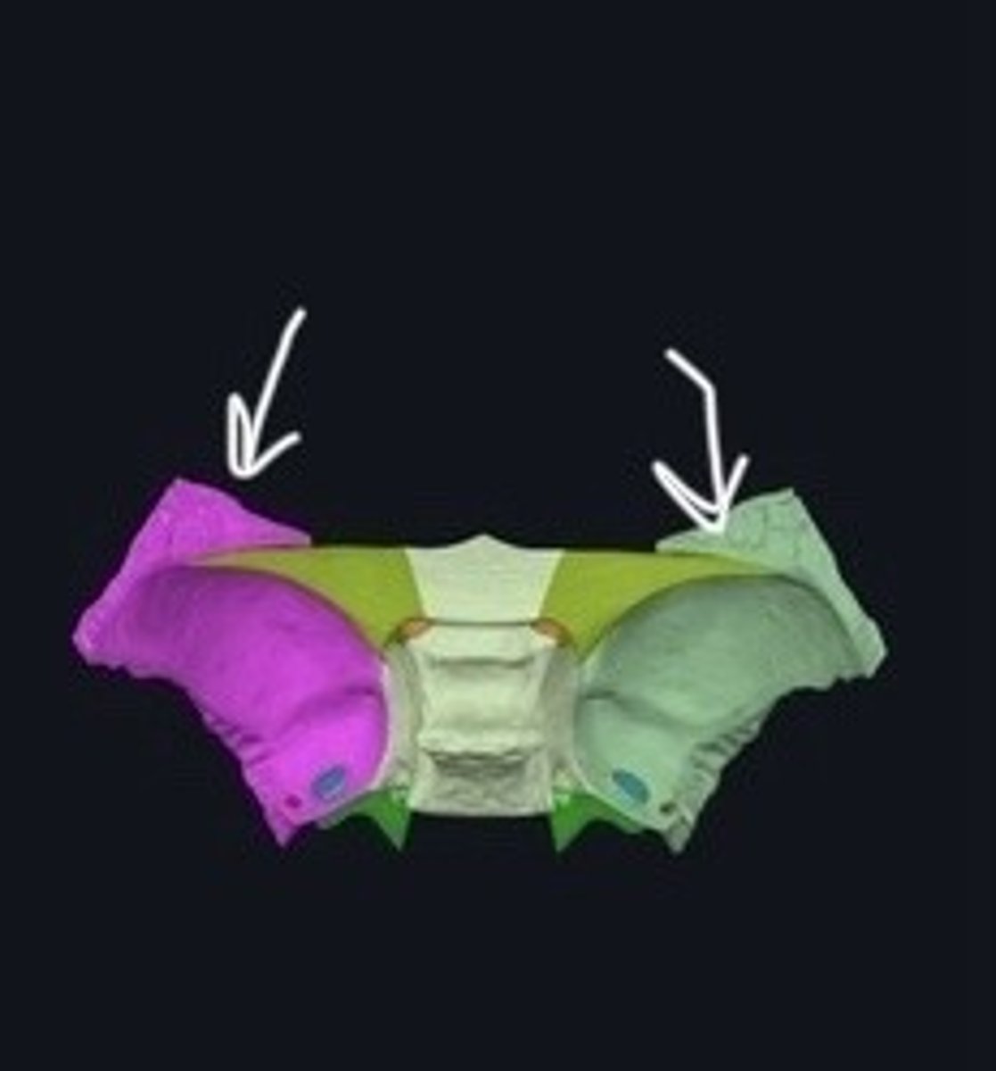
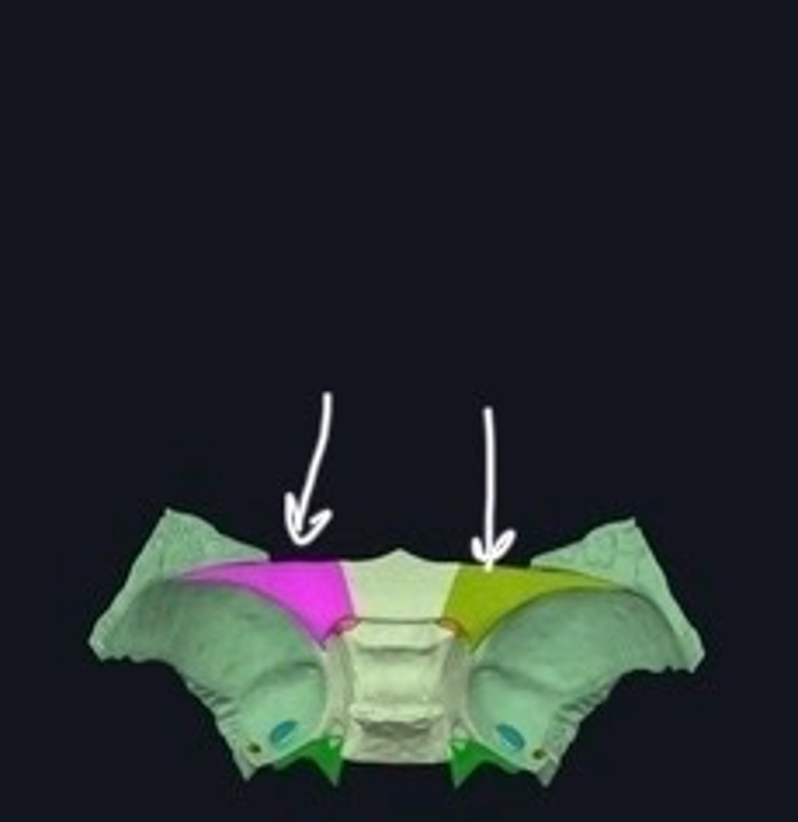
Temporal bones
Bones located at the sides of the skull.
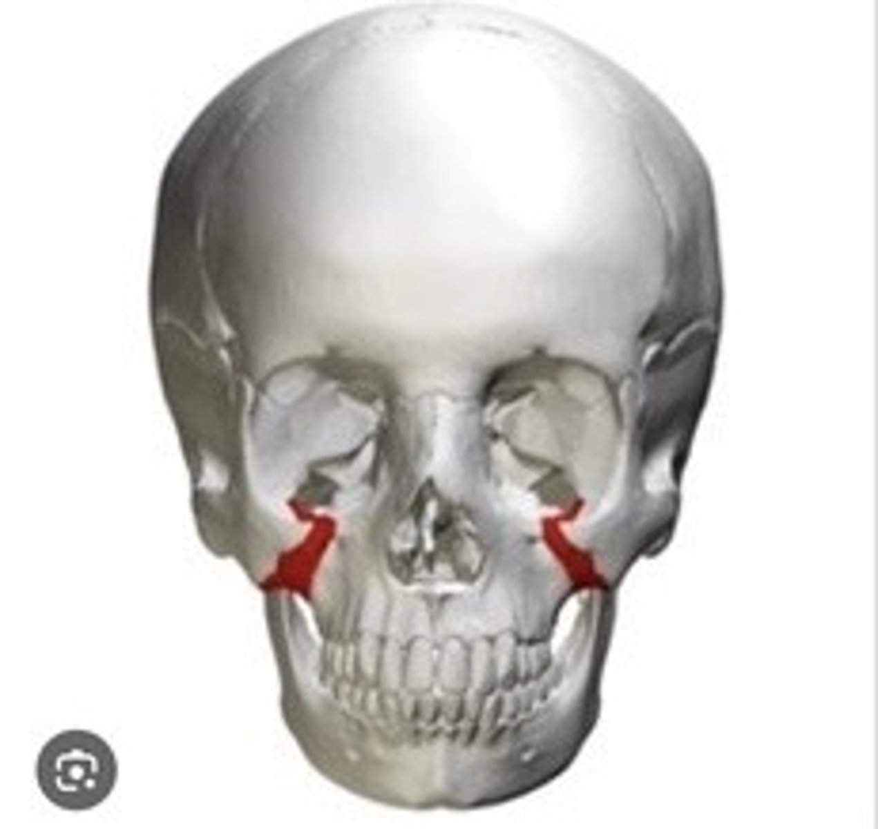
Zygomatic process
Projection of the temporal bone forming cheekbone.
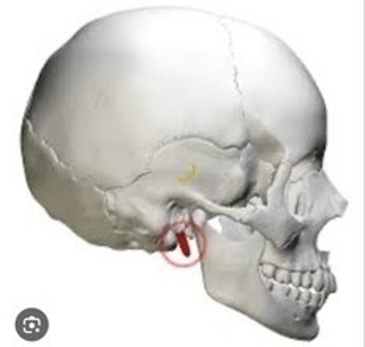
Styloid Process
Thin projection for muscle attachment beneath the skull.
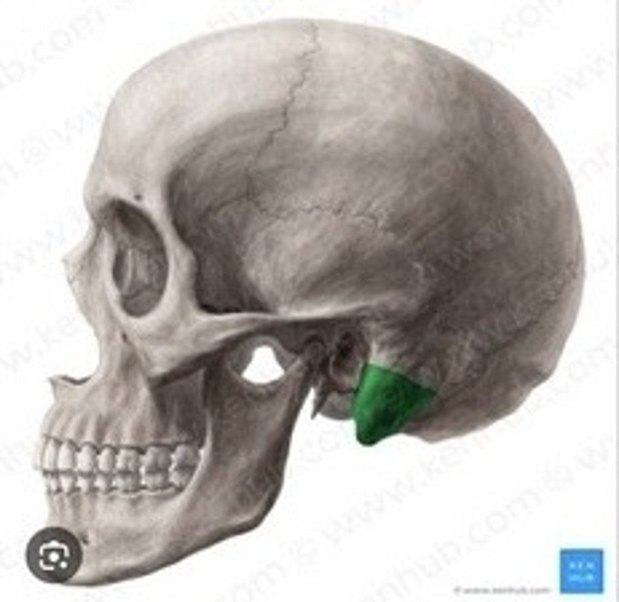
Mastoid Process
Bony prominence behind the ear for muscle attachment.
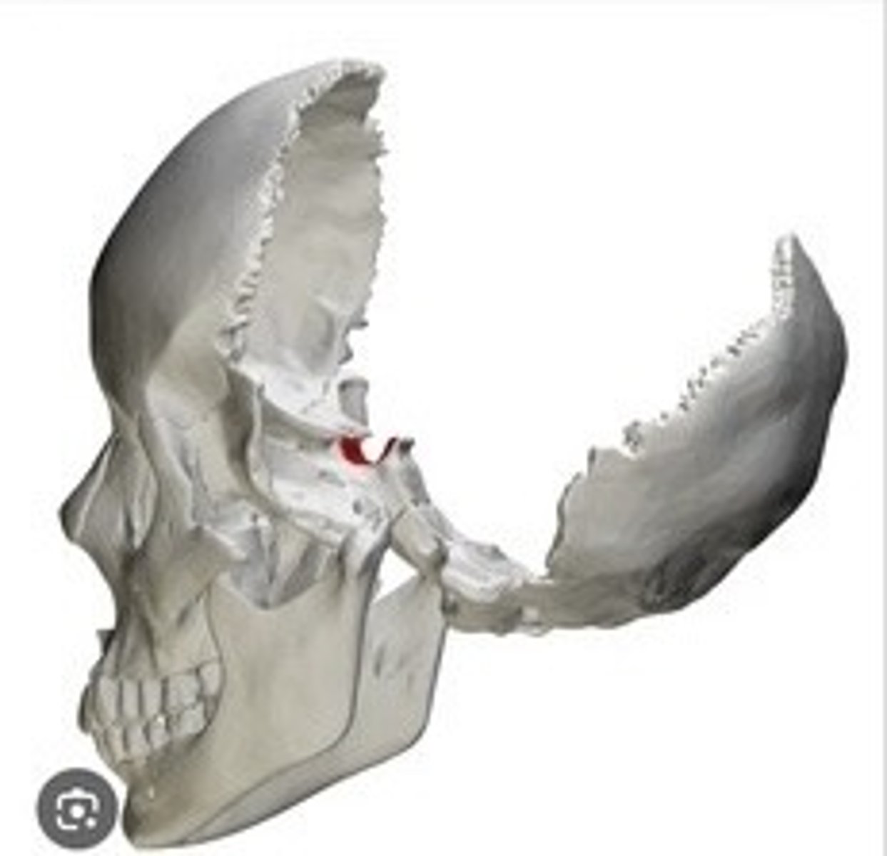
Mandibular fossa
Depression for the mandible articulation.
Sphenoid
Butterfly-shaped bone at the base of the skull.
Great wings
Part of the sphenoid bone extending laterally.
Lesser wings
Smaller extensions of the sphenoid bone.
Sella turcica
Depression housing the pituitary gland.
Ethmoid
Bone forming part of the nasal cavity.
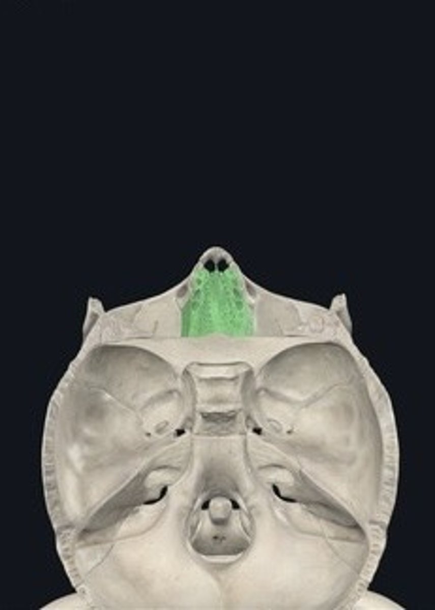
Cribriform plates
Thin bony plates with olfactory foramina.
Crista galli
Vertical projection for brain membrane attachment.
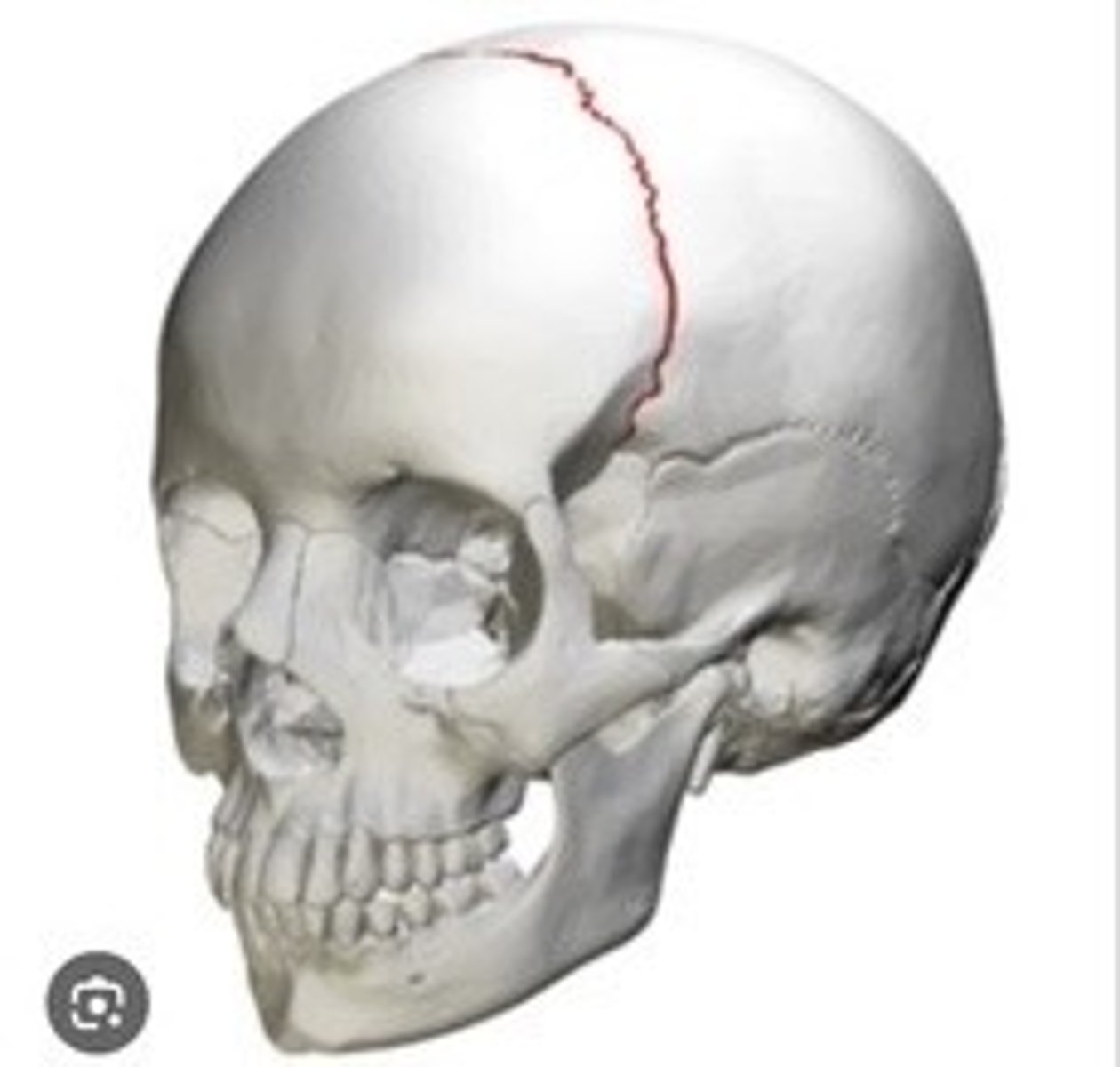
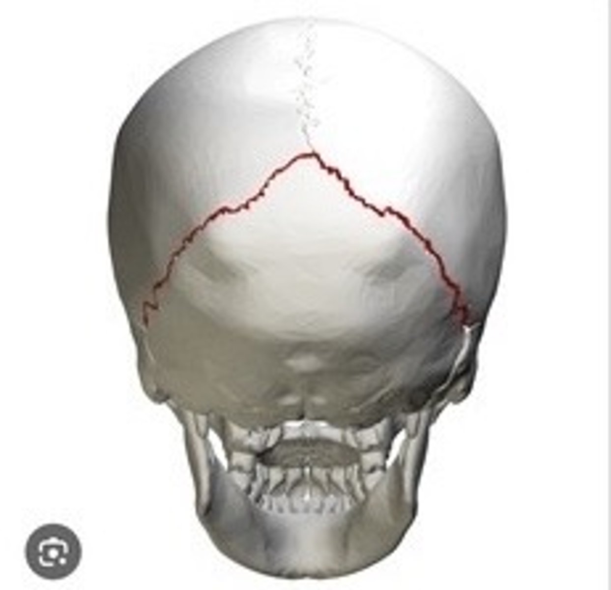
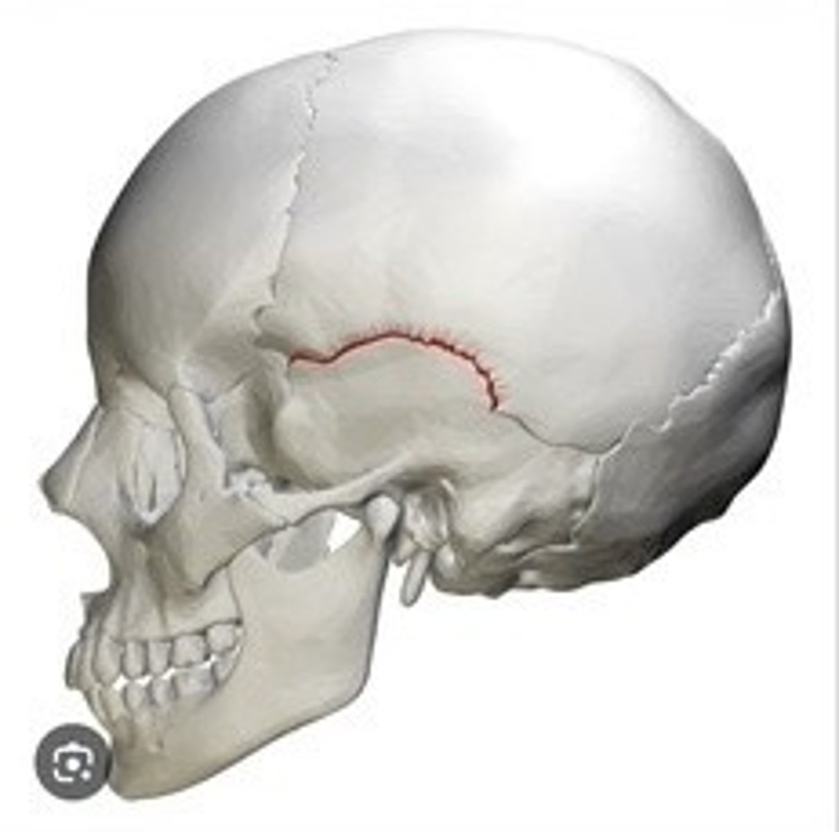
Sutures
Fibrous joints connecting skull bones.
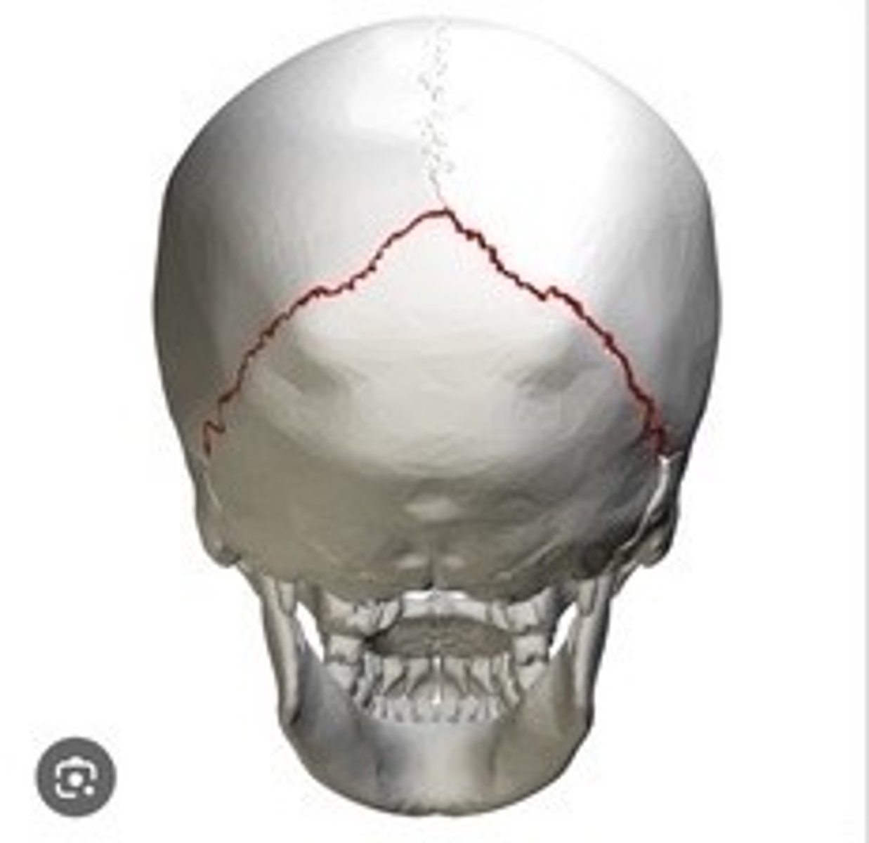
Coronal
Suture between frontal and parietal bones.
Sagittal
Suture between the two parietal bones.

Lambdoid
Suture between parietal and occipital bones.
Squamous
Suture between parietal and temporal bones.
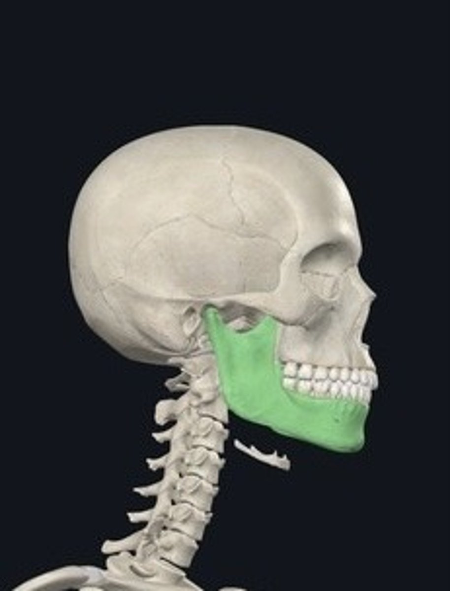
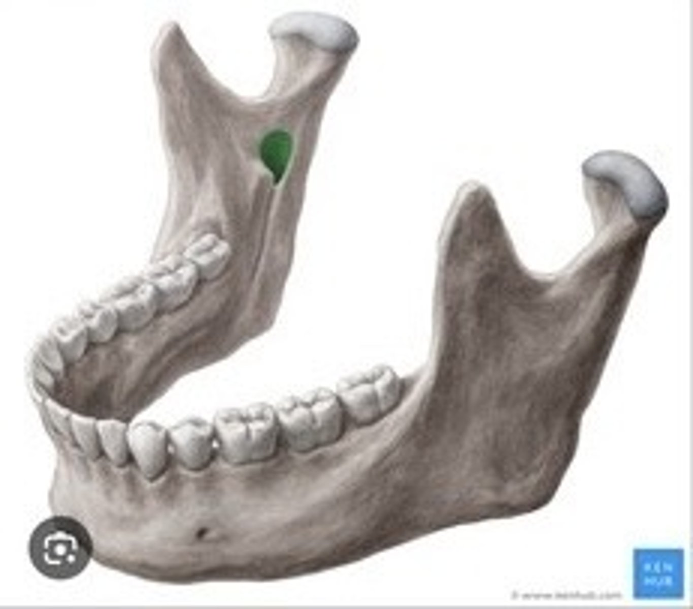
Mandible
Lower jawbone; only movable skull bone.
Mandibular notch
Indentation between the condylar and coronoid processes.
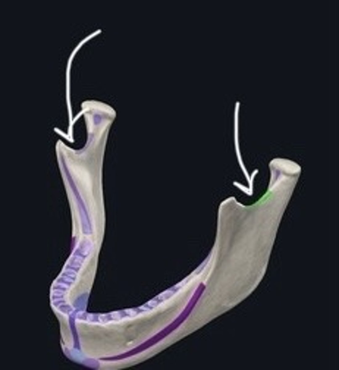
Mandibular foramina
Openings for nerves and blood vessels in the mandible.
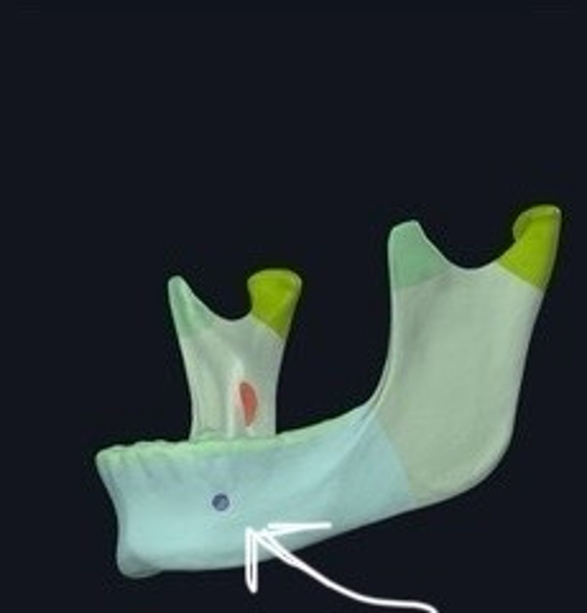
Mental foramina
Openings in the mandible for sensory nerves.
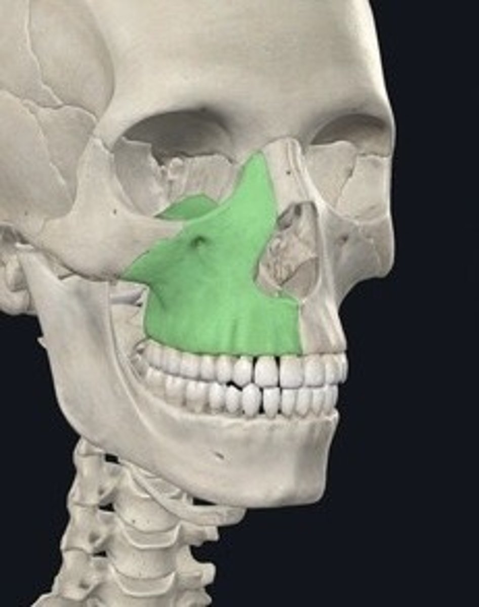
Maxillary bones
Two bones forming the upper jaw.
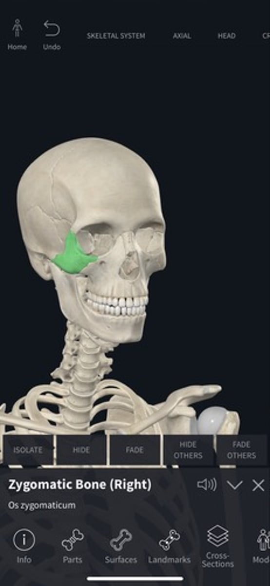
Zygomatic bones
Cheekbones; form the lateral aspect of the face.
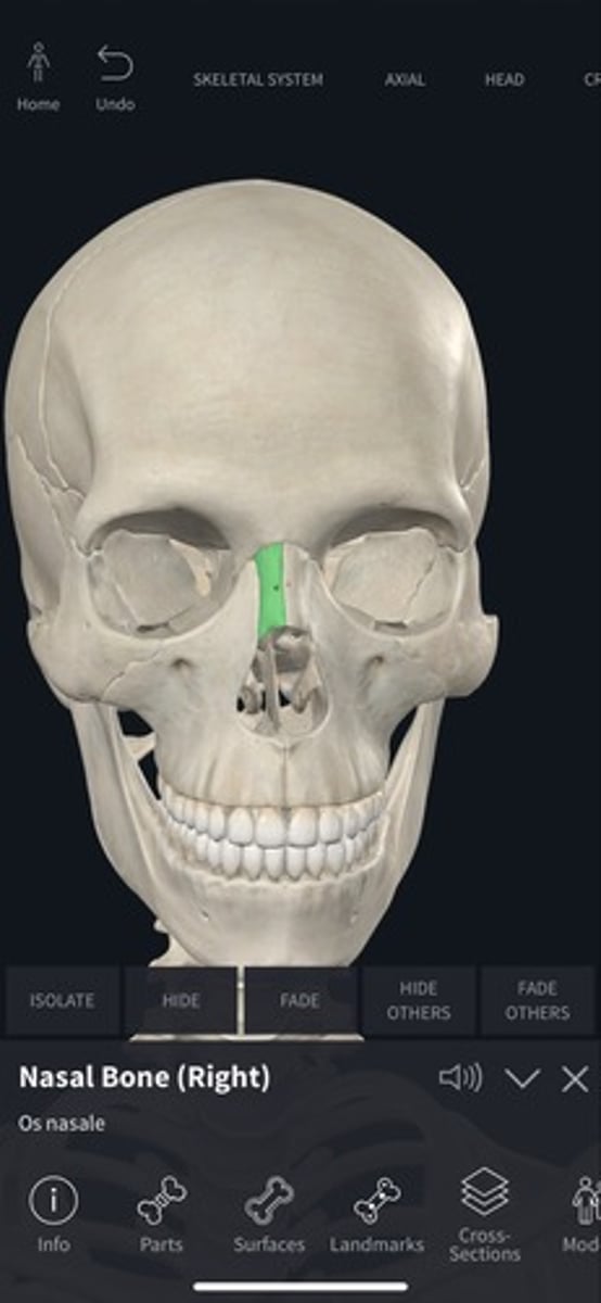
Nasal bones
Two small bones forming the bridge of the nose.
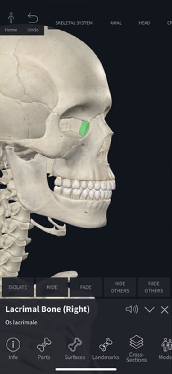
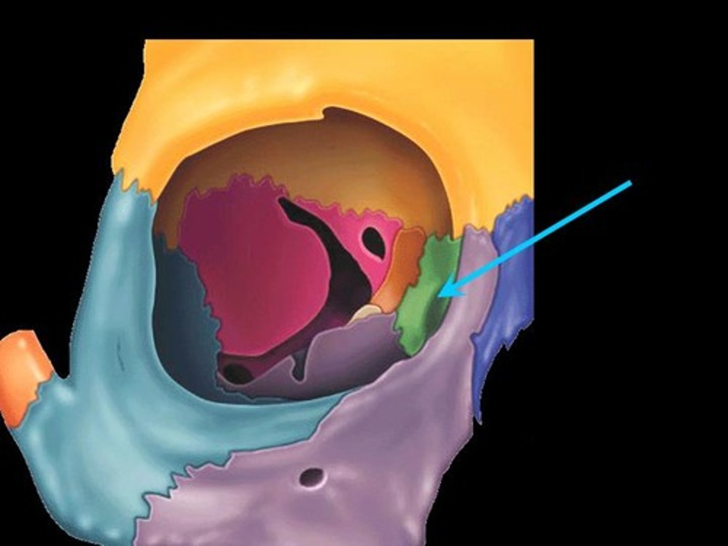
Lacrimal bones
Small bones forming part of the eye socket.
Lacrimal fossa
Depression for the lacrimal sac in the lacrimal bone.
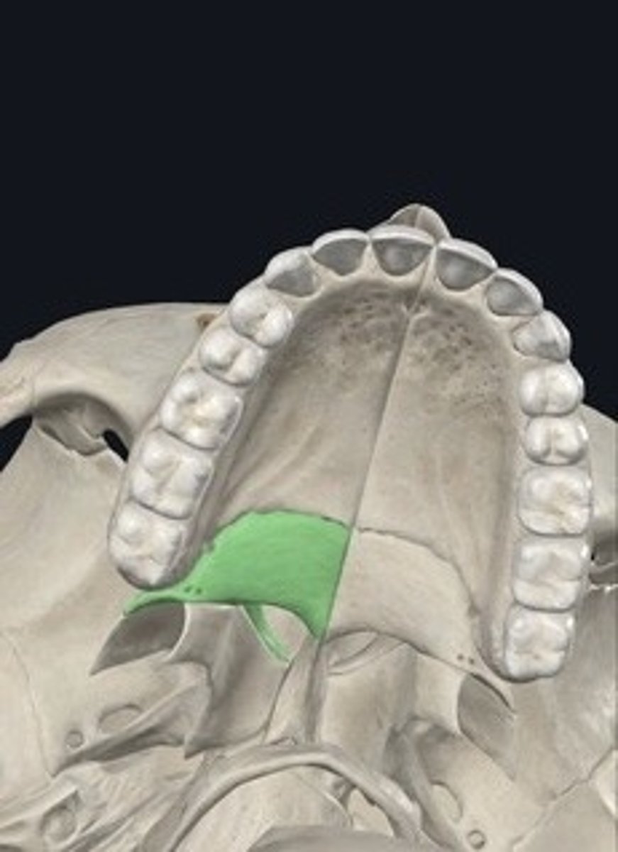
Palatine bones
Two bones forming the posterior part of the hard palate.
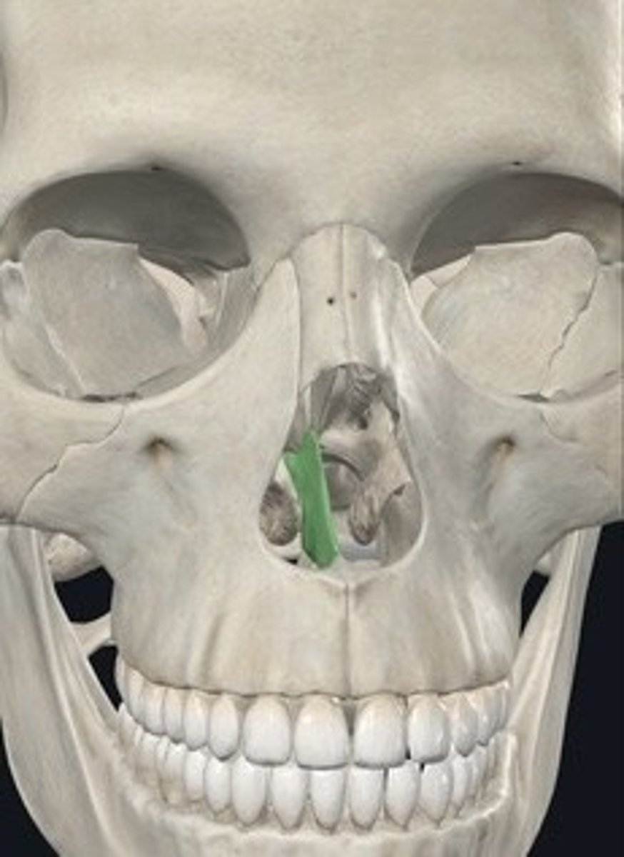
Vomer
Bone forming the inferior part of the nasal septum.
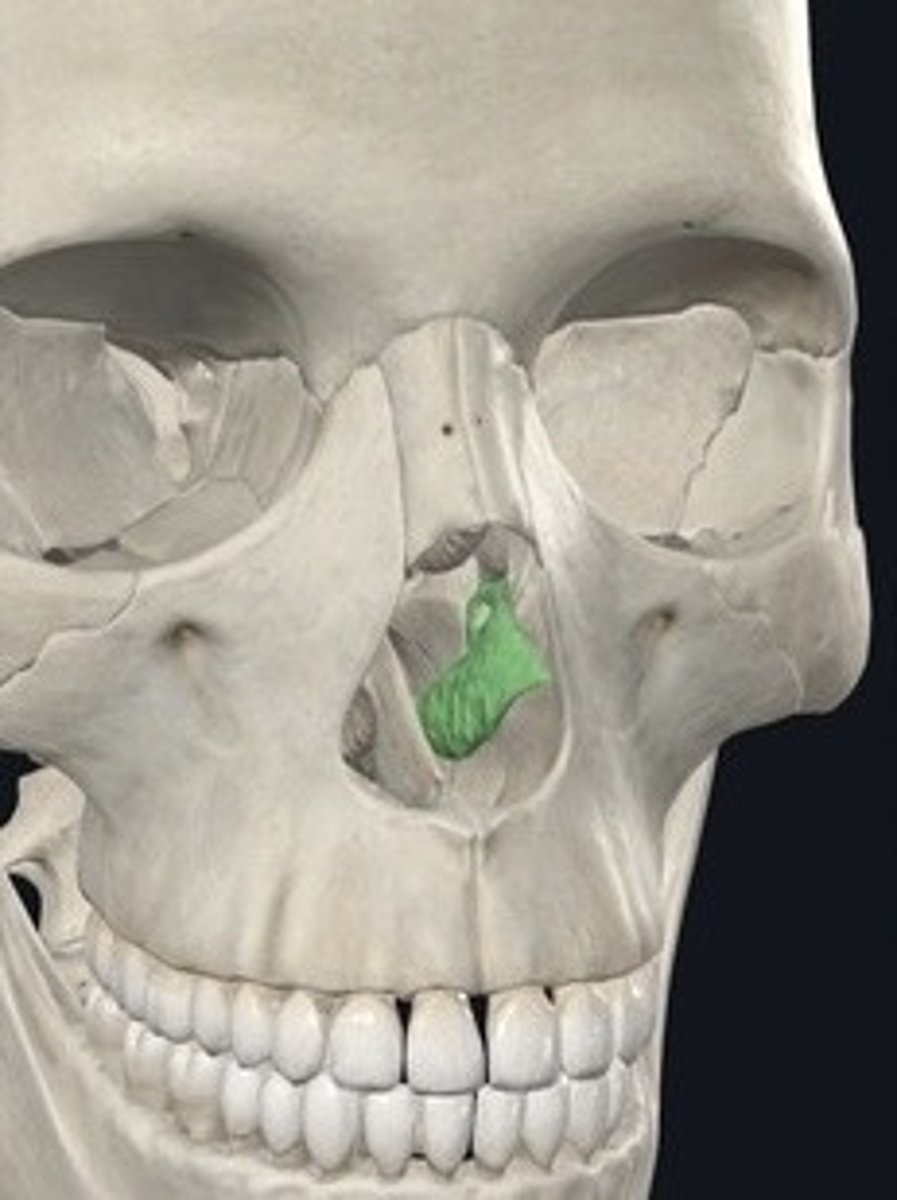
Inferior nasal conchae
Thin bones forming part of the nasal cavity.
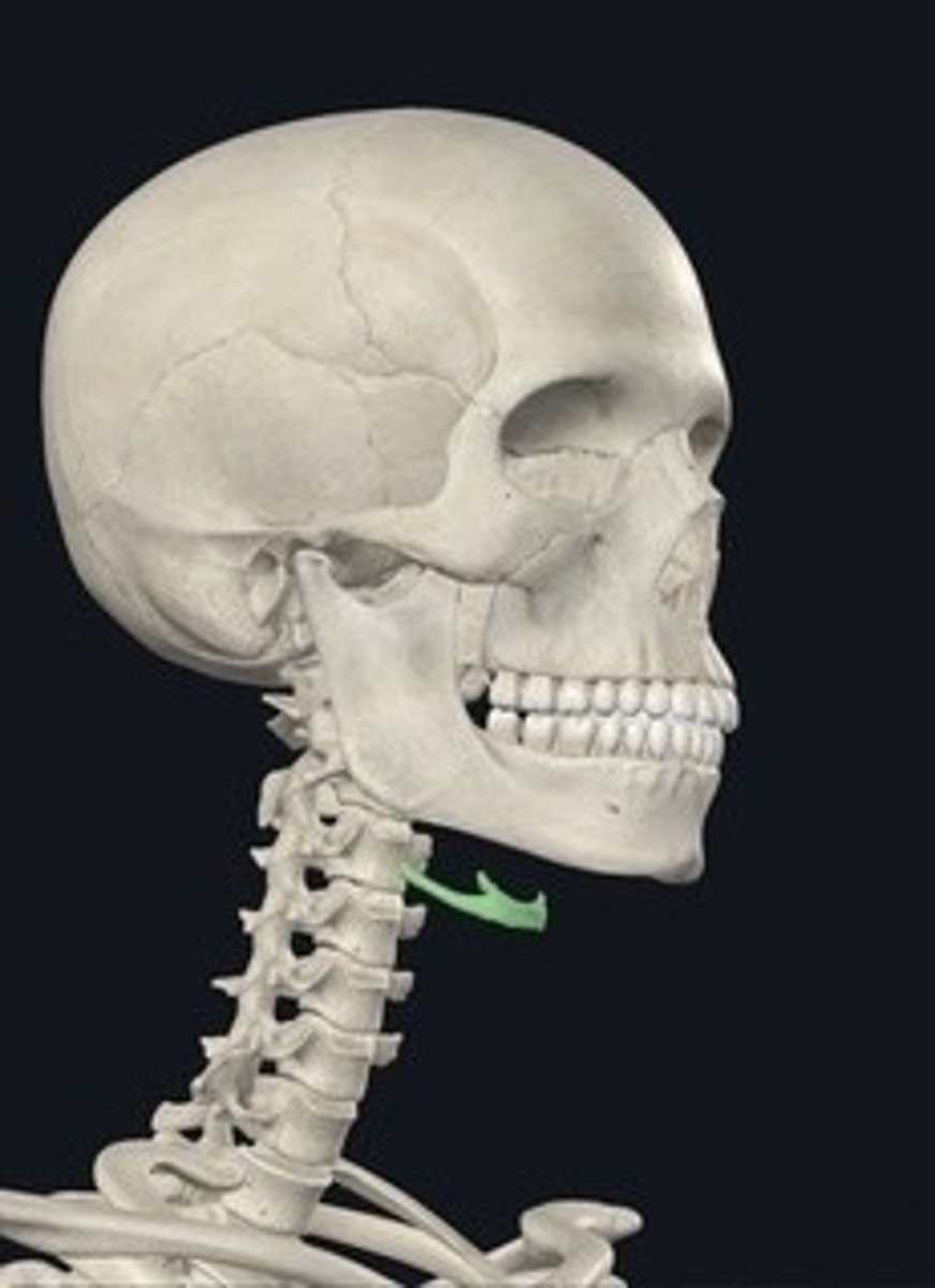
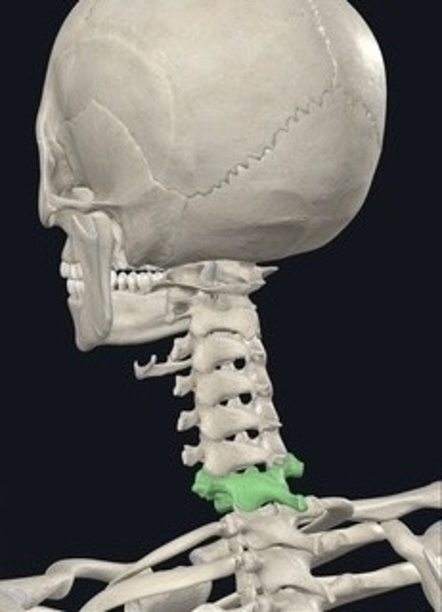
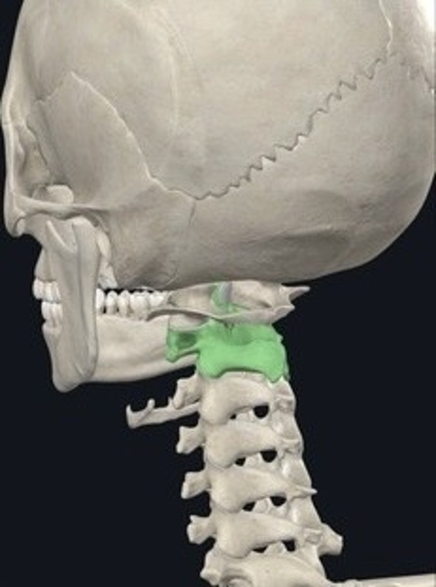
Hyoid
U-shaped bone in the neck supporting the tongue.
Vertebral Column
Series of vertebrae forming the spine.
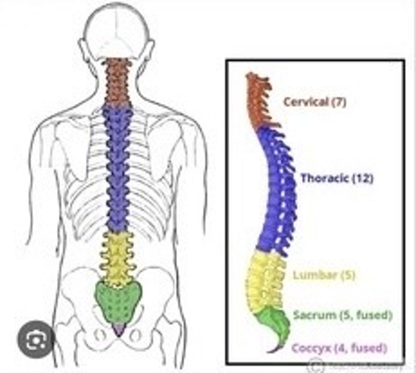
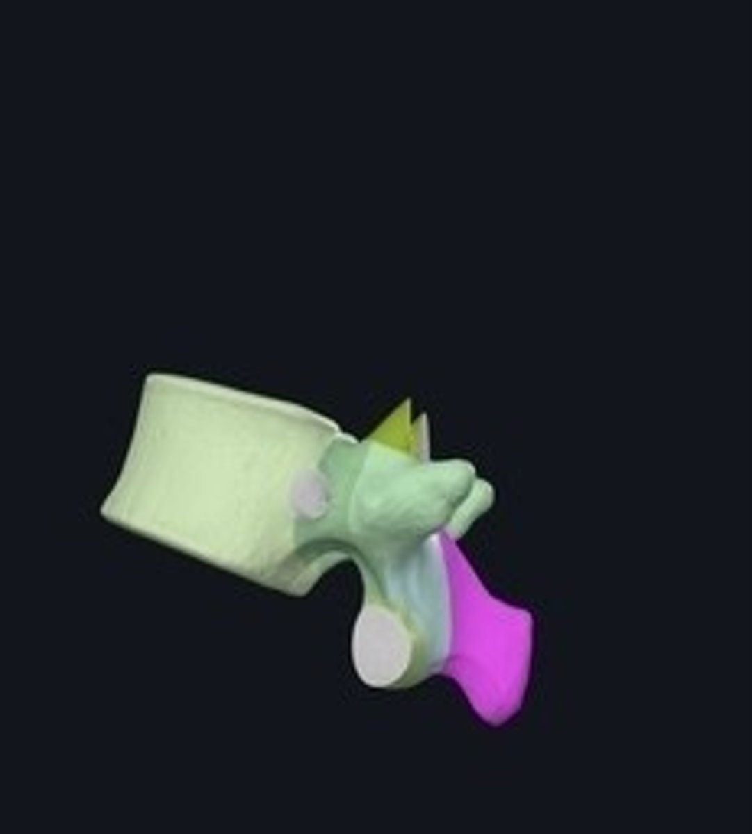
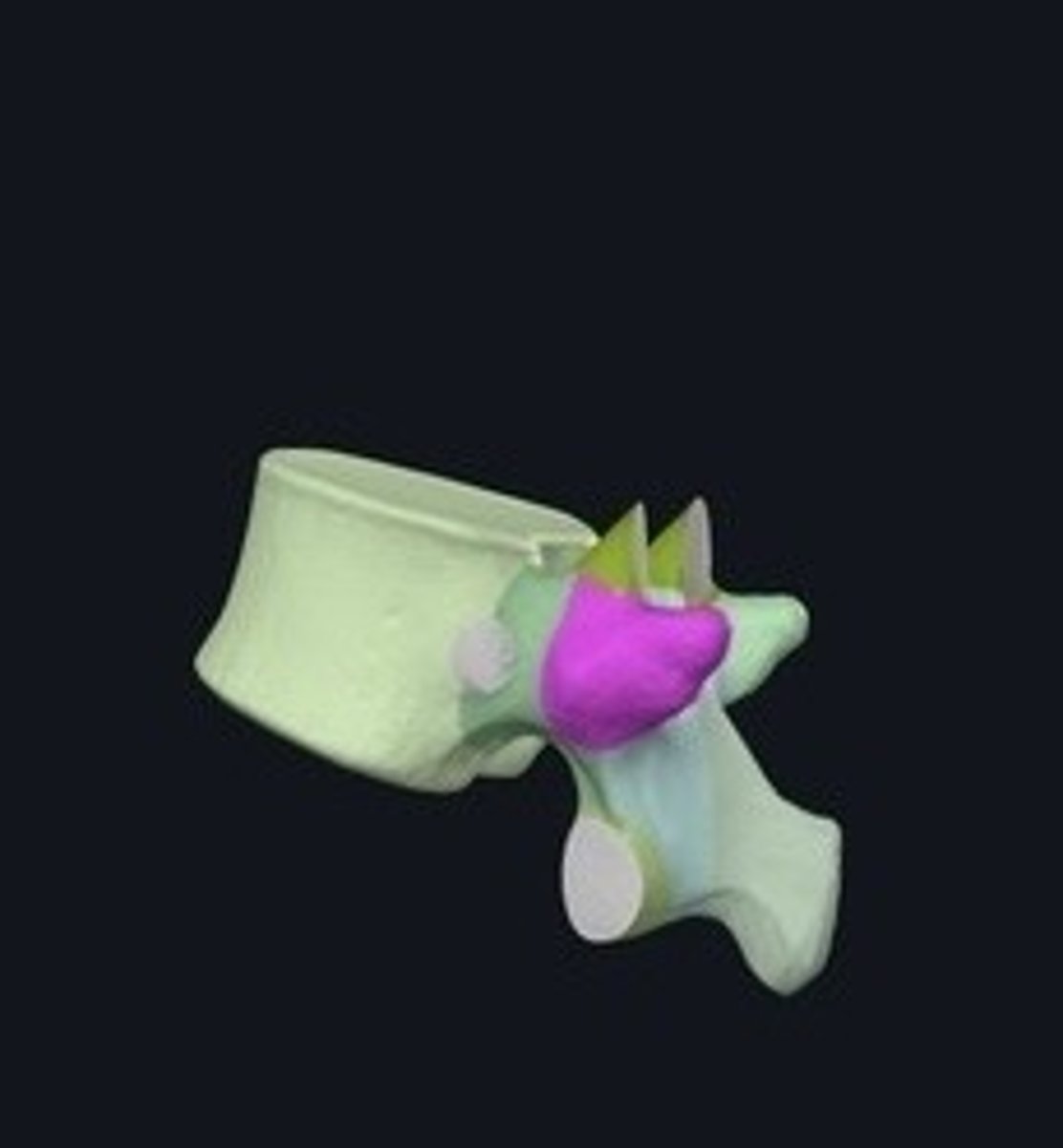
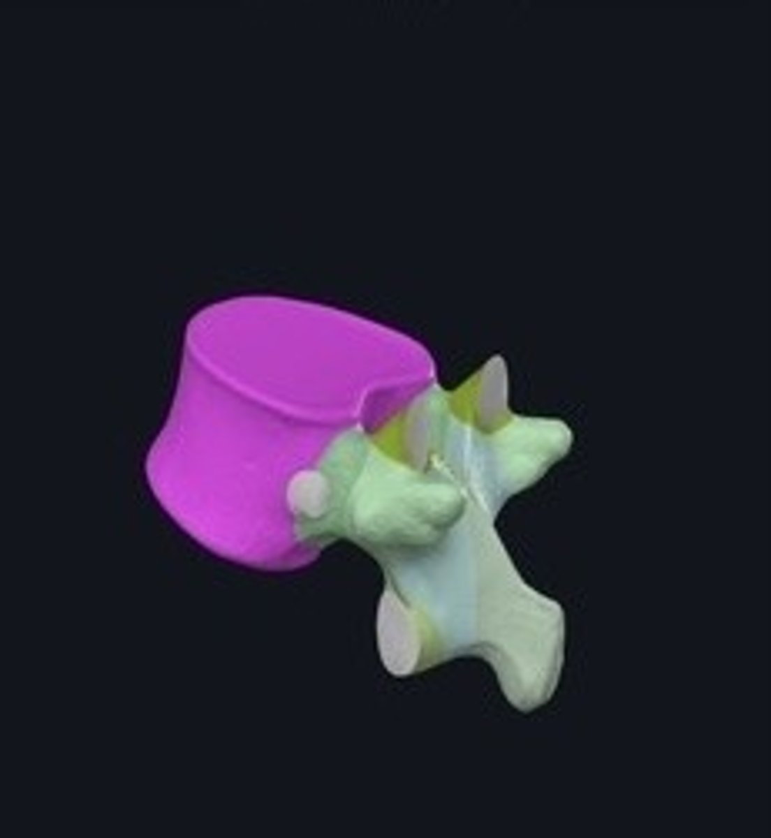
Atlas
First cervical vertebra supporting the skull.
Dens
Projection on the axis allowing head rotation.
Lumbar vertebrae
Five vertebrae in the lower back. 5PM DINNER
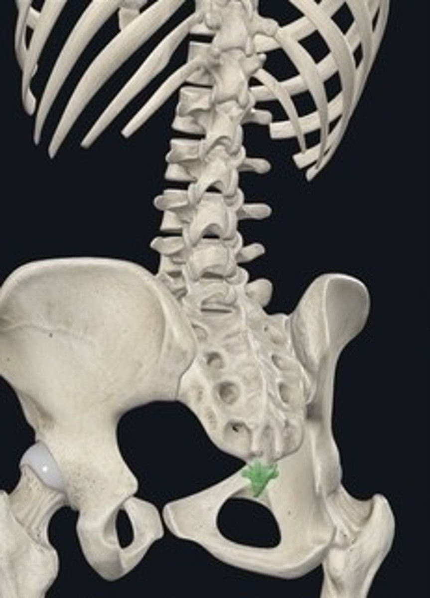
Sacral vertebrae
Five fused vertebrae forming the sacrum.
Cervical vertebrae
Seven vertebrae in the neck region. 7AM BREAKFAST
Coccyx vertebrae
Four fused vertebrae forming the tailbone.
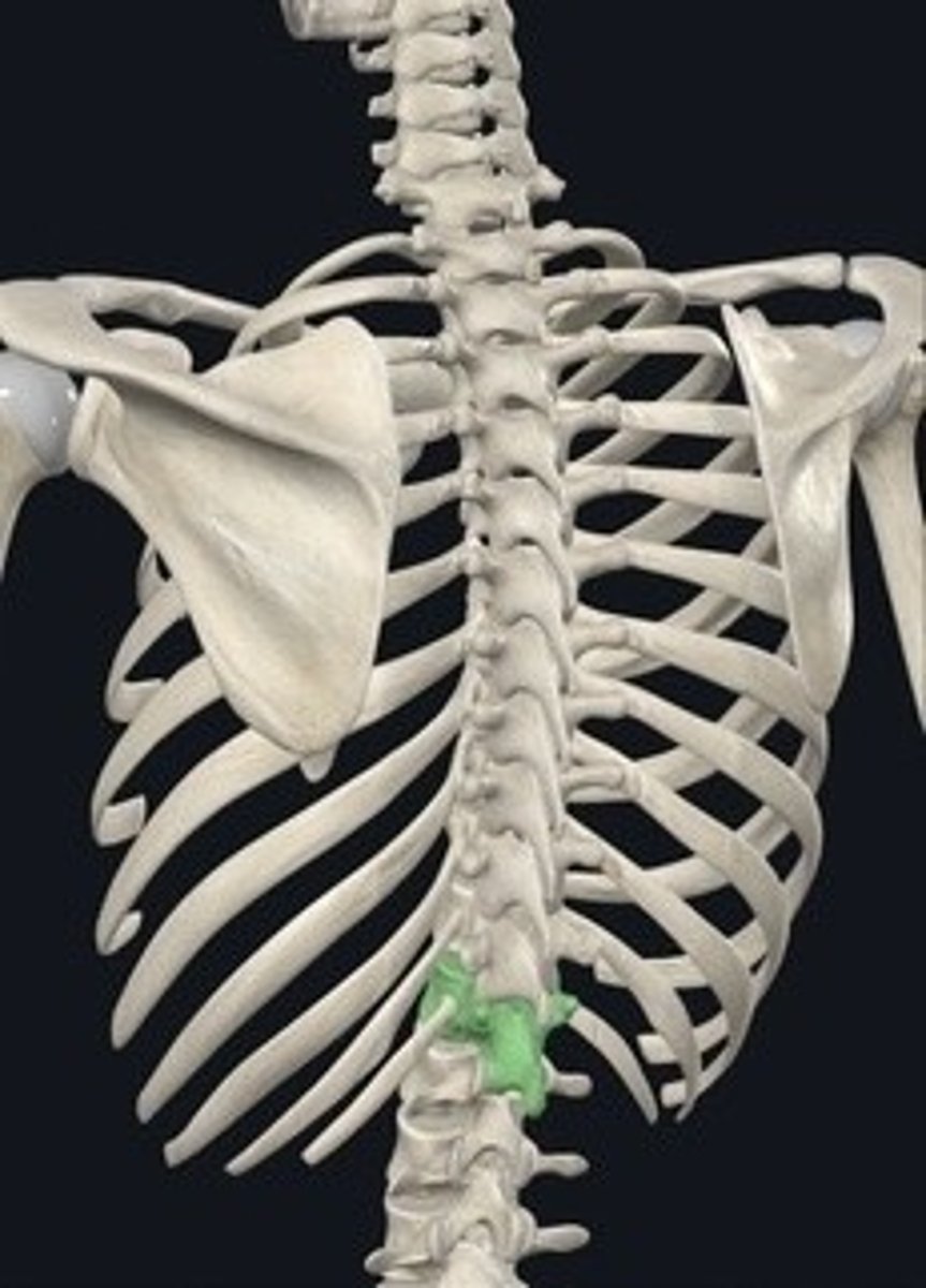
Thoracic vertebrae
Twelve vertebrae in the upper back. 12 NOON LUNCH
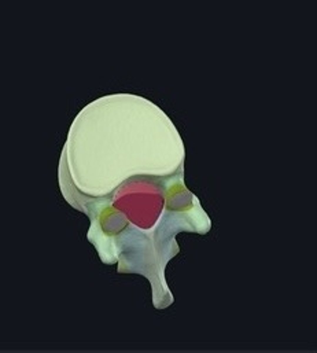
Spinous process
Bony projection on the back of a vertebra.
Transverse process
Lateral projections for muscle attachment on vertebrae.
Vertebral foramen
Opening for spinal cord in vertebrae.
Body
Main part of a vertebra providing support.
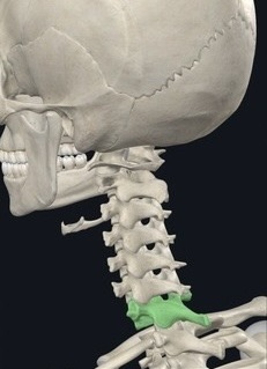
Vertebra prominens
C7 vertebra, prominent at the neck base.
Axis
Second cervical vertebra allowing head rotation.
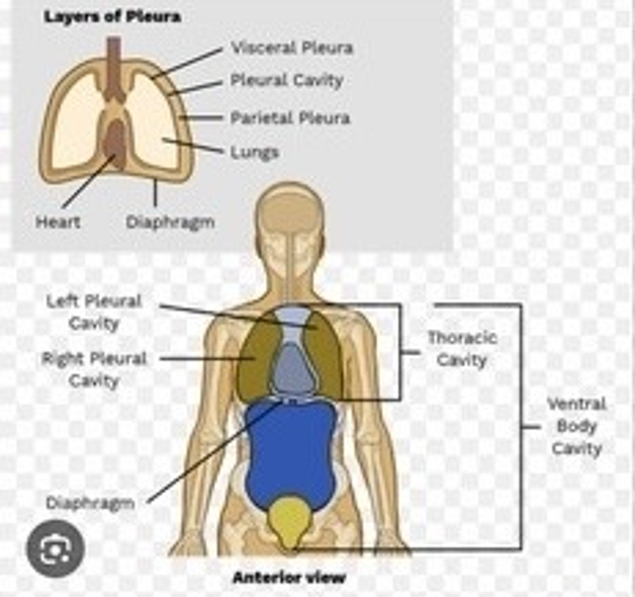
Thoracic cavity
Space housing the heart and lungs.
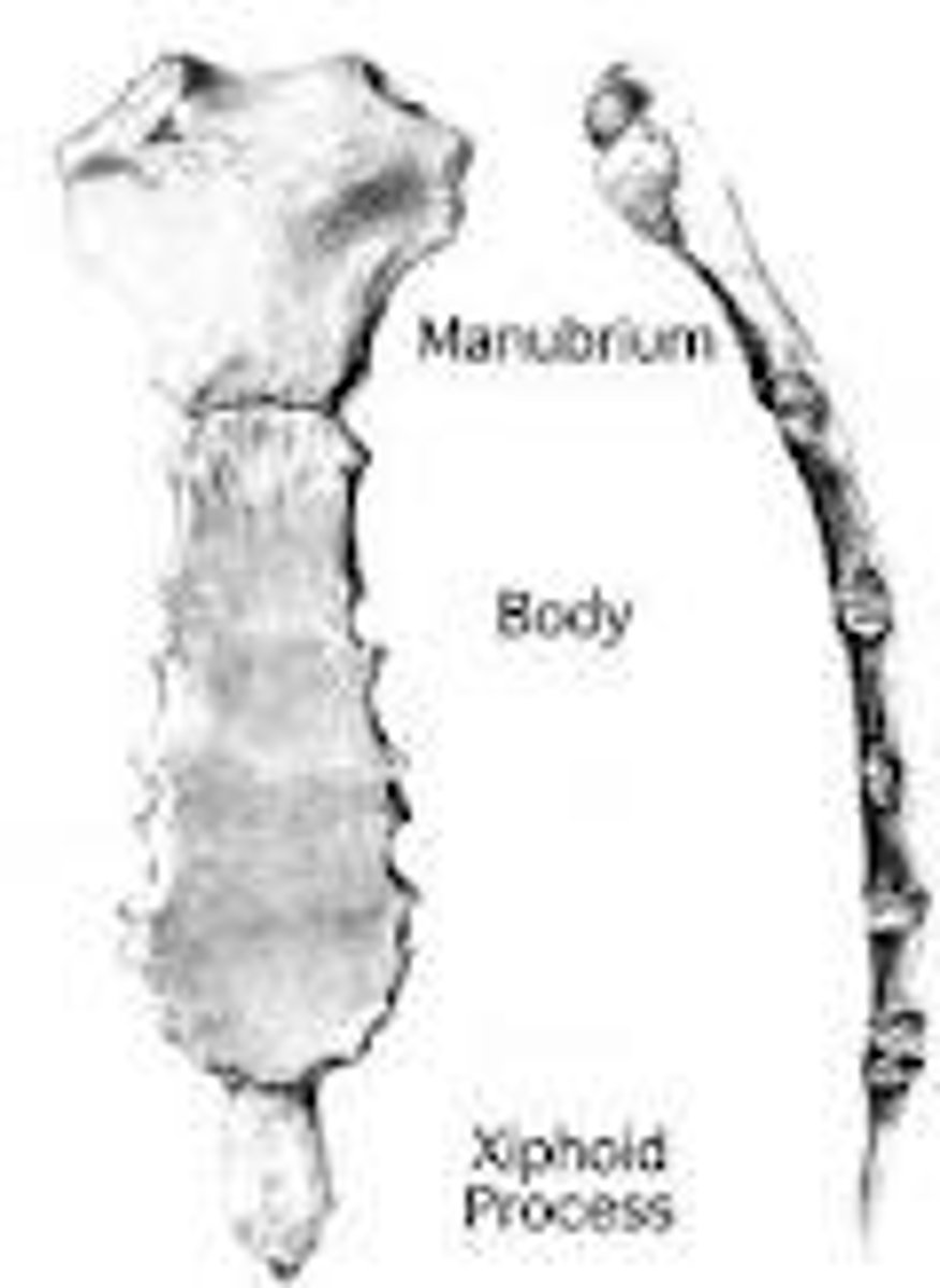
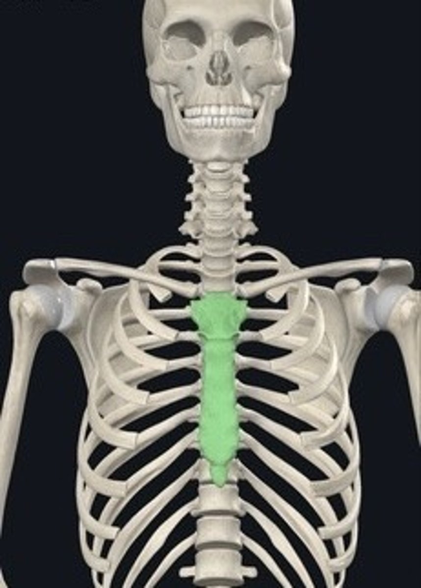
Sternum
Breastbone; consists of manubrium, body, xiphoid.
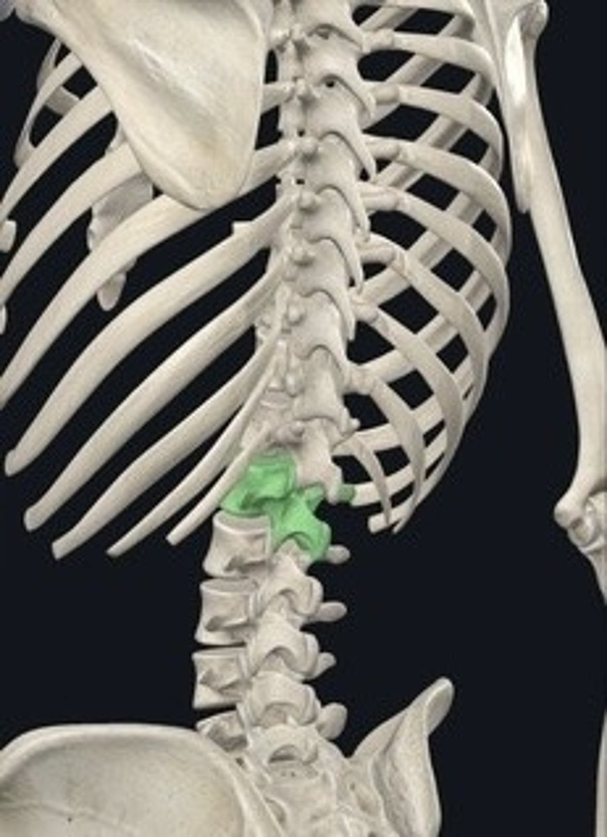
Articulation facets for ribs
Surfaces on vertebrae for rib attachment.
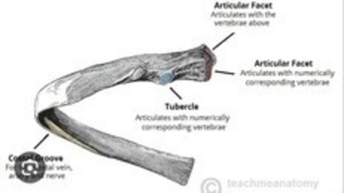
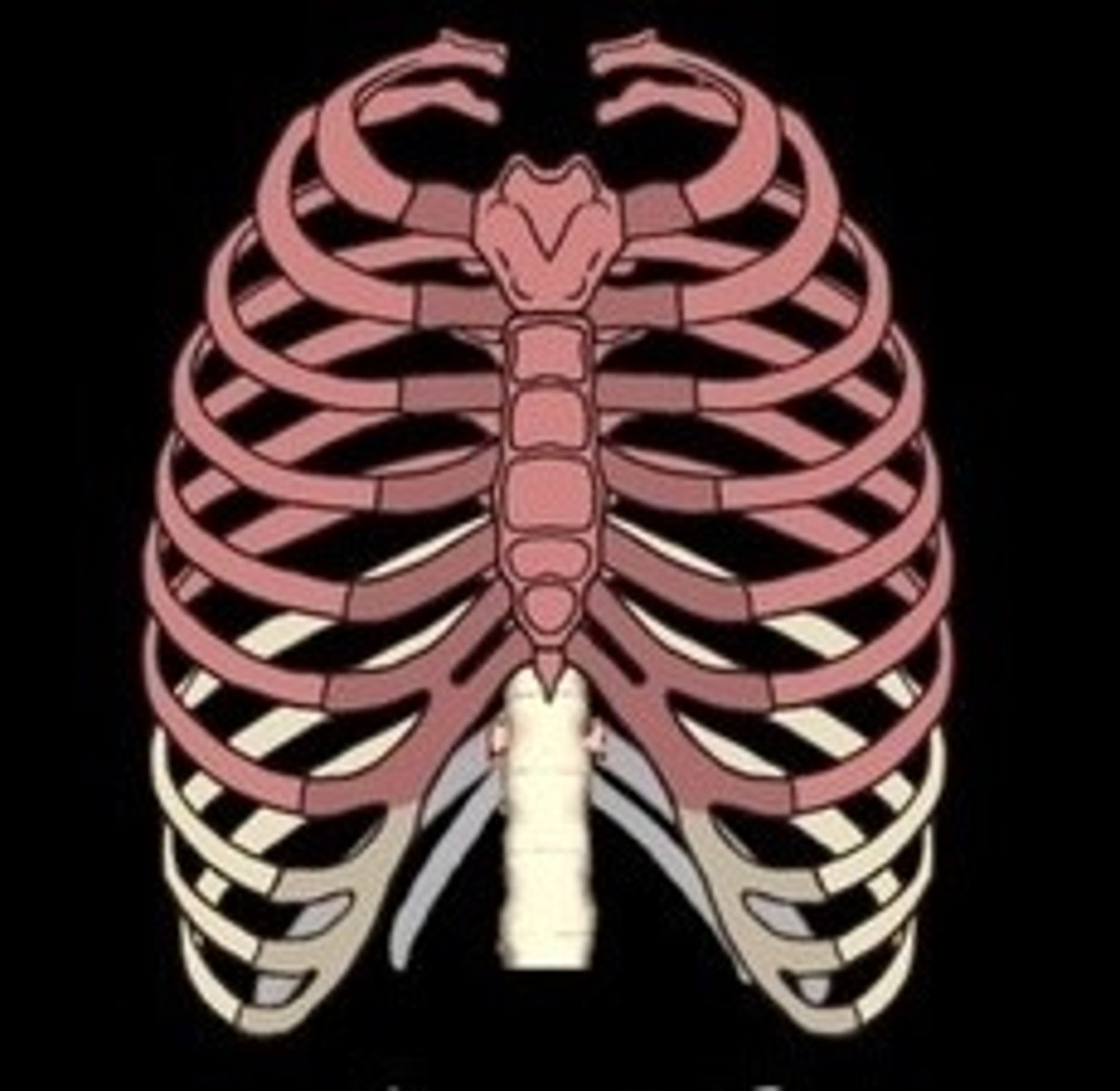
True ribs
First seven pairs of ribs directly attached to sternum.
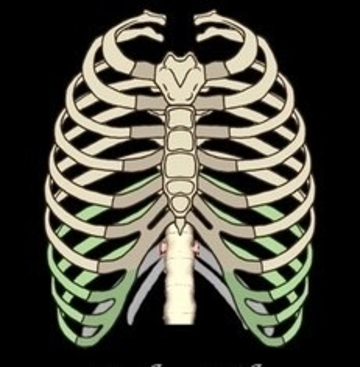
False ribs
Ribs 8-10, indirectly attached to sternum.
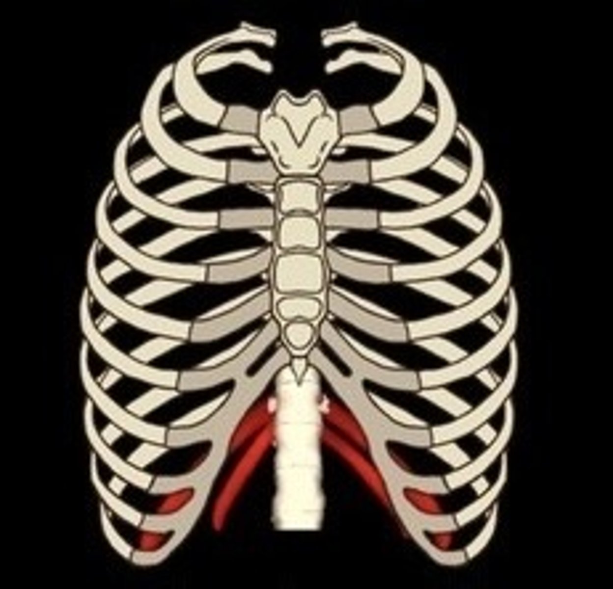
Floating ribs
Ribs 11-12, not attached to sternum.
Clavicle
Collarbone connecting arm to body.
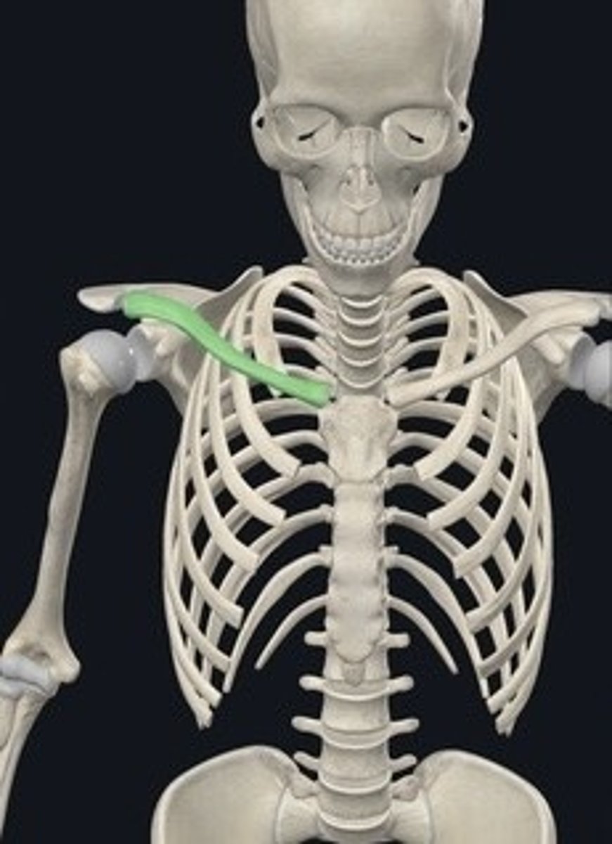
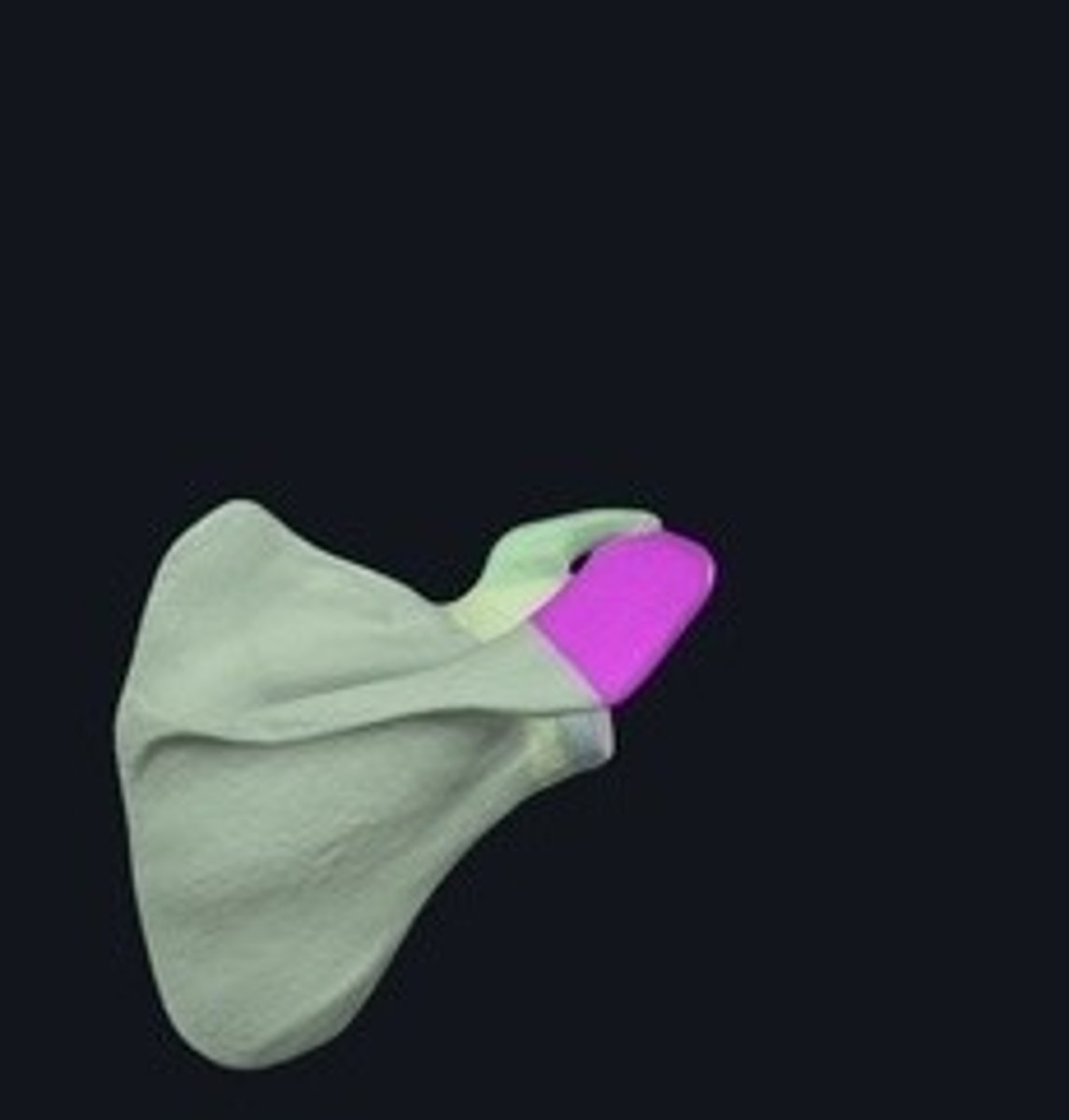
Scapula
Shoulder blade; connects humerus to clavicle.
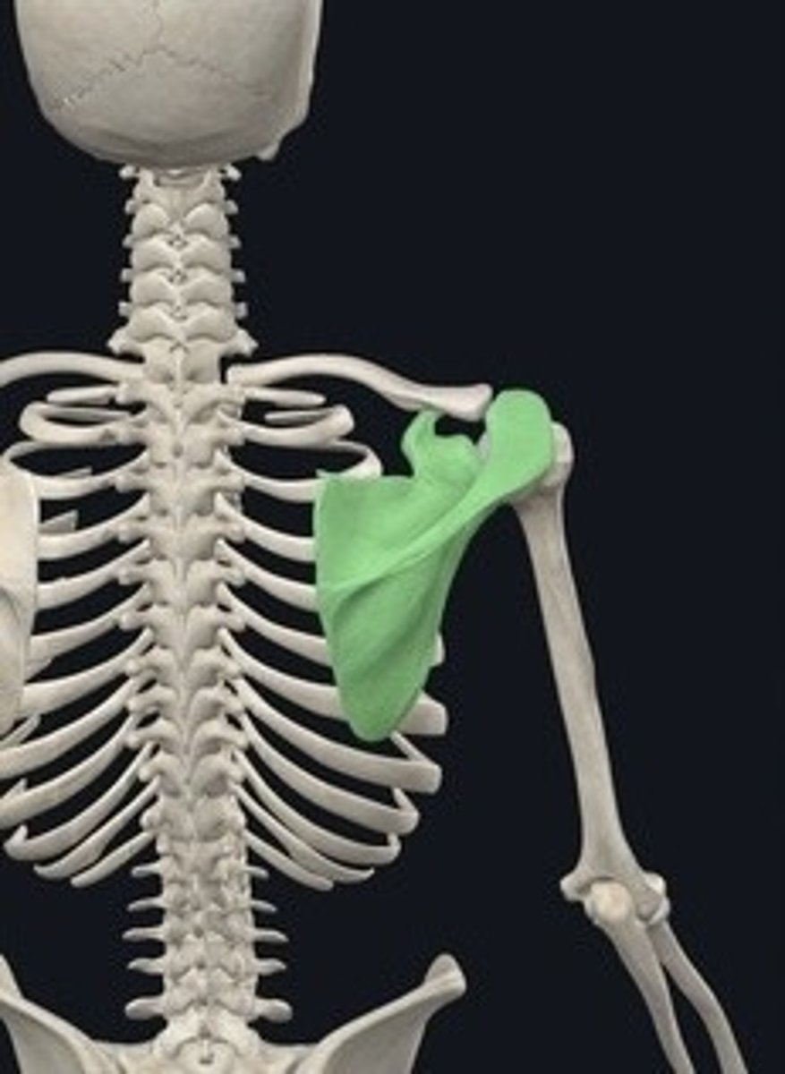
Superior angle
Topmost point of the scapula.
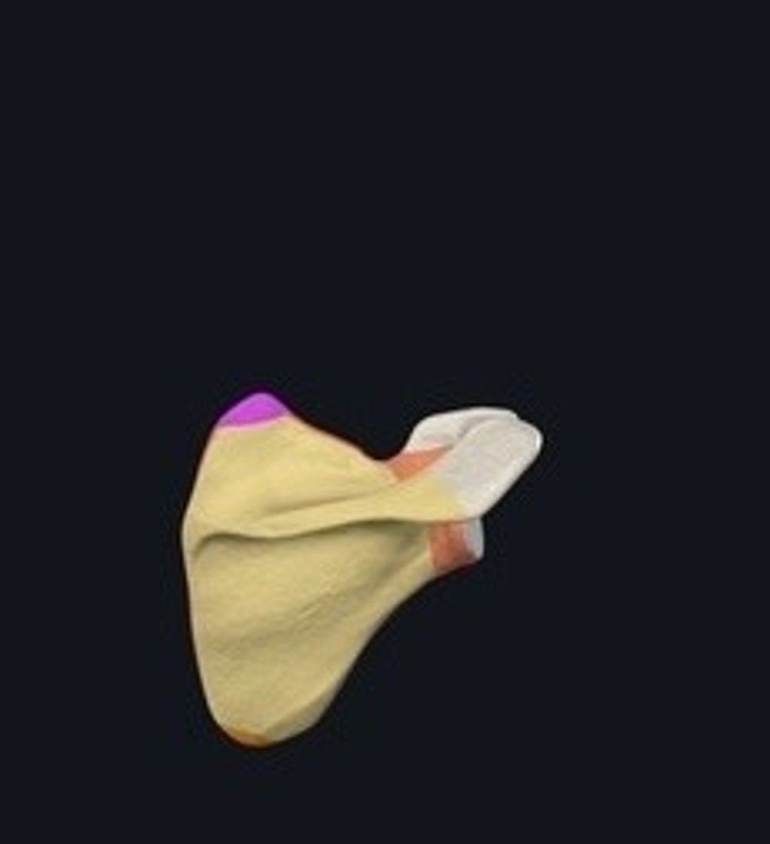
Inferior angle
Lowest point of the scapula.
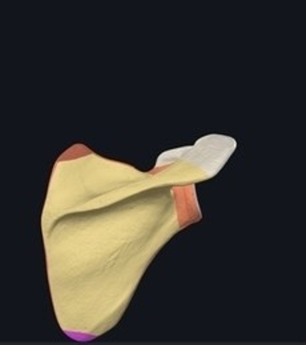
Medial border
Inner edge of the scapula.
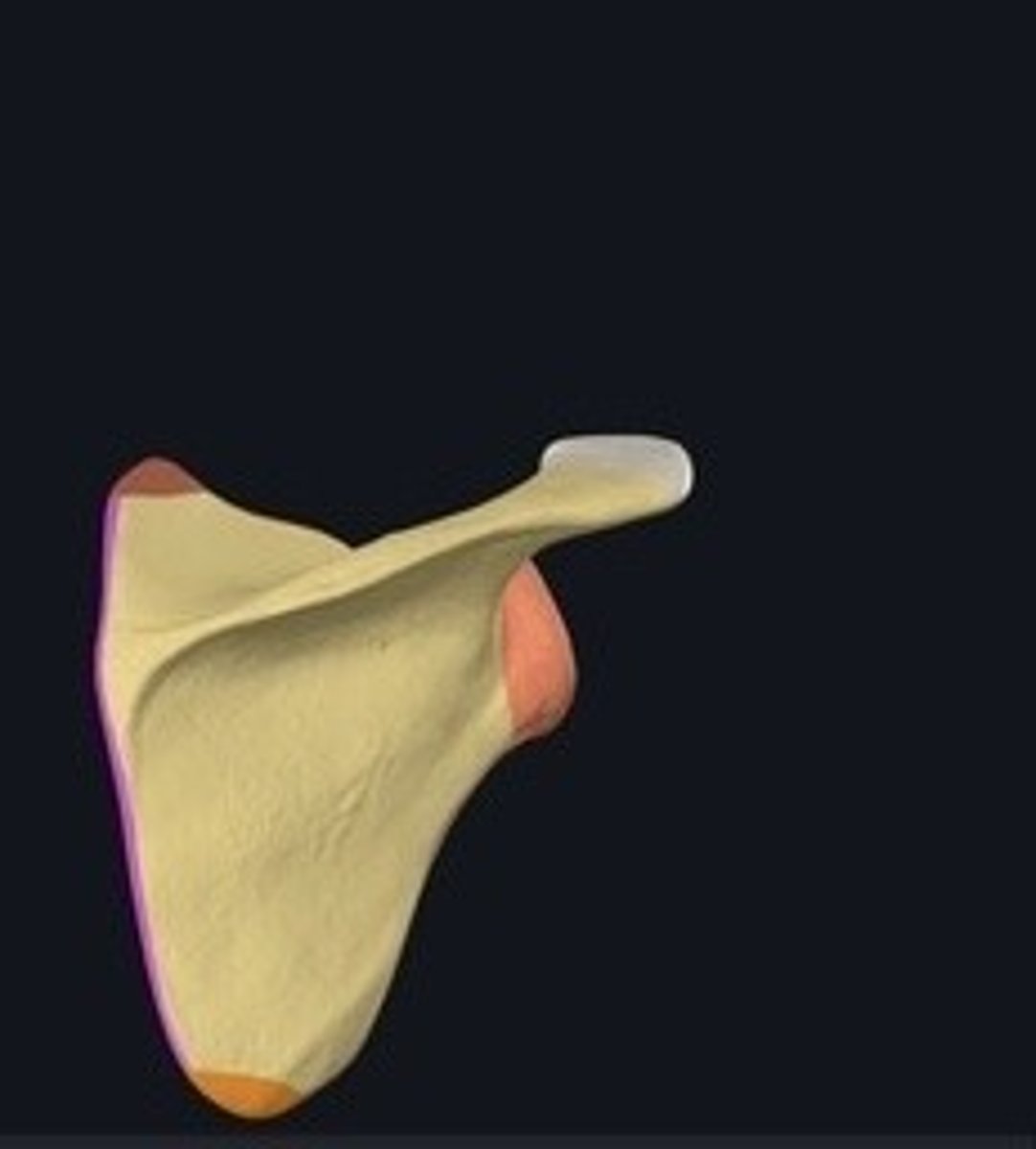
Acromion
Bony process on the scapula's superior aspect.
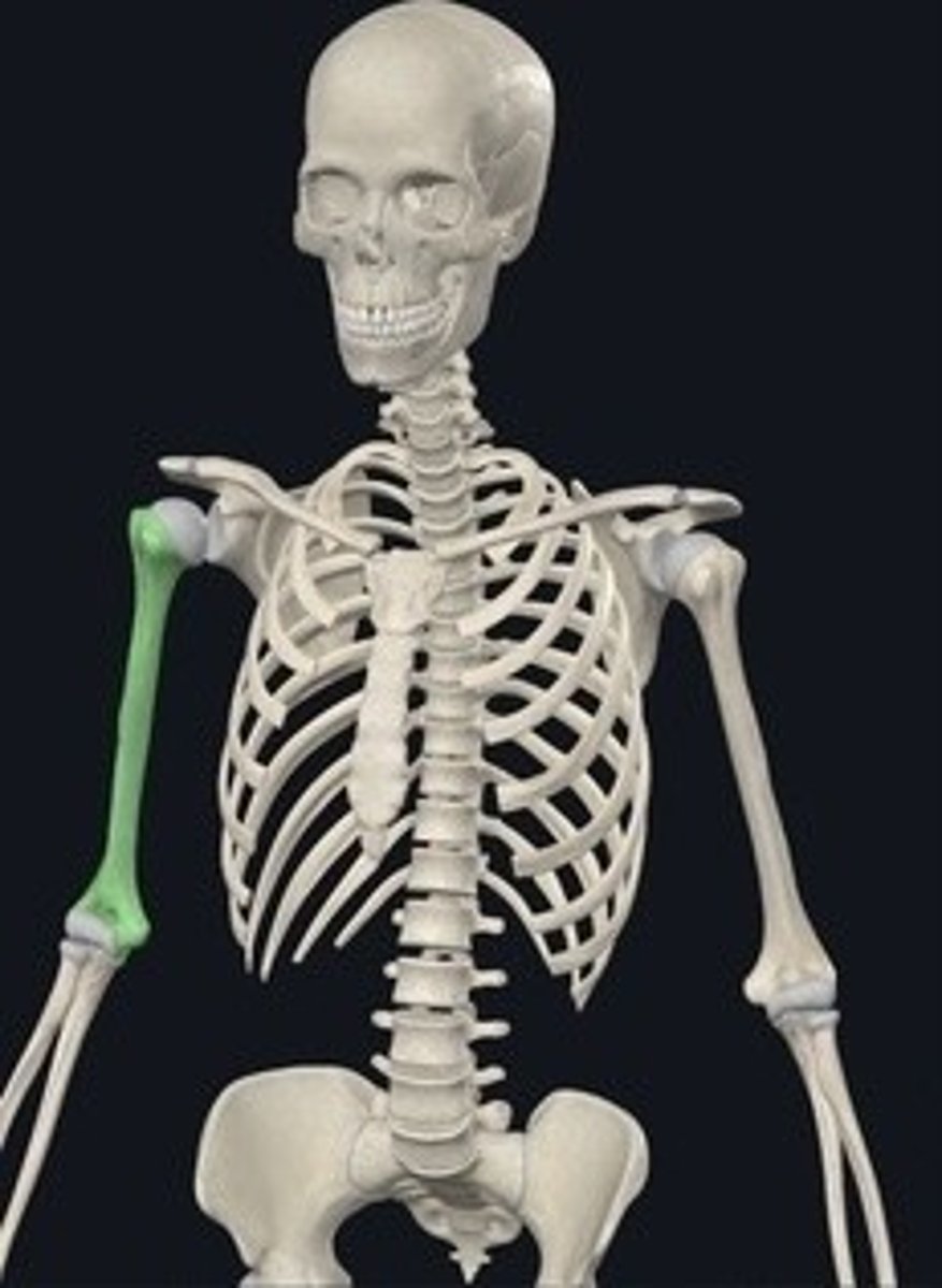
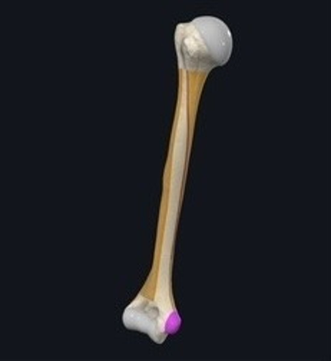
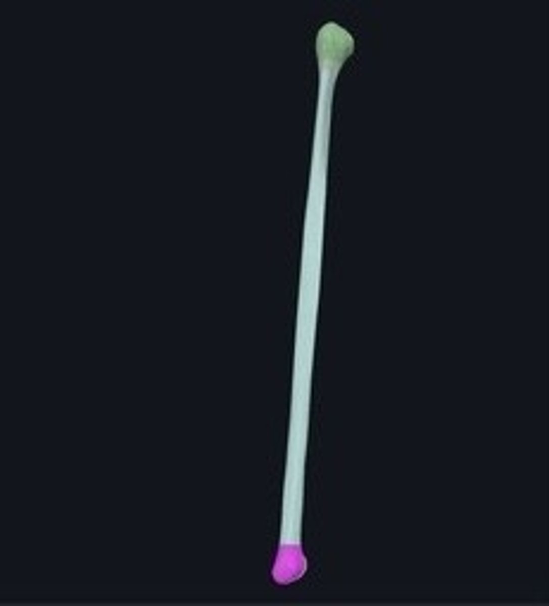
Humerus
Upper arm bone connecting shoulder to elbow.
Greater tubercle
Large projection on the humerus for muscle attachment.
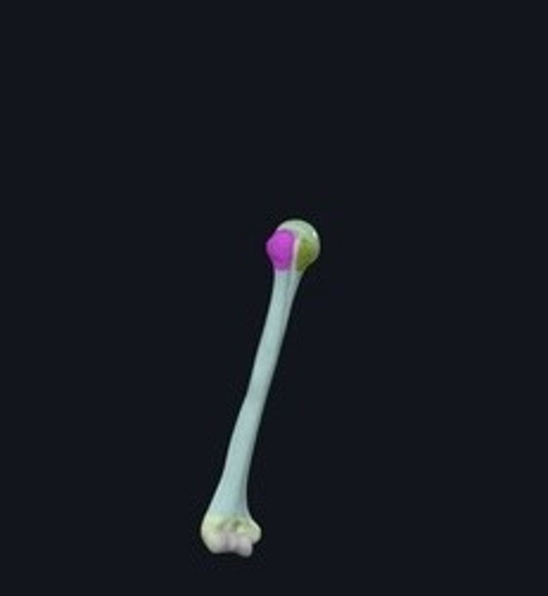
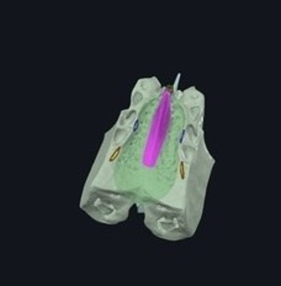
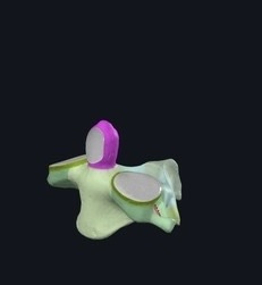
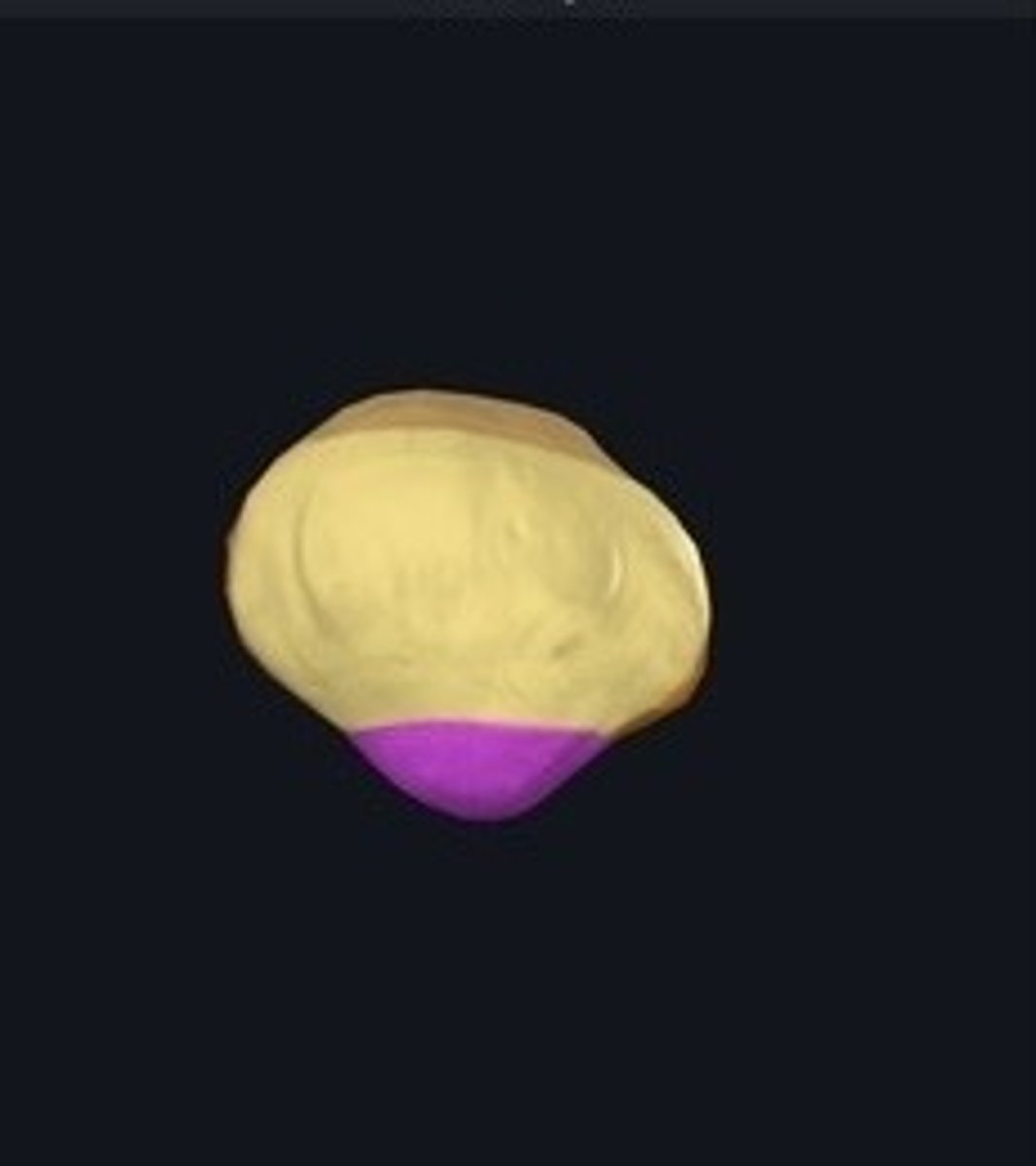
Capitulum
Rounded end of the humerus articulating with the radius. (Green)
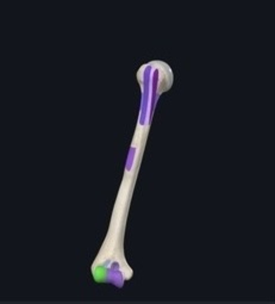
Trochlea
Pulley-shaped structure on the humerus for ulna articulation.
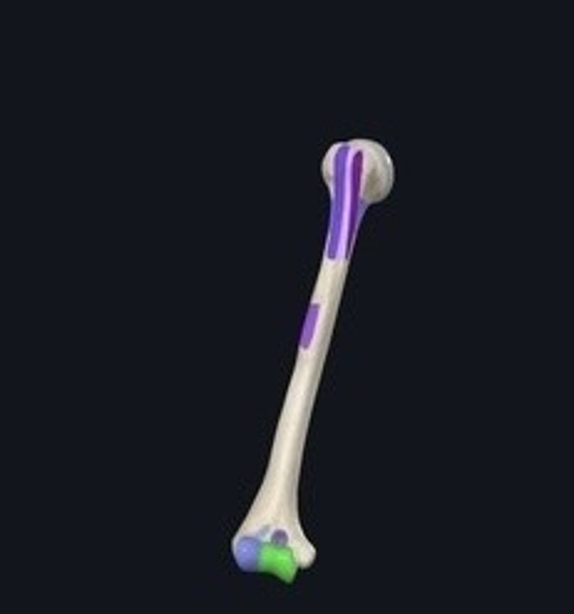
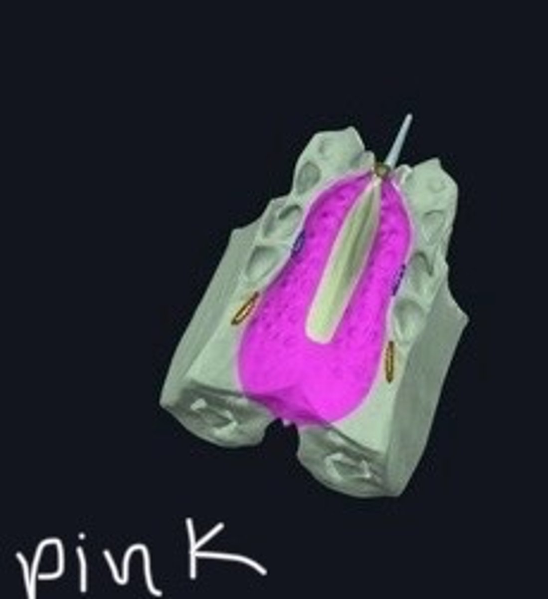
Medial epichonyle
Bony prominence on the humerus for muscle attachment. (Pink)
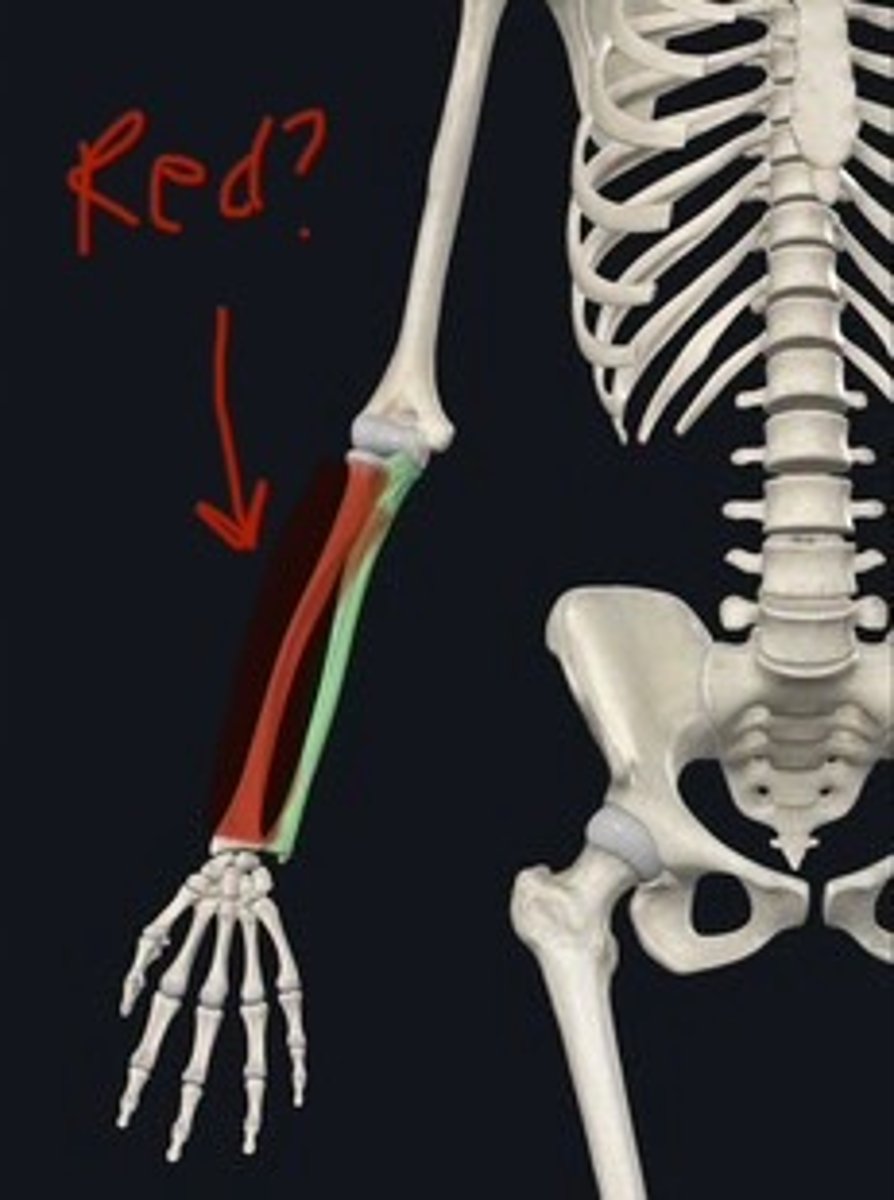
Ulna
Forearm bone on the inner side.
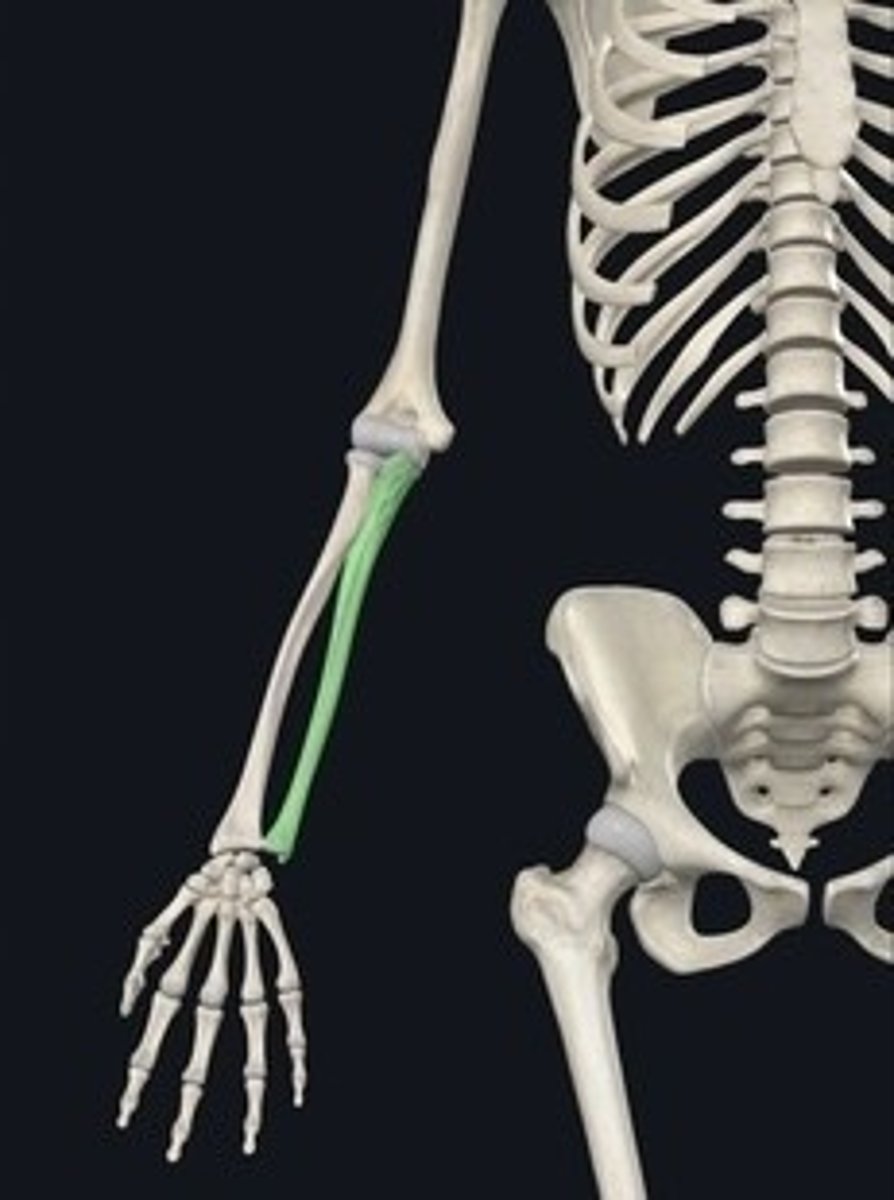
Radius
Forearm bone on the outer side.
Carpus
Wrist composed of eight small bones.
Scaphoid
Lunate
Triquetrum
Pisiform
Hamate
Capitate
Trapezoid
Trapezium
(So Long To Pinky Here Comes The Thumb)
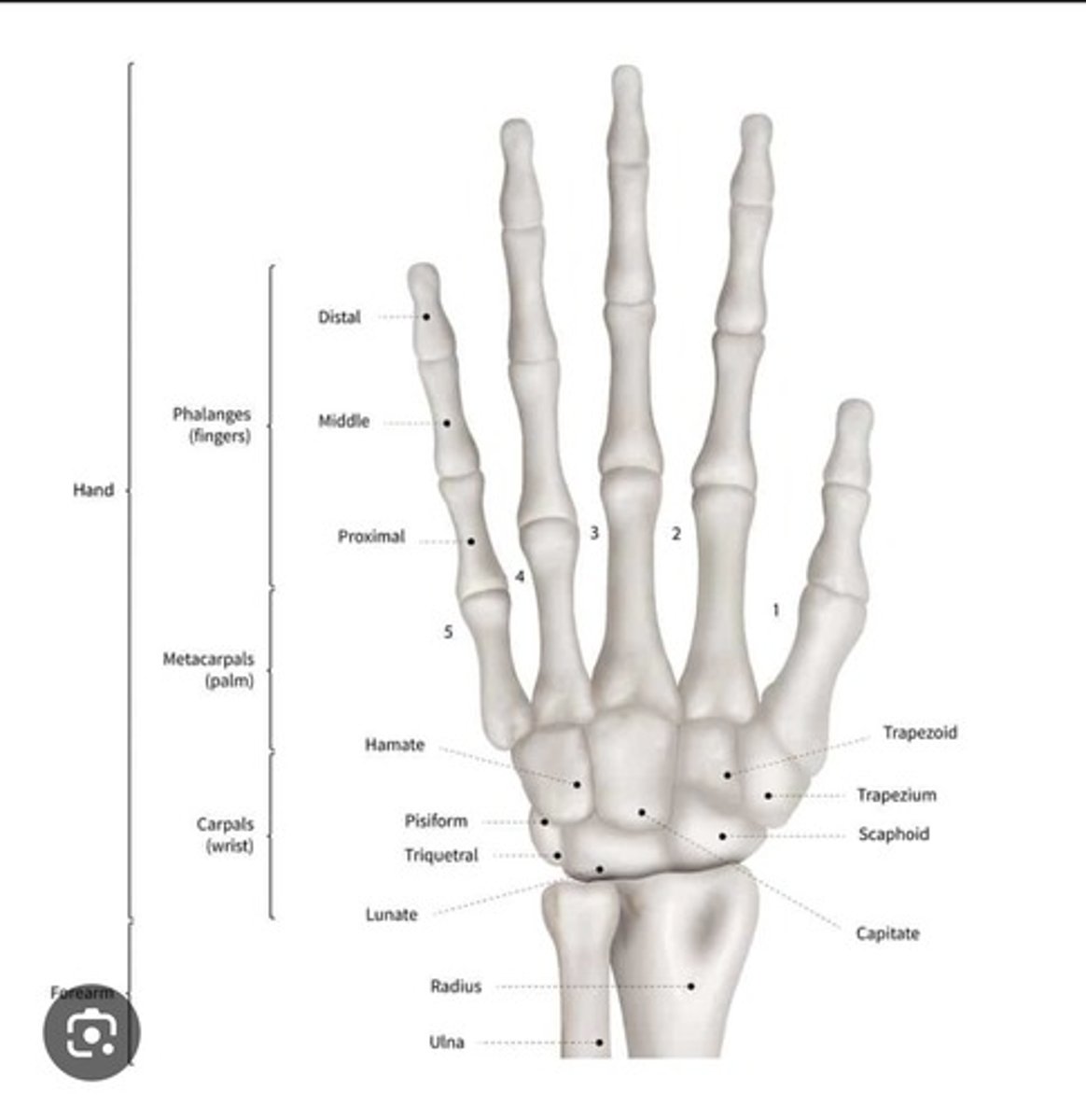
Metacarpals
Five bones forming the hand's palm.
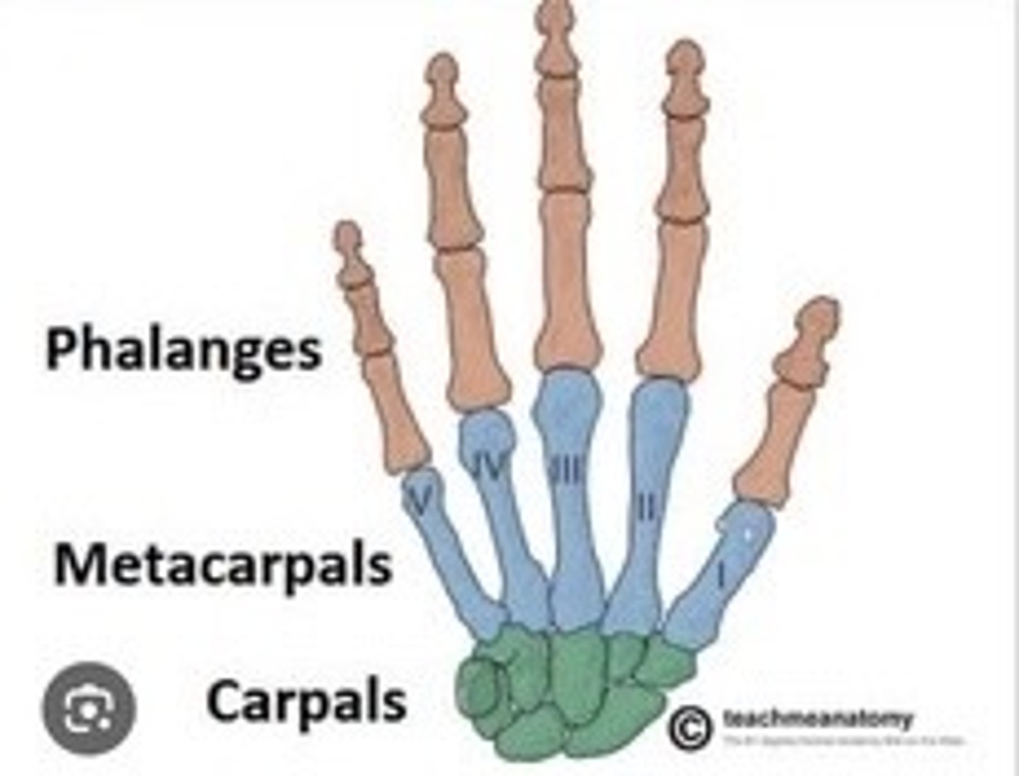
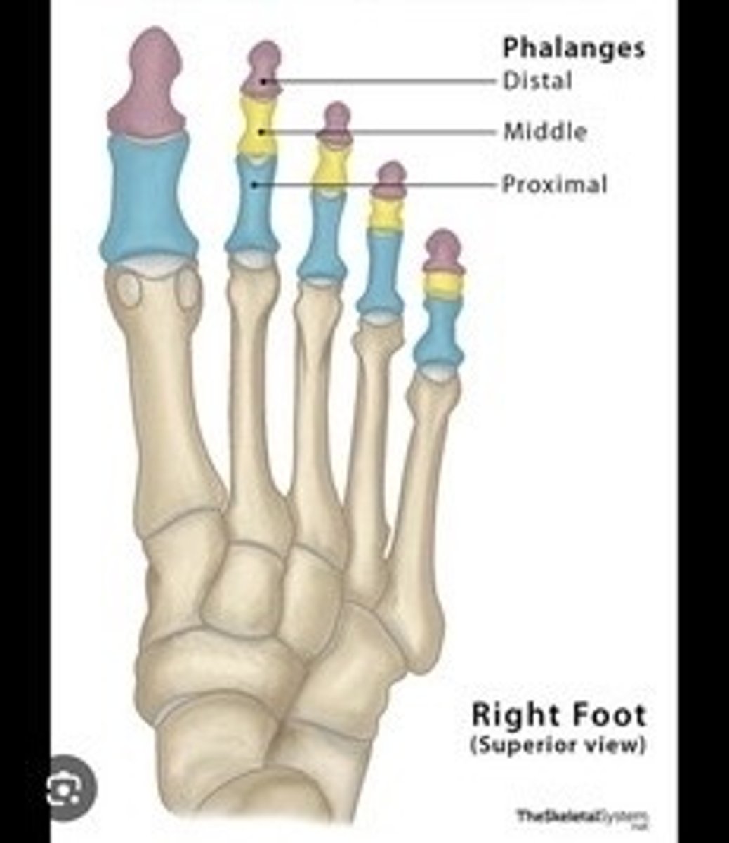
Phalanges
Bones of the fingers; distal, middle, proximal.
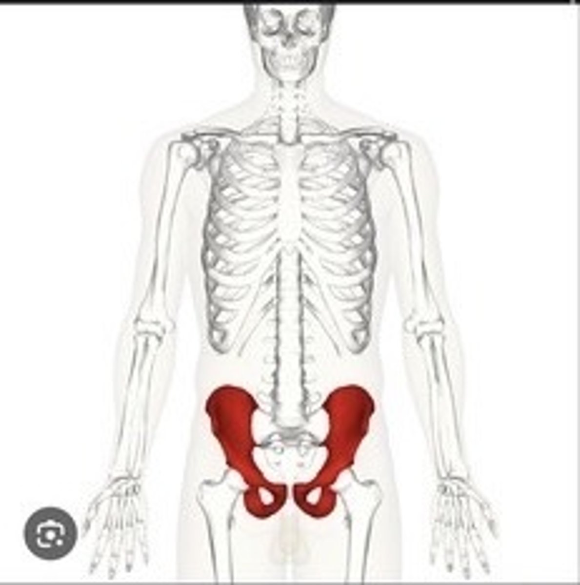
Pelvic Girdle
Structure connecting spine to lower limbs.

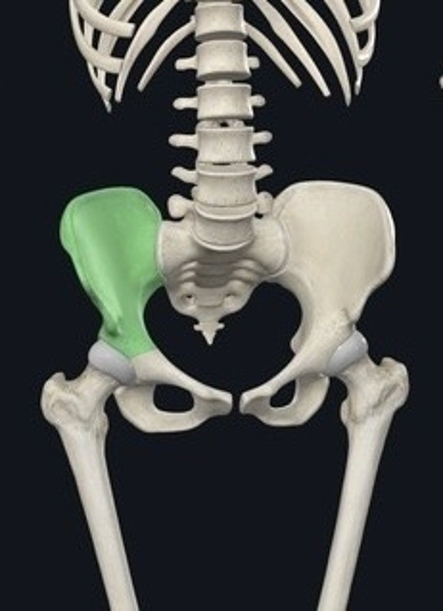
Coxal bones
Hip bones forming the pelvic girdle.
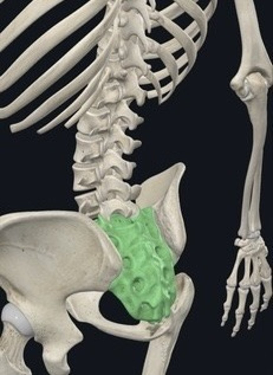
Ilium
Largest part of the coxal bone.
Iliac crest
Top border of the ilium.
Iliac fossa
Concave surface of the ilium.
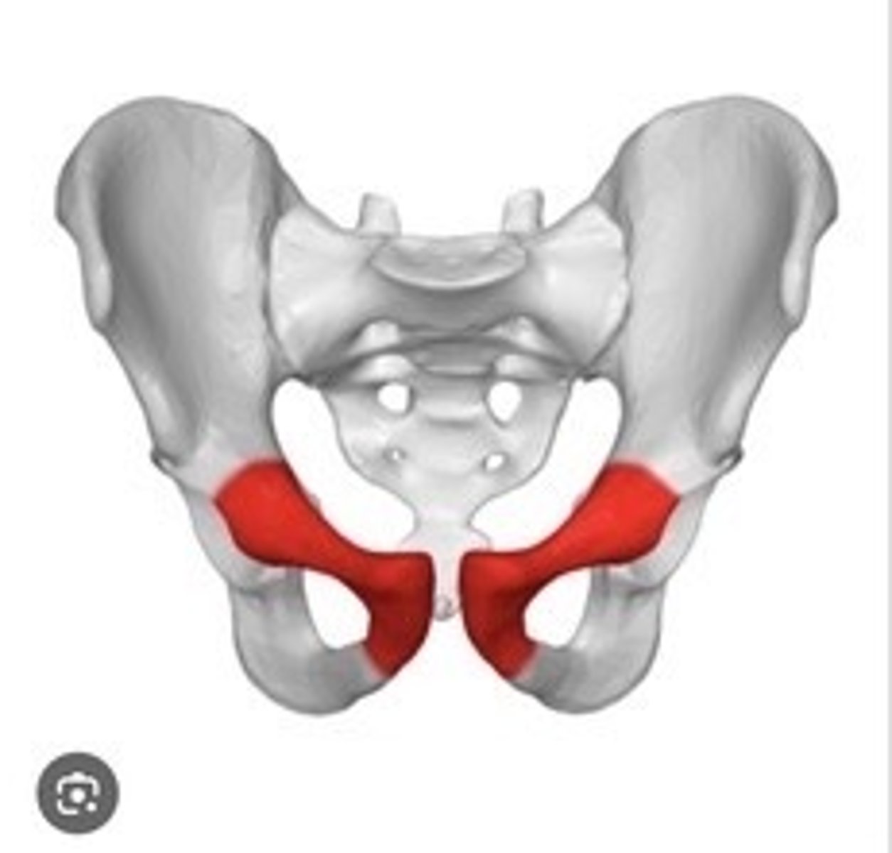
Ischium
Lower part of the coxal bone.
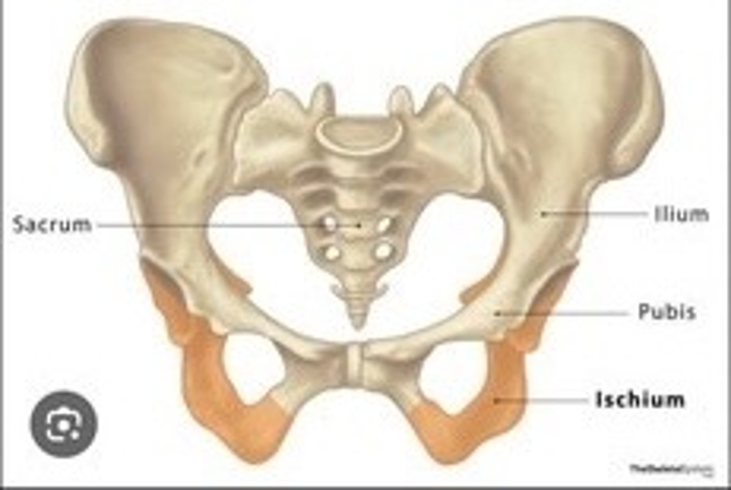
Pubis
anterior part of the coxal bone
Obturator foramen
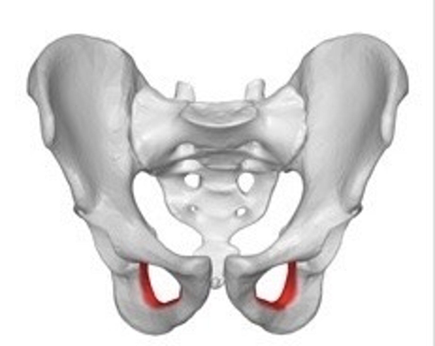
Large opening in the coxal bone.
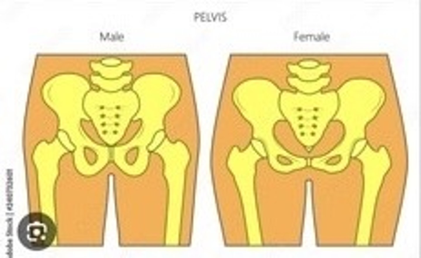
Difference between male and female pelvis
Female pelvis is wider and has a larger inlet.
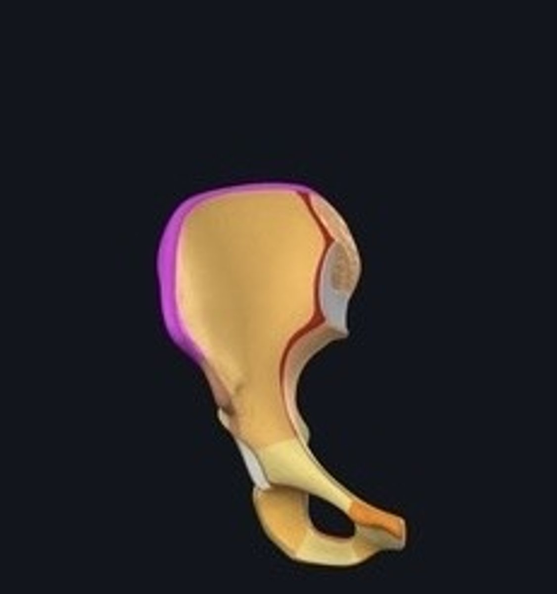
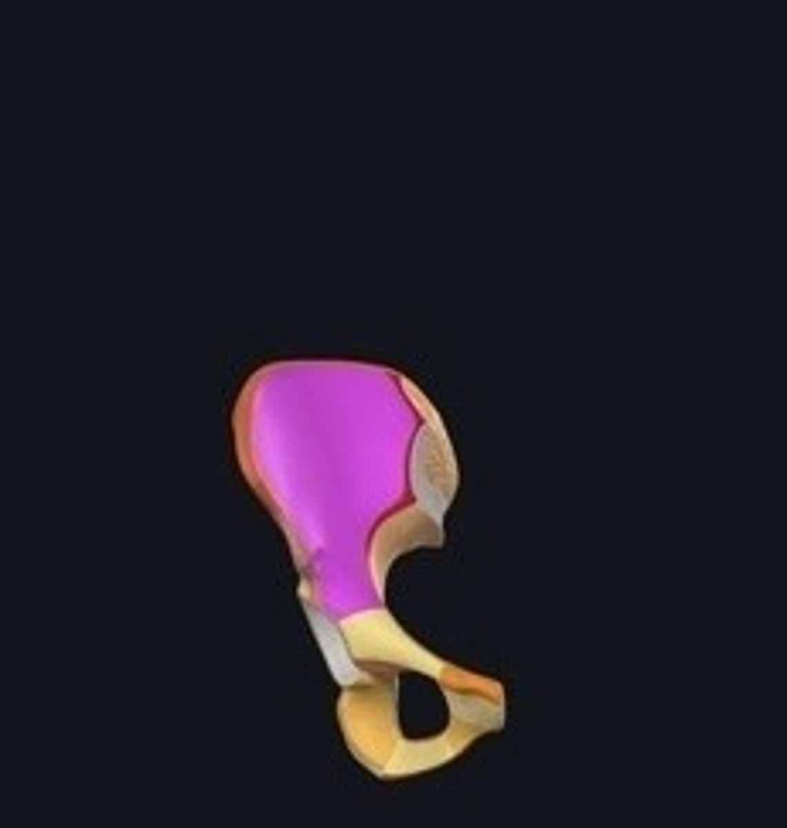
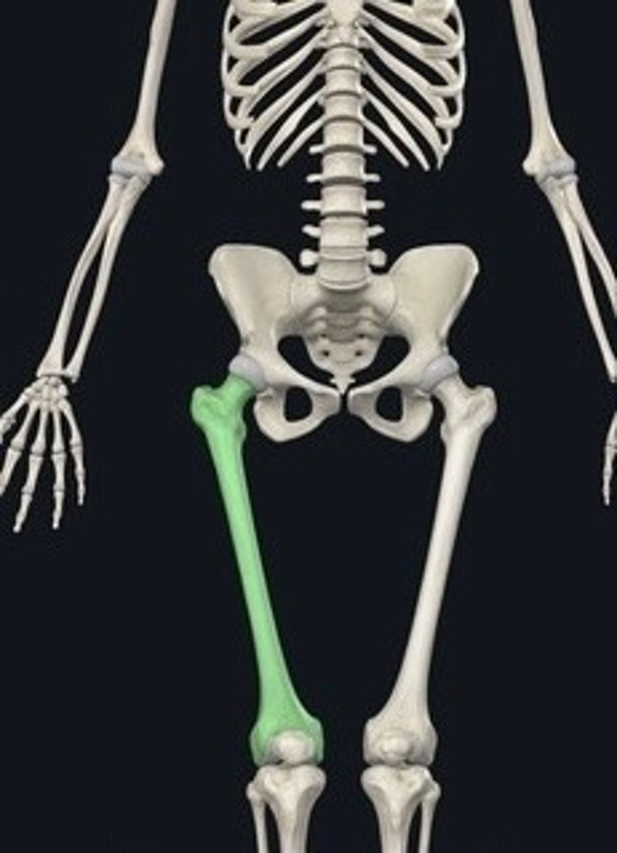
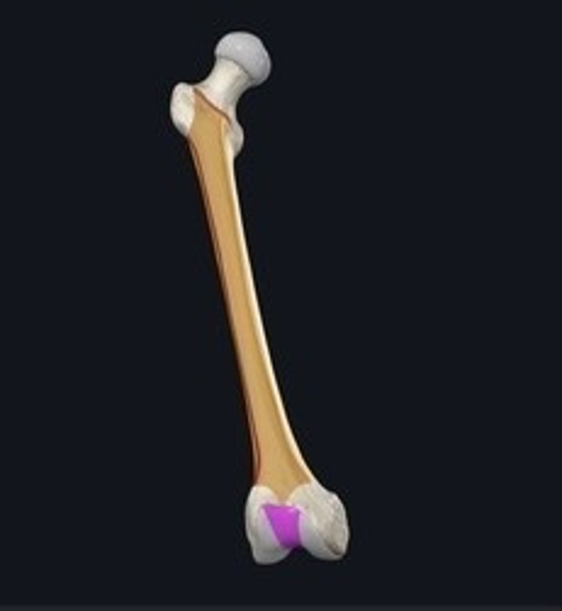
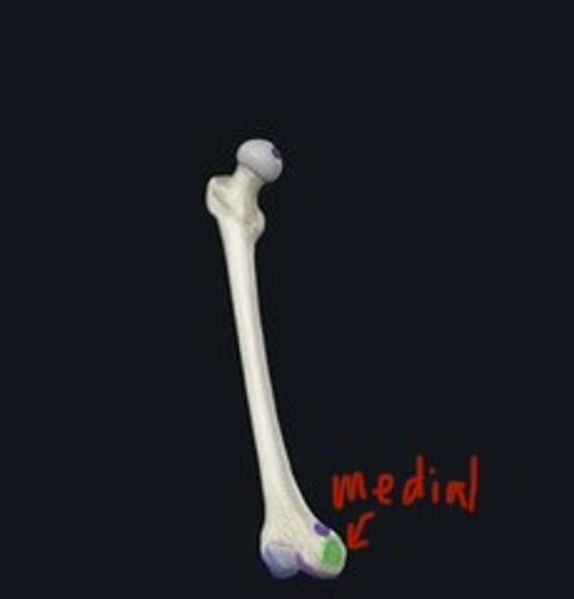
Femur
Thigh bone; longest bone in the body.
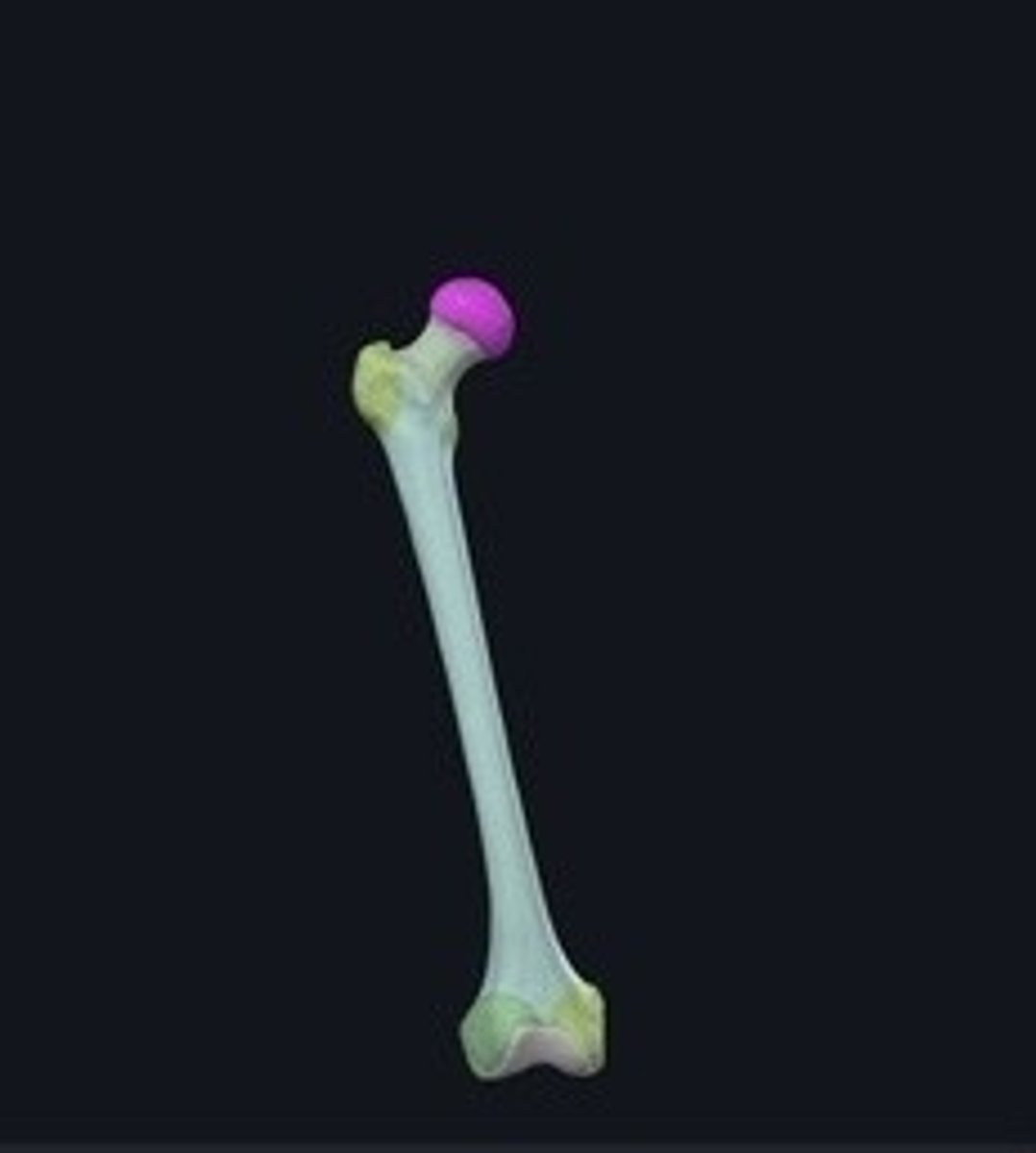
Head
Rounded proximal end of the femur.
Patellar surface
Articulating surface for the patella on femur.
Medial and lateral condyle
Rounded ends of femur for knee joint.
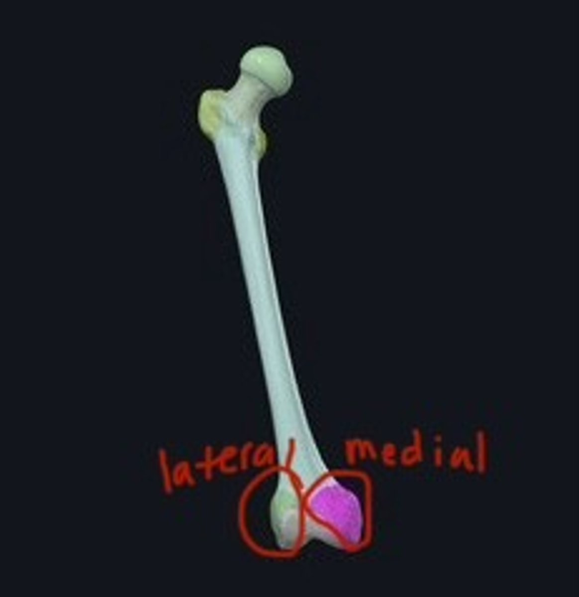
Medial and lateral epicondyle
Bony projections above the condyles on femur.
Patella
Kneecap; protects knee joint.
Apex
Pointed end of the patella.
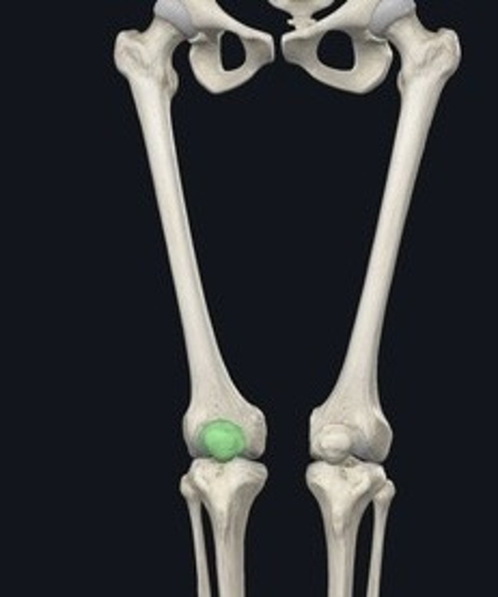
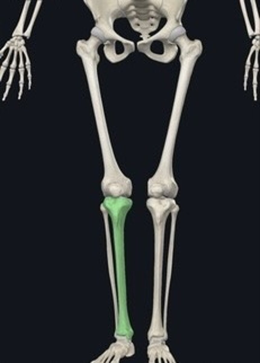
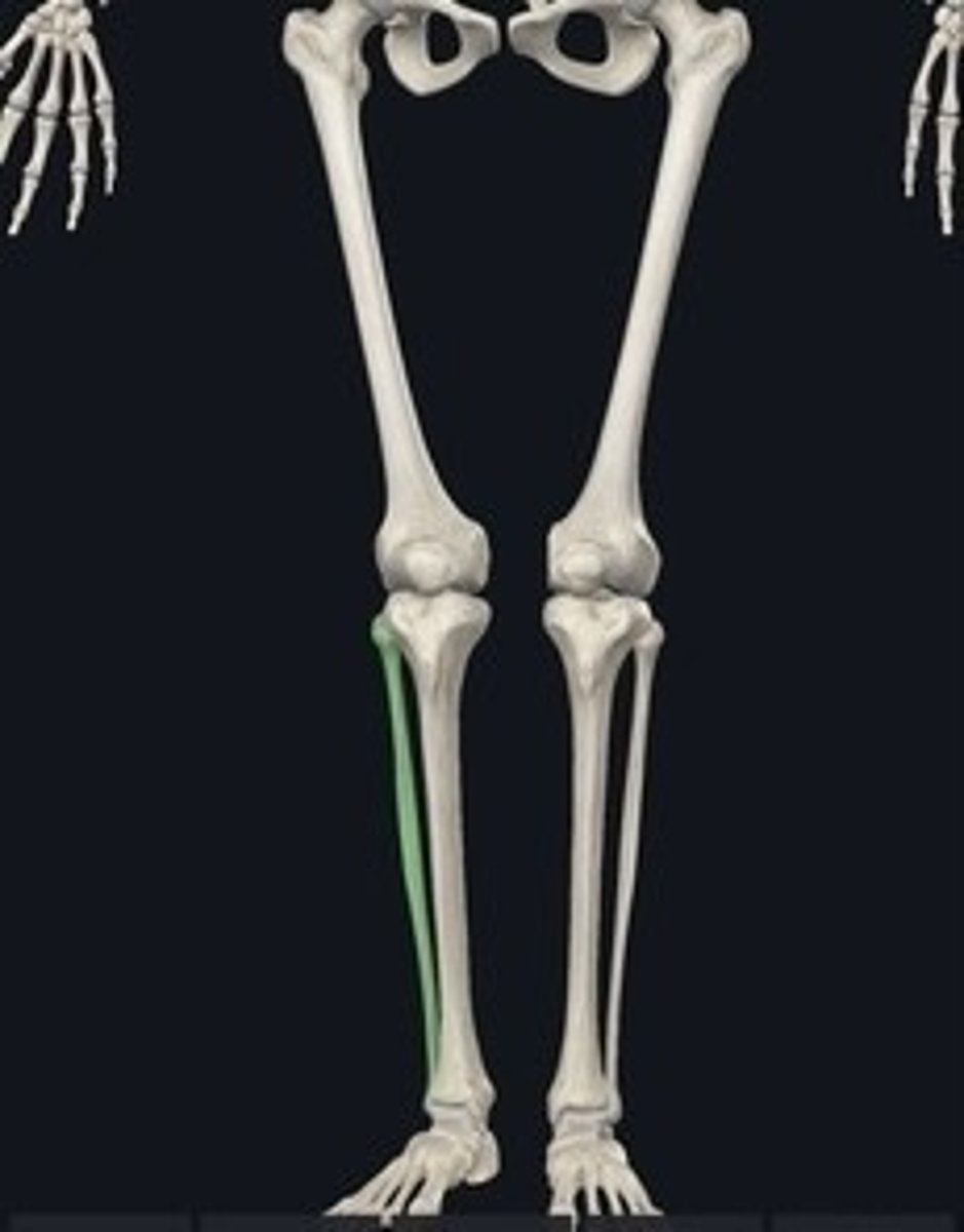
Tibia
Shin bone; larger lower leg bone.
Medial malleolus
Bony prominence on the tibia's distal end.
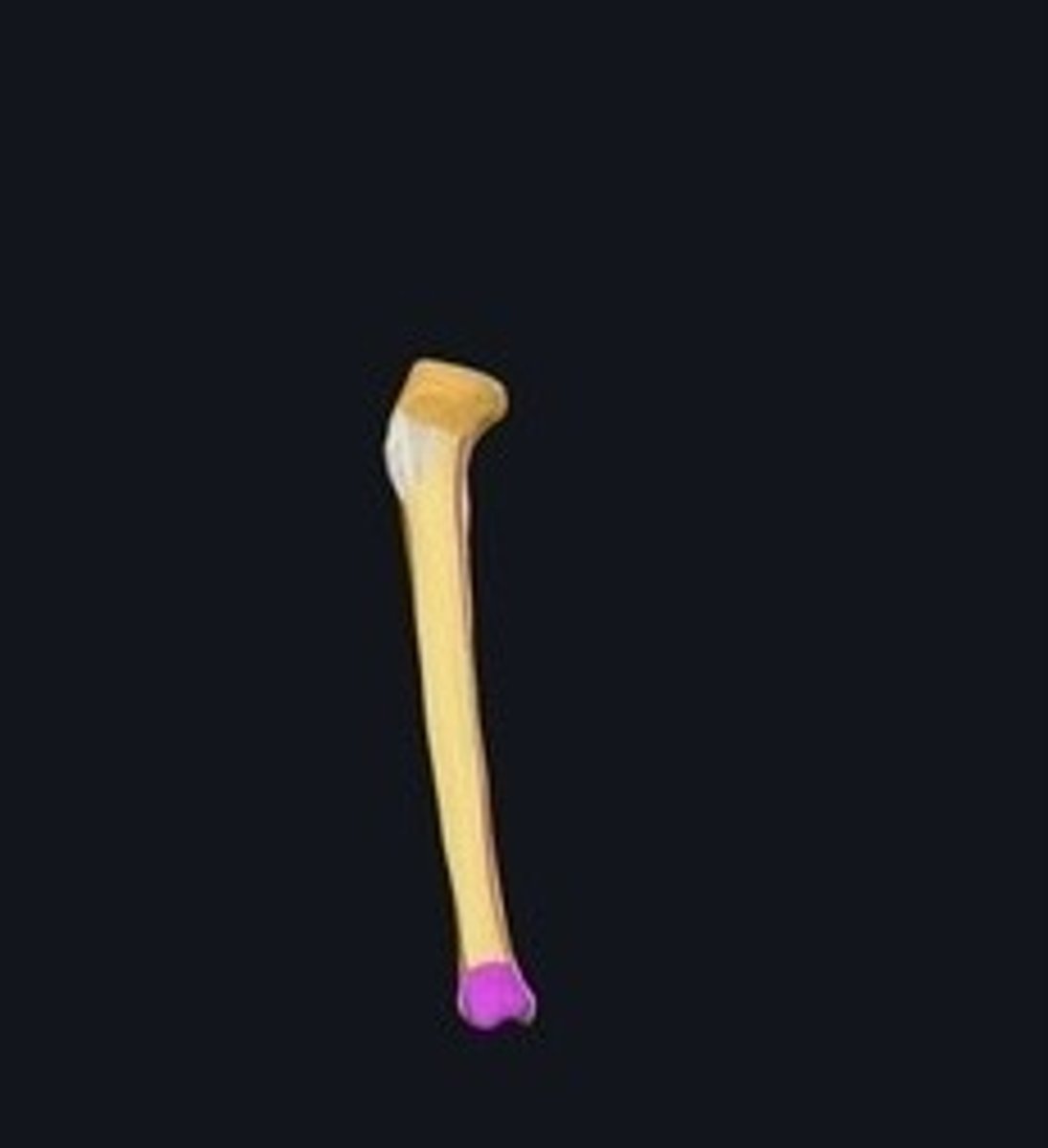
Fibula
Thinner lower leg bone parallel to tibia.
Lateral malleolus
Bony prominence on the fibula's distal end.
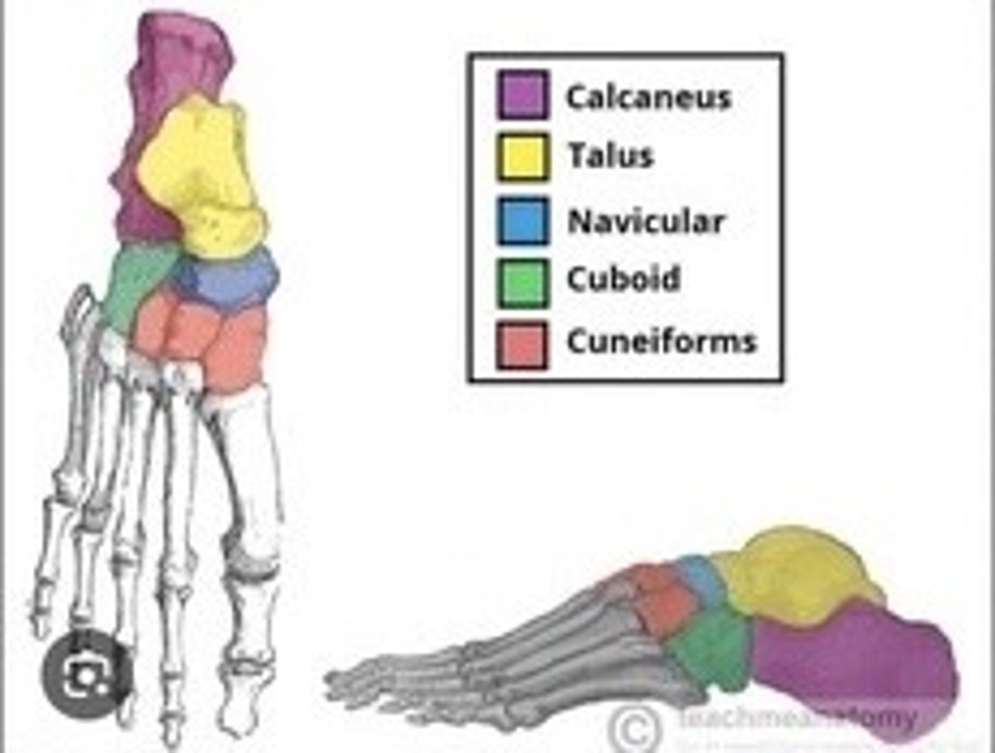
Tarsus
Ankle composed of seven bones.
Calcaneus
Talus
Navicular
Cuneiforms (medial, intermediate, lateral)
Cuboid
Metatarsals
Five bones forming the foot's arch.
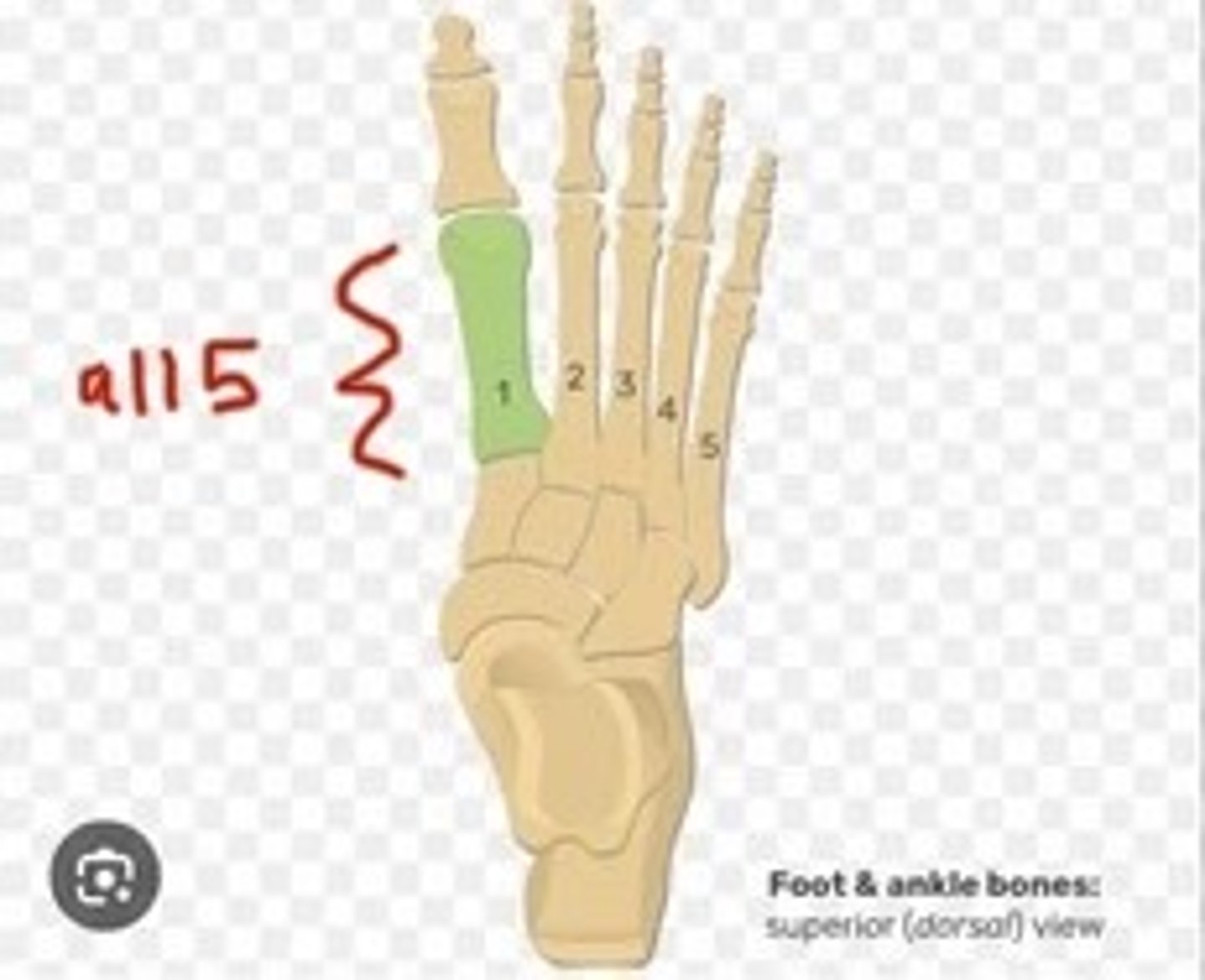
Phalanges
Bones of the toes; distal, middle, proximal.
parts of the sternum
top: manubrium
middle: body
bottom: xiphoid process