Muscle Anatomy
1/83
There's no tags or description
Looks like no tags are added yet.
Name | Mastery | Learn | Test | Matching | Spaced | Call with Kai |
|---|
No analytics yet
Send a link to your students to track their progress
84 Terms
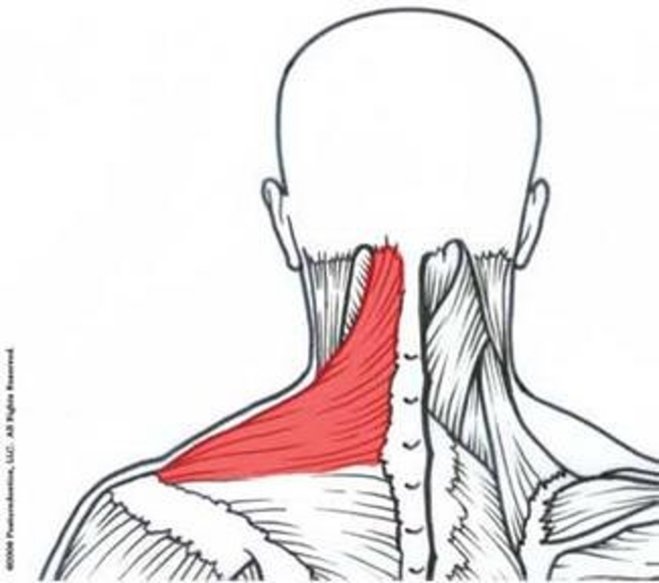
upper trapezius
-Nerve:accessory nerve (motor)
-Actions: scapula eleavtion
-origin occipital bone, nuchal ligament on upper cervical spinous processes
-insertion outer third of clavicle, acrominon process
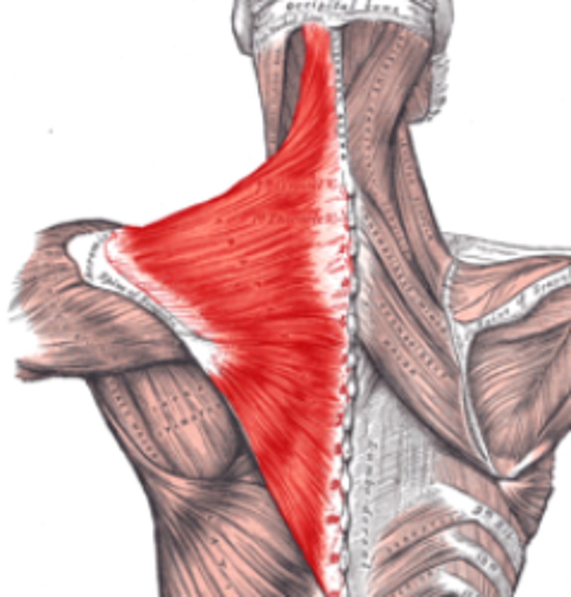
trapezius
-Origin spinous processes of vertebrae C1-T12
-Insertion external occipital protuberance, nuchal ligament, medial superior nuchal line, posterior border of the lateral third of the clavicle, acromion process, and spine of scapula
-Nerve accessory nerve (motor)
cervical spinal nerves C3 and C4 (motor and sensation)
-Actions rotation, retraction, elevation, and depression of scapula
-Antagonist
=serratus anterior muscle,
=Latissimus dorsi
levator scapulae
-Origin:Posterior tubercles of transverse processes of C1 - C4 vertebrae
-Insertion:Superior part of medial border of scapula
-Nerve cervical nerve; (C3, C4) and dorsal scapular nerve (C5)
-Actions: Elevates scapula and downward rotation
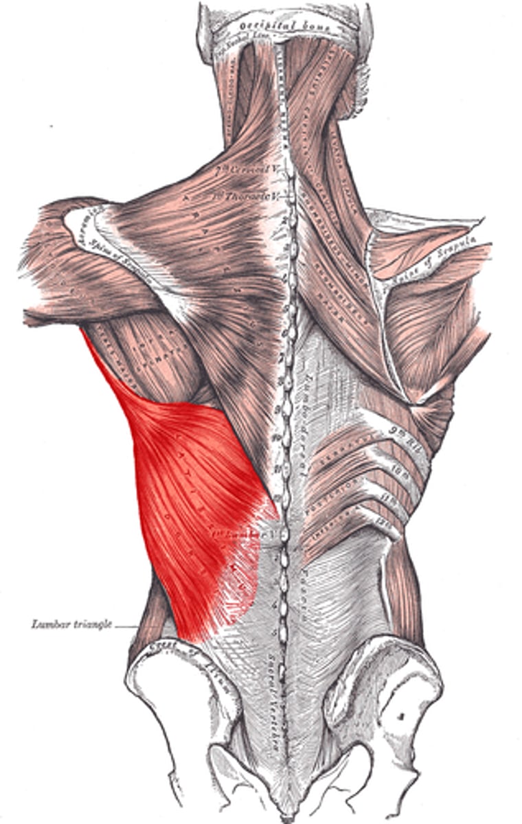
latissimus dorsi
-Origin: Spinous processes of vertebrae T7-L5, thoracolumbar fascia, iliac crest, inferior 3 or 4 ribs and inferior angle of scapula
-Insertion:Floor of intertubercular groove of the humerus
-Nerve: Thoracodorsal nerve
-Actions: Adducts, extends and internally rotates the arm when the insertion is moved towards the origin.
-Antagonist: Deltoid and trapezius muscle
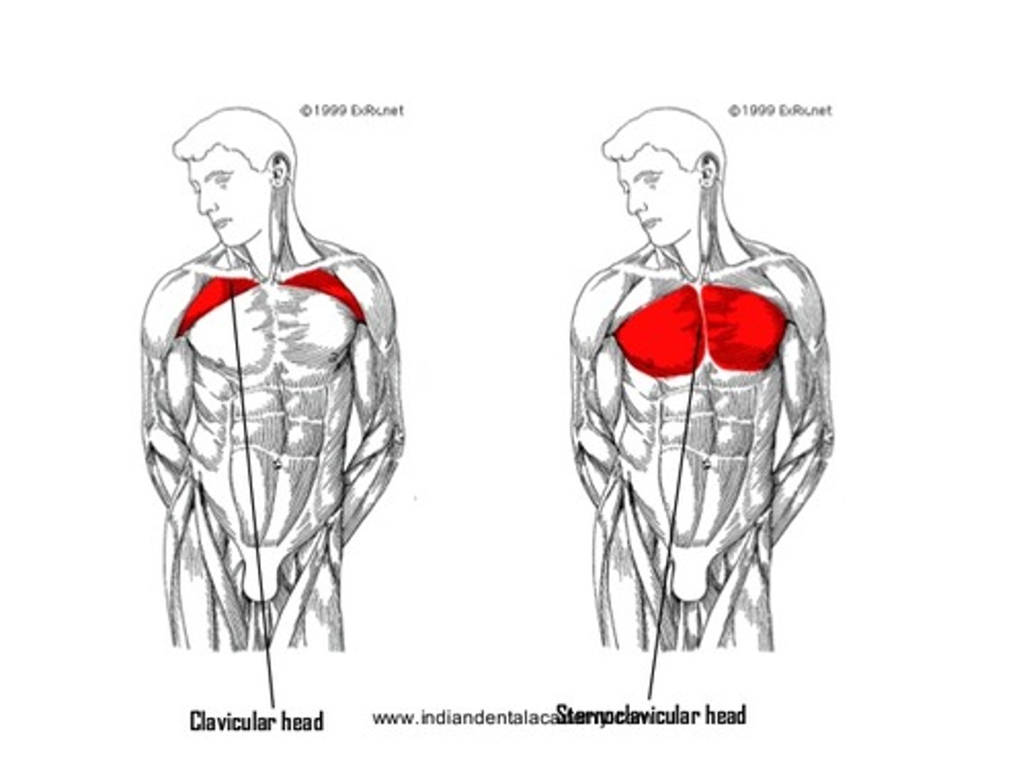
pectoralis major
-Origin:
=Clavicular head: anterior border of the medial half of the clavicle.
=Sternocostal head: anterior surface of the sternum, the superior six costal cartilages, and the aponeurosis of the external oblique muscle
-Insertion: Lateral lip of the bicipital groove of the humerus
(anteromedial proximal humerus)
-Nerve: lateral pectoral nerve and medial pectoral nerve
=Clavicular head: C5 and C6
=Sternocostal head: C7, C8 and T1
-Actions:
=Clavicular head: flexes the humerus
=Sternocostal head: extends the humerus. As a whole, adducts and medially rotates the humerus. It also draws the scapula anteriorly and inferiorly.
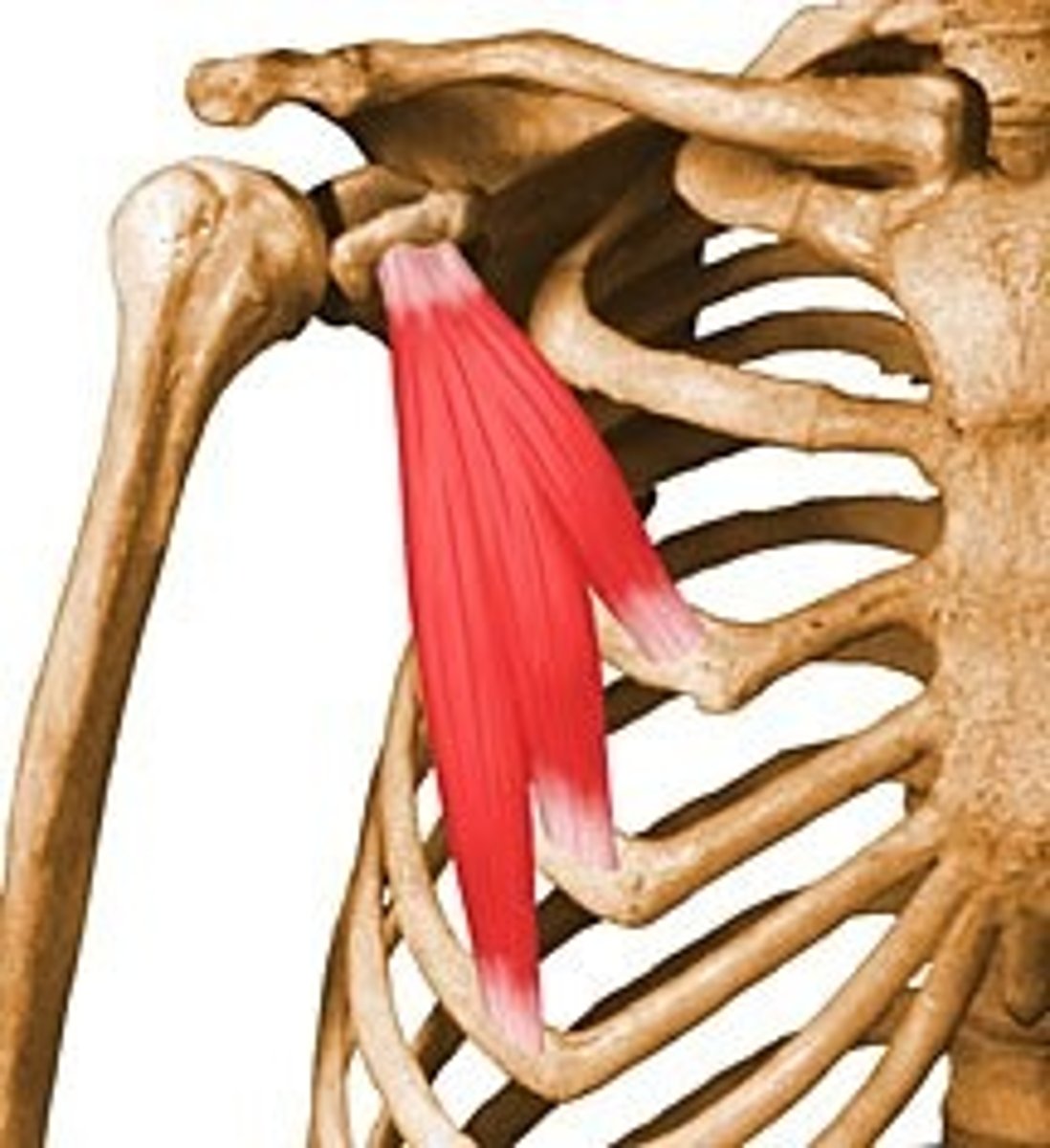
pectoralis minor
-Origin; Third to fifth ribs, near their costal cartilages
-Insertion: Medial border and superior surface of the coracoid process of the scapula
-Nerve: Medial pectoral nerve (C8, T1)
-Actions: Stabilizes the scapula by drawing it inferiorly and anteriorly against the thoracic wall, raises ribs in inspiration
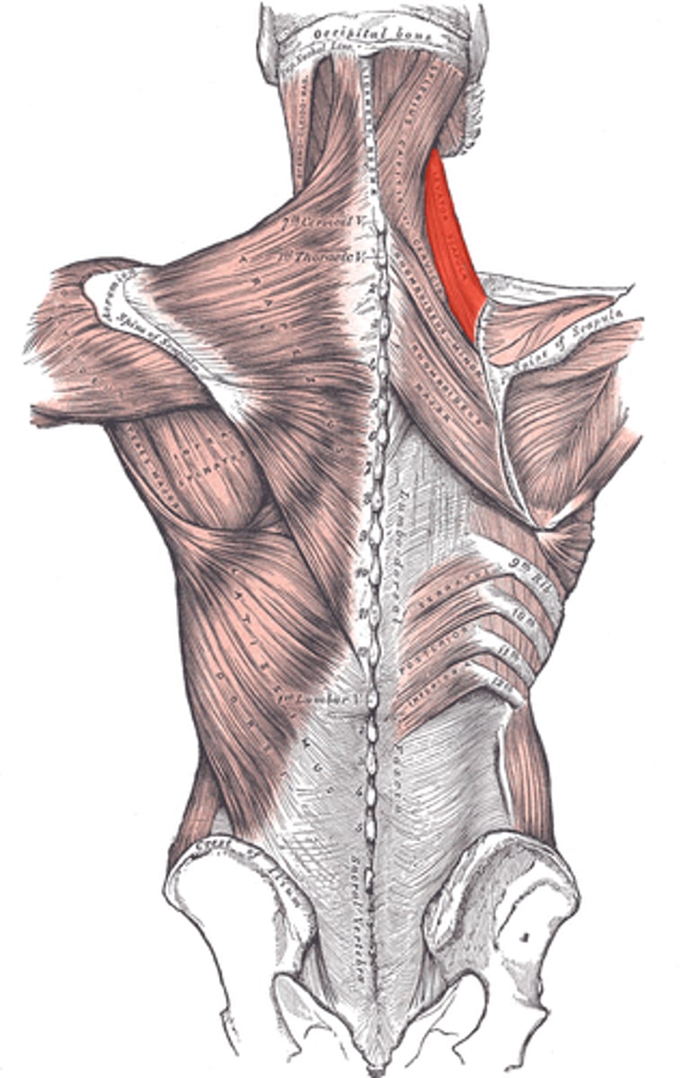
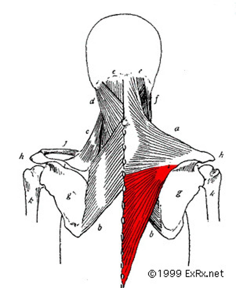
lower trapezius
-O: spinous processes of middle and lower thoracic vertebra
-I:spine medial nagle
-F: depression of scapula
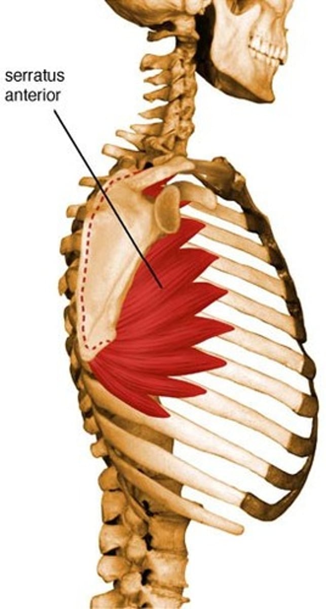
serratus anterior
-Origin: fleshy slips from the outer surface of upper 8 or 9 ribs
-Insertion: costal aspect of medial margin of the scapula
-Nerve: long thoracic nerve (from roots of brachial plexus C5, 6, 7)
-Actions: protracts and stabilizes scapula, assists in upward rotation.
-Antagonist: Rhomboid major, Rhomboid minor, Trapezius
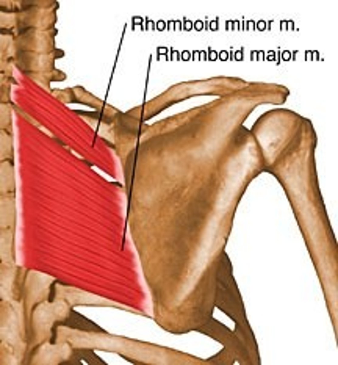
rhomboids
-Origin nuchal ligaments, spinous processes of the C7 to T5 vertebrae
-Insertion medial border of the scapula
-Nerve dorsal scapular nerve
-Actions
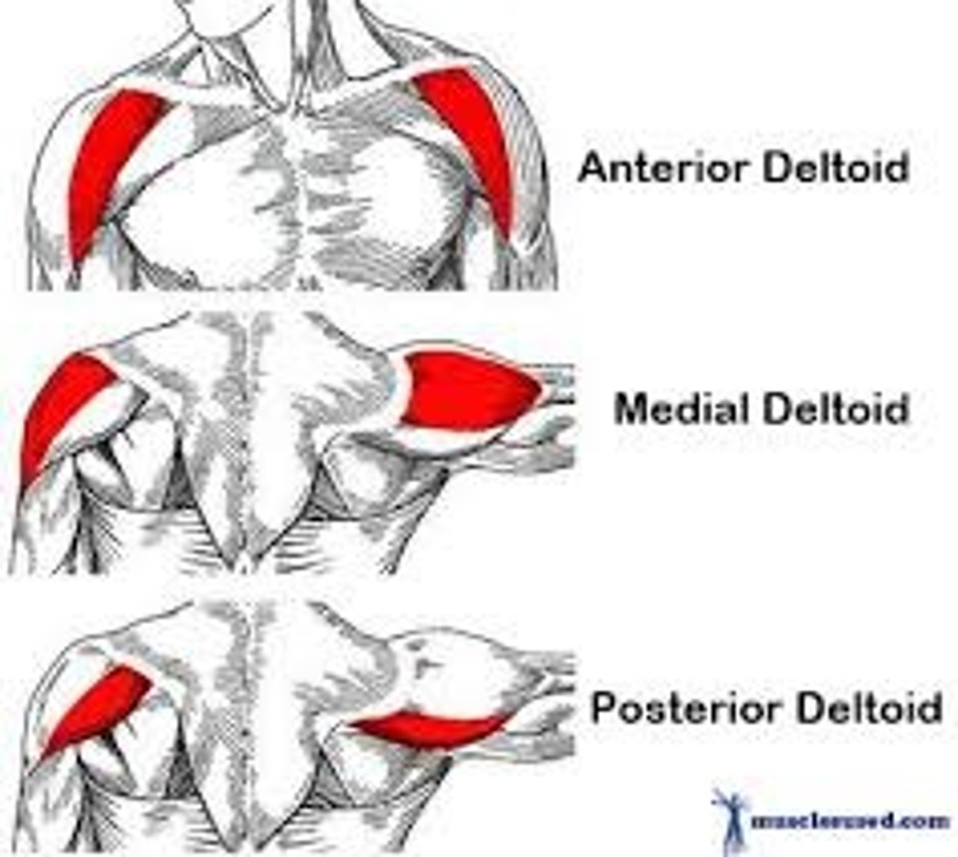
deltoid
-Origin the anterior border and upper surface of the lateral third of the clavicle, acromion, spine of the scapula
-Insertion deltoid tuberosity of humerus
-Nerve Axillary nerve
-Actions shoulder abduction by middle deltoid, flexion by anterior deltoid and extension by posterior deltoid
-Antagonist Latissimus dorsi
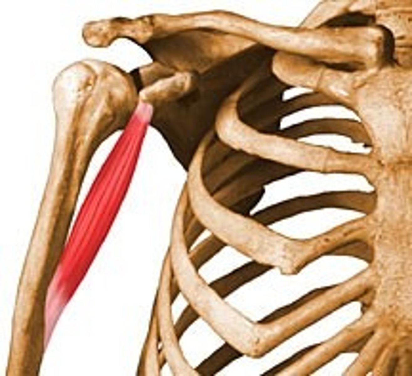
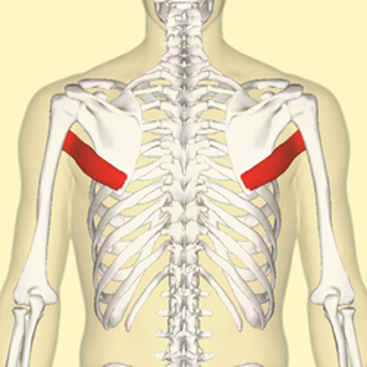
coracobrachialis
-Origin Coracoid process of scapula
-Insertion Medial humerus
-Nerve Musculocutaneous nerve (C5, C6, and C7)
-Actions adducts humerus , flexes the arm at glenohumeral joint
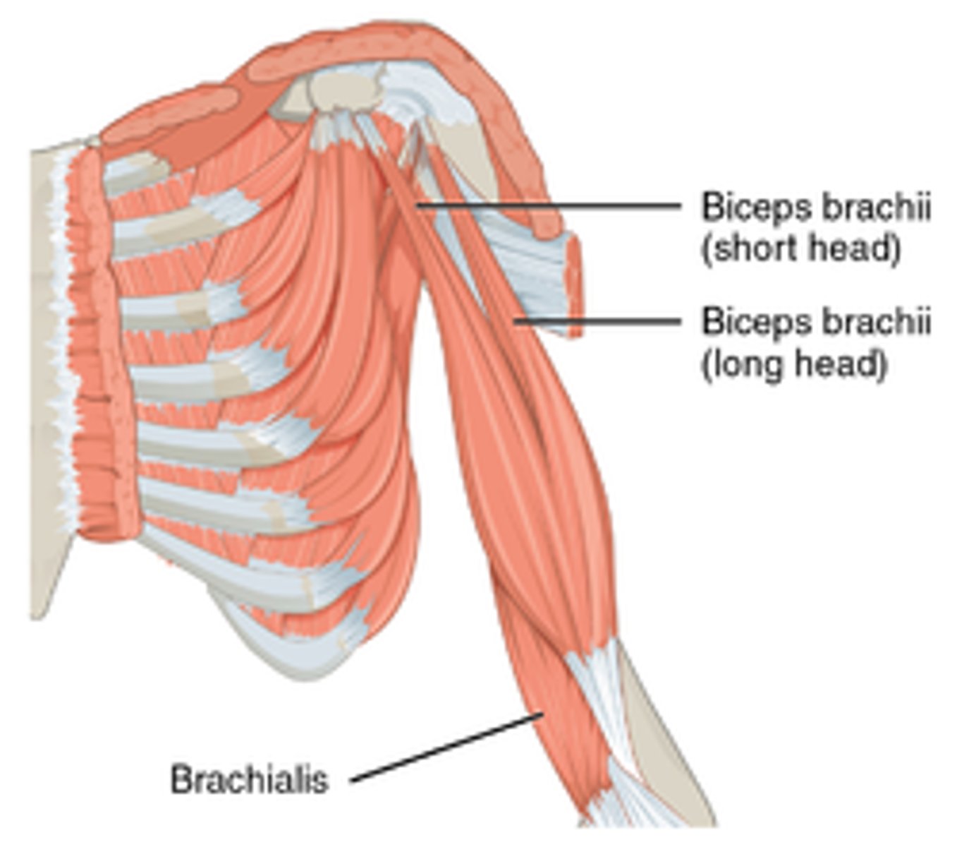
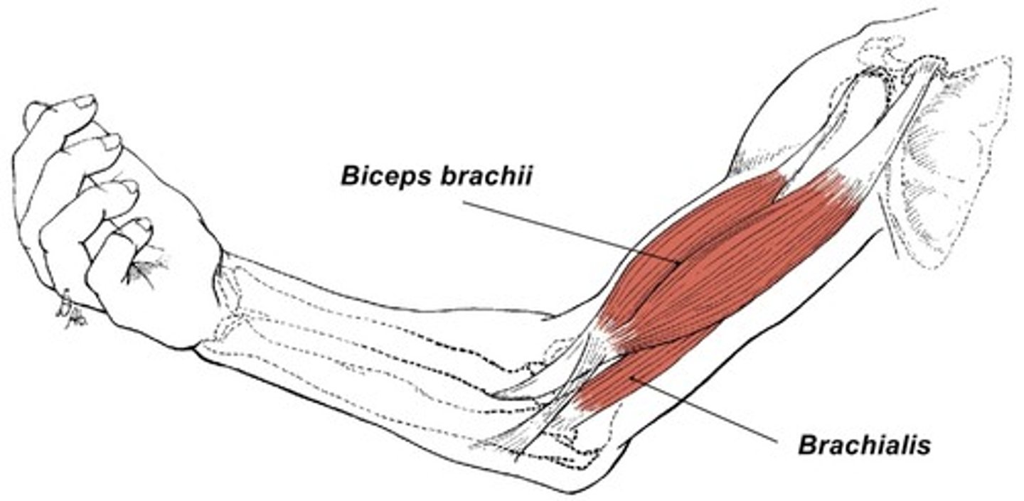
biceps brachii
-Origin
=Short head: coracoid process of the scapula.
=Long head: supraglenoid tubercle
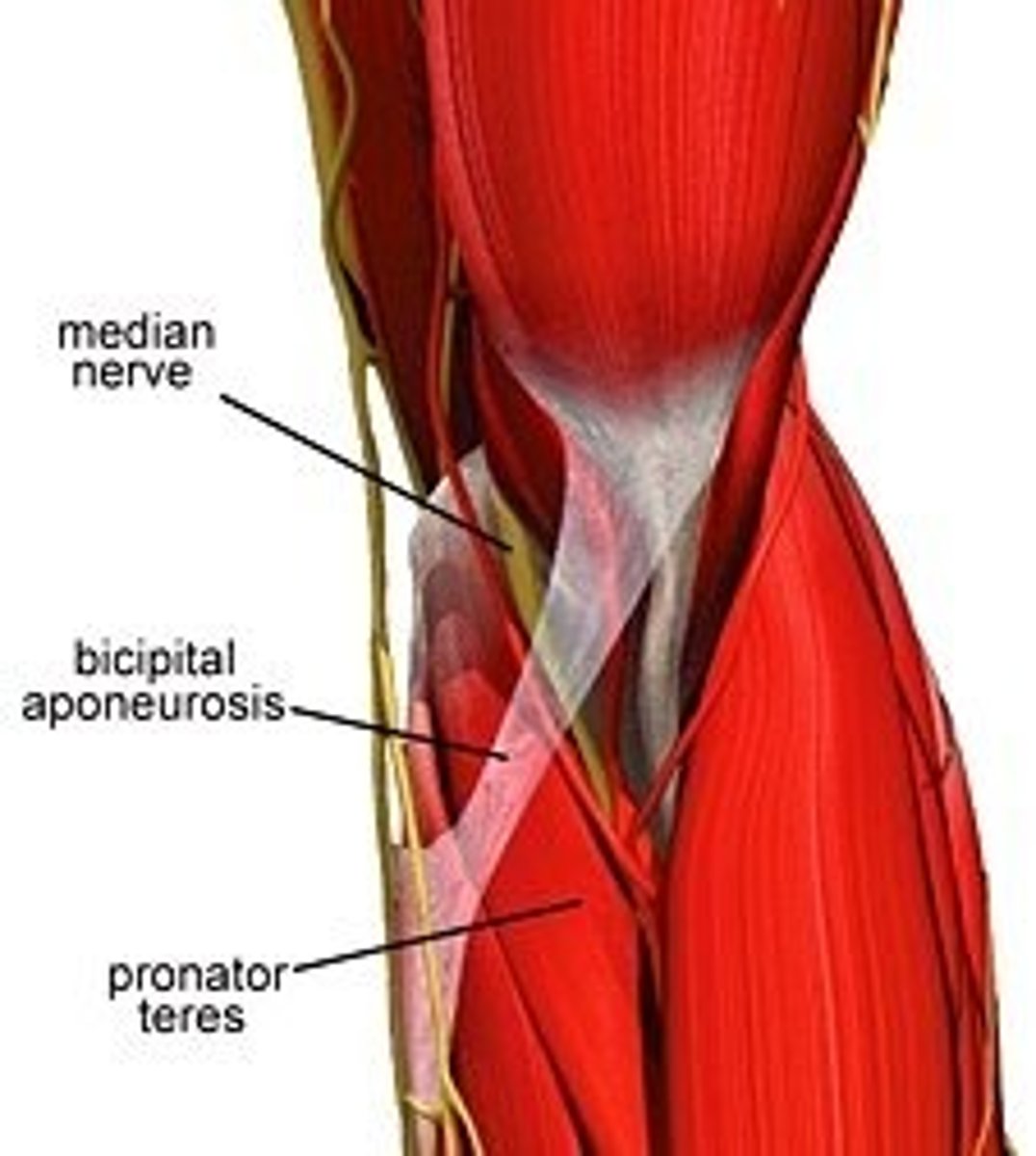
-Insertion Radial tuberosity and bicipital aponeurosis into deep fascia on medial part of forearm
-Nerve Musculocutaneous nerve (C5-C7)
-Actions
=Flexes elbow,
=supinates radioulnar joint in the forearm
-Antagonist Triceps brachii muscle
bicipital aponeurosis
triceps
-Origin
=Long head: infraglenoid tubercle of scapula
=Lateral head: above the radial sulcus
=Medial head: below the radial sulcus
-Insertion Olecranon process of ulna
-Nerve Radial nerve
-Actions
= Extends forearm,
= long head extends,adducts arm, Extends shoulder
-Antagonist Biceps brachii muscle
teres major
-Origin Posterior aspect of the inferior angle of the scapula
-Insertion Medial lip of the intertubercular sulcus of the humerus
-Nerve Lower subscapular nerve (segmental levels C5 and C6)
-Actions
= adduct the humerus,
= Internal rotation (medial rotation) of the humerus
= extend the humerus from flexed position,
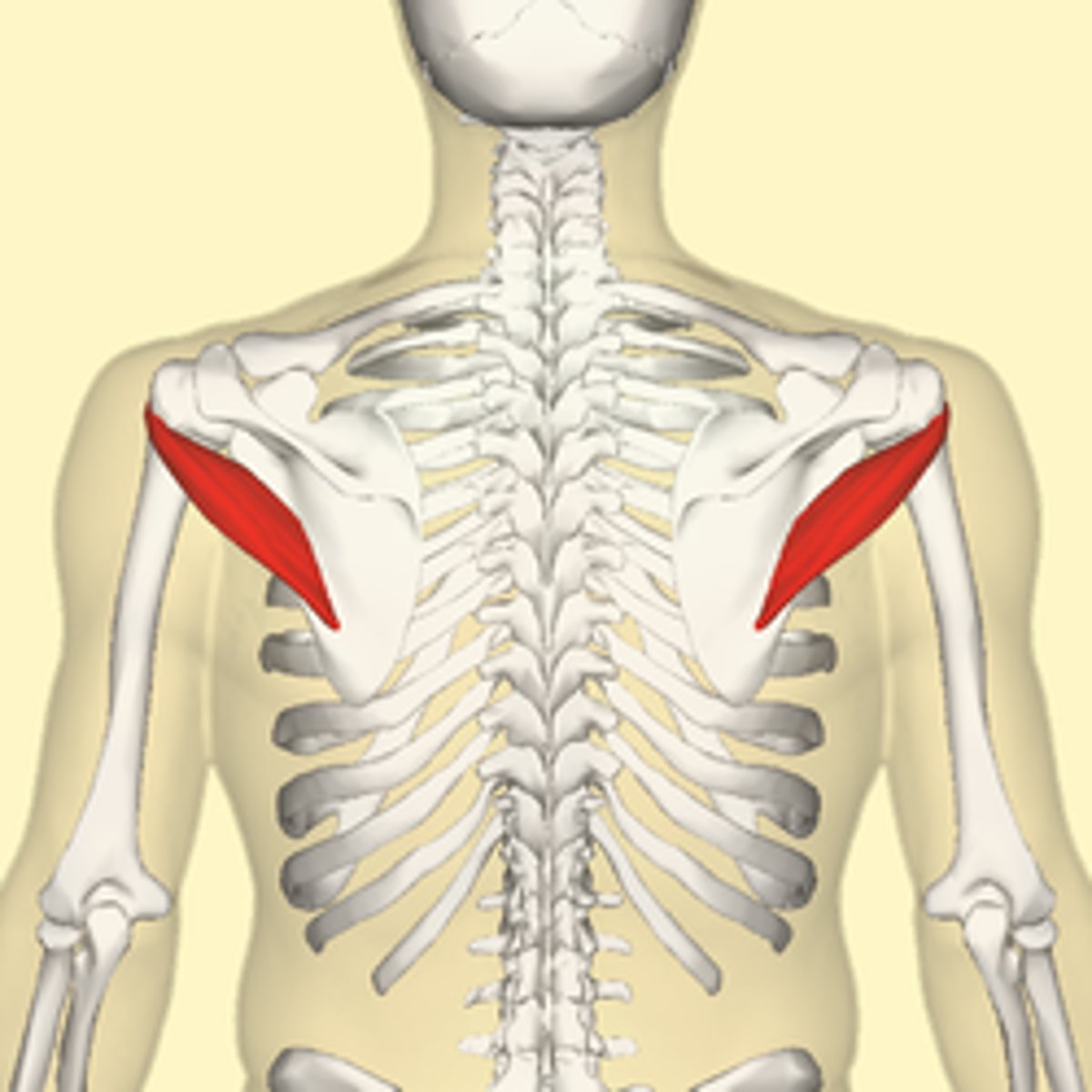
teres minor
-Origin lateral border of the scapula
-Insertion inferior facet of greater tubercle of the humerus
-Nerve axillary nerve
-Actions laterally rotates the arm, stabilizes humerus
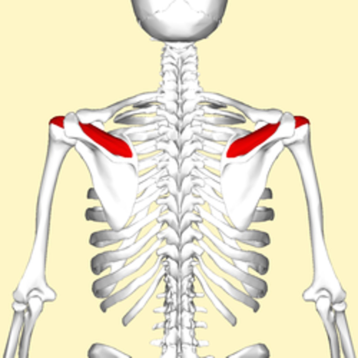
supraspinatus
-Origin supraspinous fossa of scapula
-Insertion superior facet of greater tubercle of humerus
-Nerve suprascapular nerve
-Actions abduction of arm and stabilizes humerus
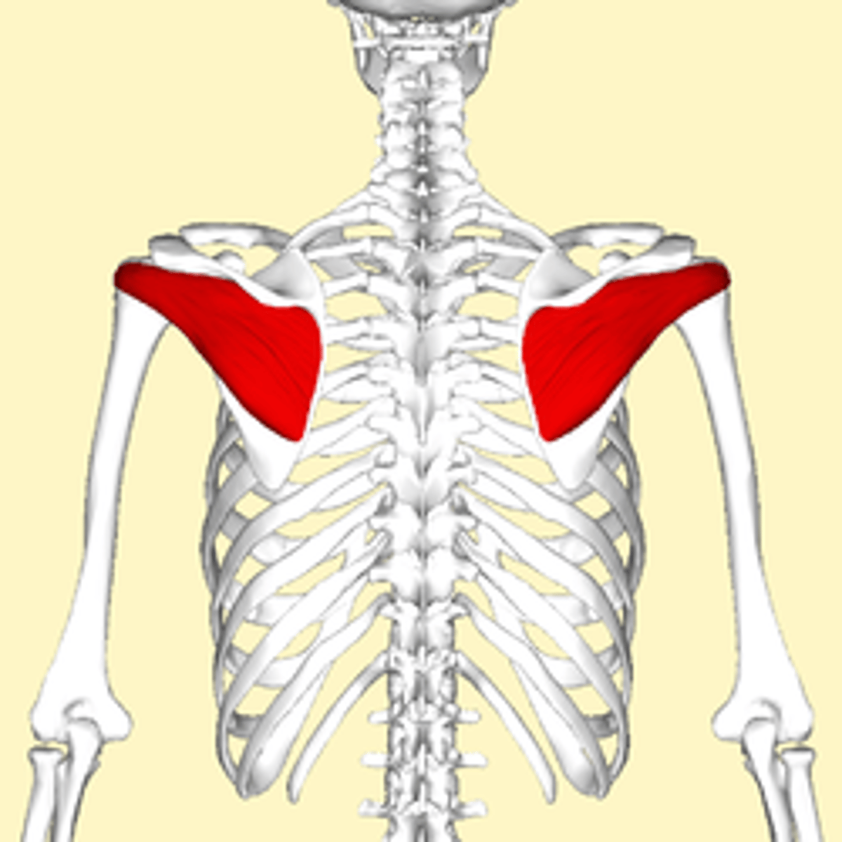
infraspinatus
-Origin infraspinous fossa of the scapula
-Insertion middle facet of greater tubercle of the humerus
-Nerve suprascapular nerve
-Actions Lateral rotation of arm and stabilizes humerus
subscapularis
-Origin Subscapular fossa
-Insertion Lesser tubercle of humerus
-Nerve Upper subscapular nerve, lower subscapular nerve (C5, C6)
-Actions Internally (medially) rotates humerus and stabilizes shoulder
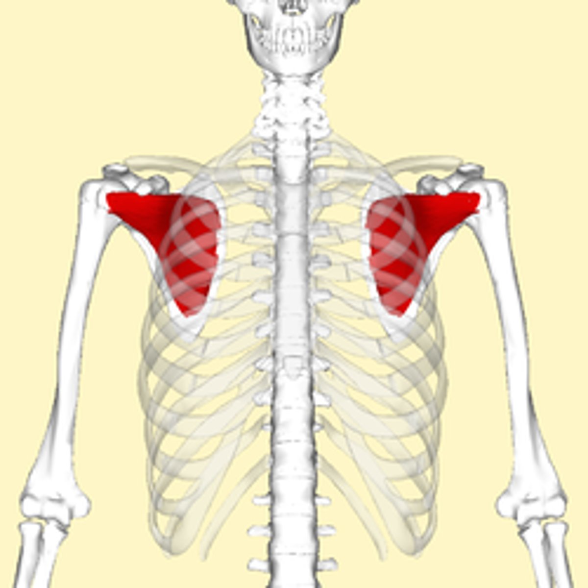
Subscapular fossa
subscapular fossa is a feature on the scapula bone, which is also known as the shoulder bone (or shoulder blade)
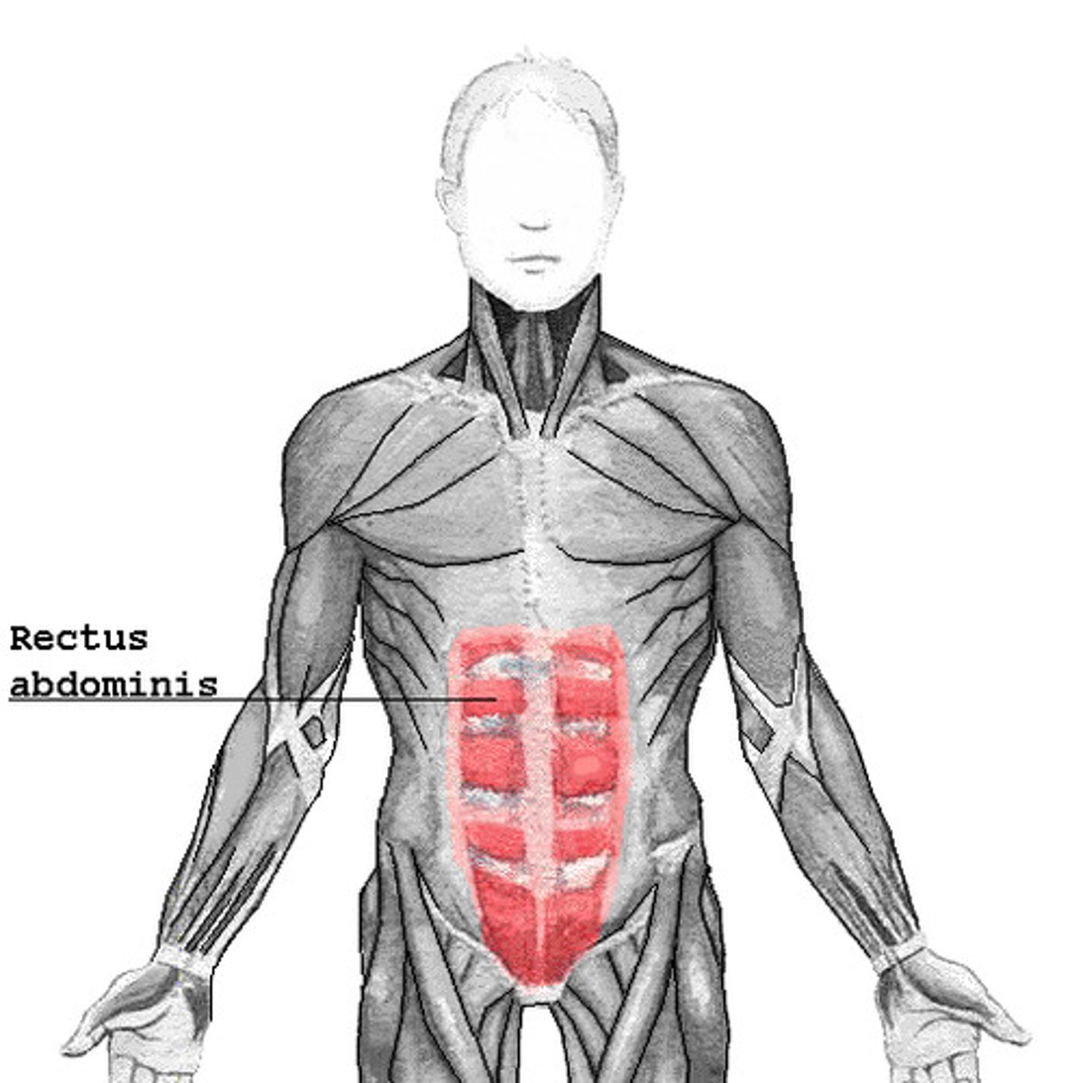
rectus abdominis
-Origin crest of pubis
-Insertion
=Costal cartilage of ribs 5-7,
=xiphoid process of sternum
-Nerve segmentally by thoraco-abdominal nerves (T7 to T11)
-Actions Flexion of the lumbar spine
-Antagonist Erector spinae
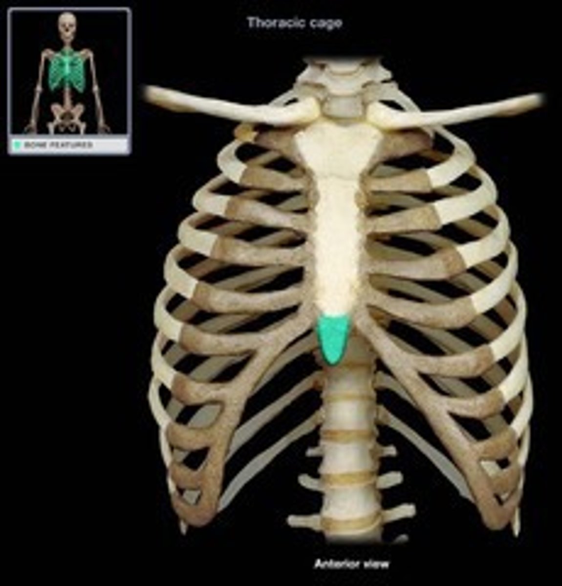
xiphoid process of sternum
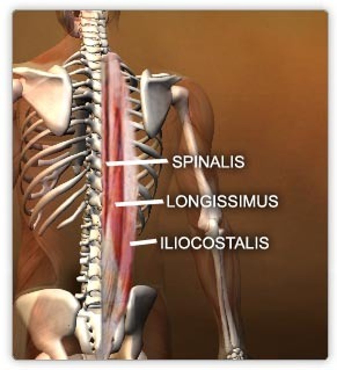
erector spinae
-Origin Spinous processes of T9-T12 thoracic vertebrae, medial slope of the dorsal segment of illiac crest
-Insertion spinous processes of T1 and T2 thoracic vertebrae and the cervical vertebrae
-Actions extends the vertebral column
-Antagonist rectus abdominis muscle
internal oblique
-Origin Inguinal ligament, Iliac crest and the Lumbodorsal fascia.
-Insertion Linea alba, Pecten Pubis (via Conjoint tendon) and ribs 10-12.
-Actions
= Compresses abdomen
=unilateral contraction rotates vertebral column to same side.

external oblique
-Origin Ribs 5-12
-Insertion Iliac crest, Pubic tubercle, Linea alba
-Actions Contralateral rotation of torso
multifidus
-Origin Sacrum, Erector spinae Aponeurosis, PSIS, and Iliac crest
-Insertion spinous process
-Actions
=Bilateral backward extension,
= unilateral side-bending to the ipsilateral side
=rotation to the contralateral side

brachialis
-Origin anterior surface of the humerus, particularly the distal half of this bone
-Insertion coronoid process and the tuberosity of the ulna
-Nerve musculocutaneous nerve (C5, C6)
-Actions flexion at elbow joint
brachioradialis
-Origin Lateral supracondylar ridge of the humerus
-Insertion Distal radius (radial styloid process)
-Nerve :Radial nerve
-Actions :Flexion of elbow and supination
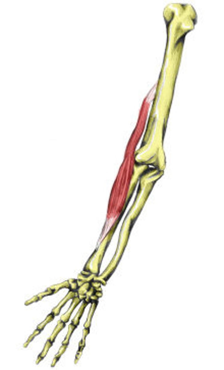
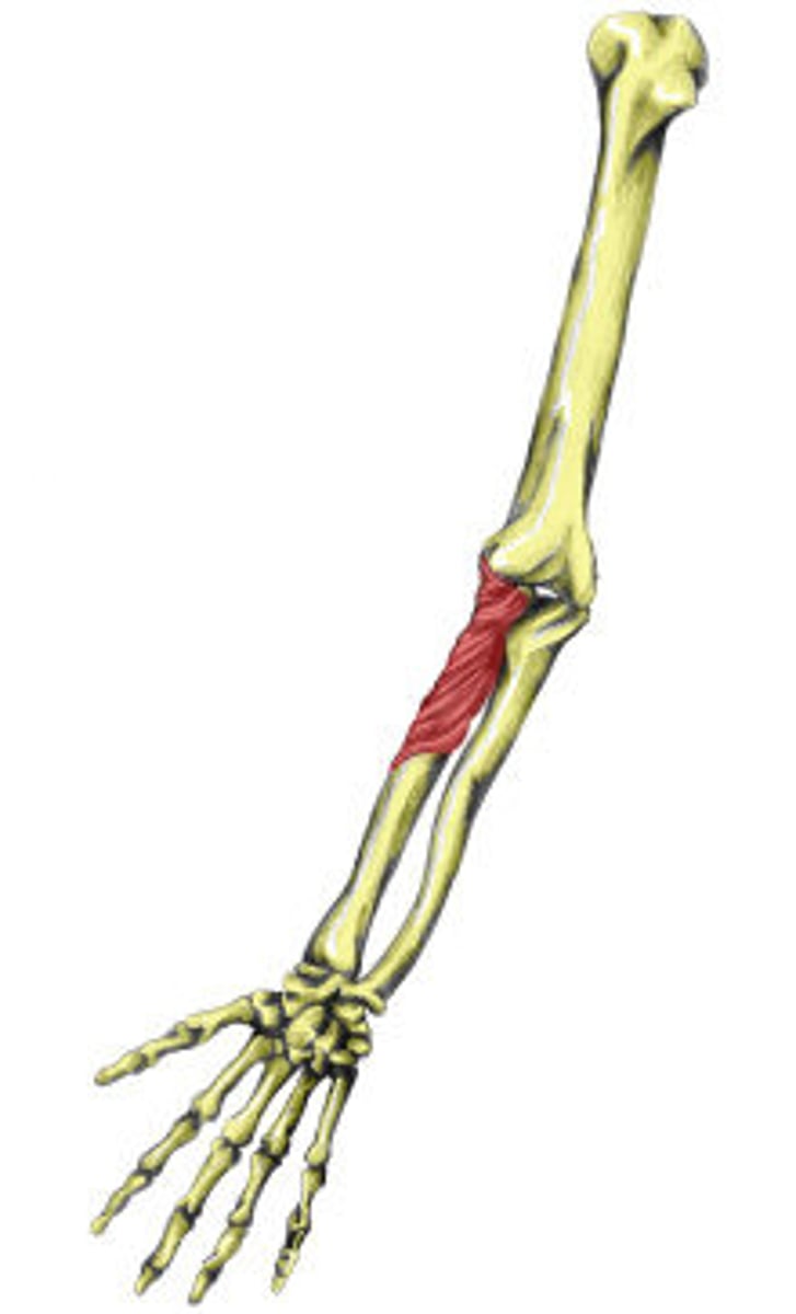
supinator
-Origin Lateral epicondyle of humerus, supinator crest of ulna, radial collateral ligament, annular ligament
-Insertion Lateral proximal radial shaft
-Nerve Deep branch of the radial nerve
-Actions Supinates forearm
-Antagonist Pronator teres, pronator quadratus
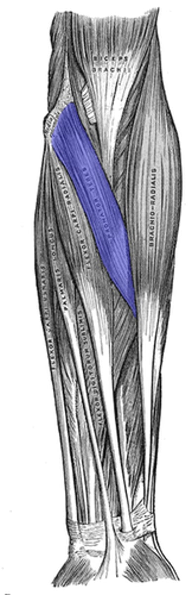
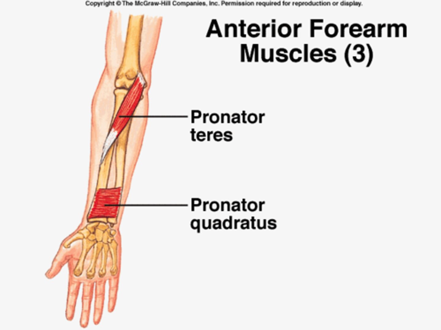
pronator teres
-Origin
=humeral head: medial supracondylar ridge of humerus (common flexor tendon)
=ulnar head: coronoid process of ulna
-Insertion Middle of the lateral surface of the body of the radius
-Nerve median nerve
-Actions pronation of forearm, flexes elbow
-Antagonist Supinator muscle
pronator quadratus
-Origin medial, anterior surface of the ulna
-Insertion lateral, anterior surface of the radius
-Nerve median nerve (anterior interosseous nerve)
-Actions pronates the forearm
-Antagonist Supinator muscle
flexor carpi radialis
-Origin medial epicondyle of humerus (common flexor tendon)
-Insertion Bases of second and third metacarpal bones
-Nerve Median nerve
-Actions Flexion and abduction at wrist
-Antagonist Extensor carpi ulnaris muscle
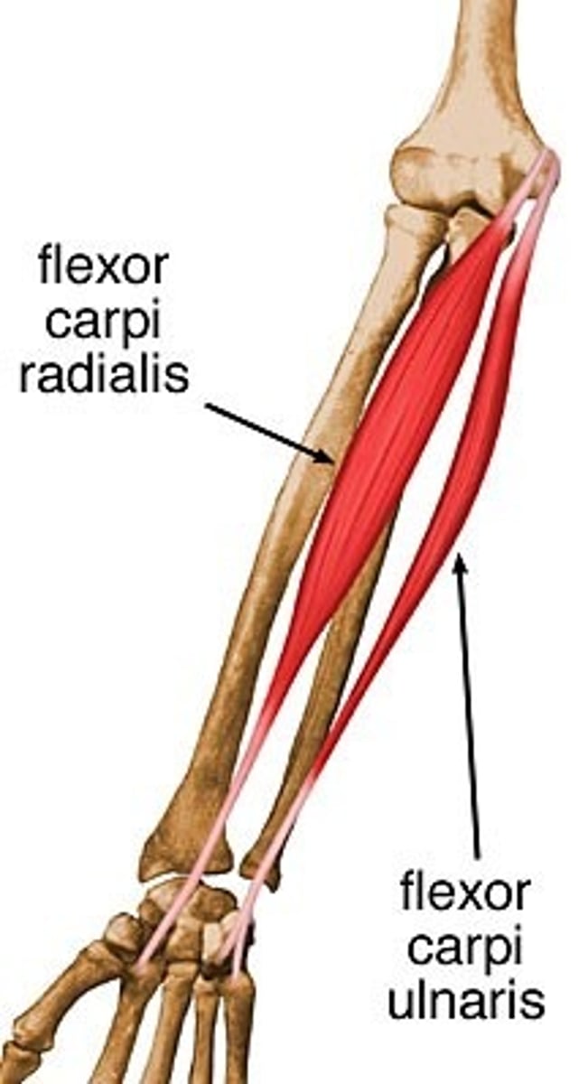
flexor carpi ulnaris
-Origin Medial epicondyle (common flexor tendon) and medial margin on olecranon of ulna
-Insertion Pisiform, hook of the hamate, base of the fifth metacarpal bone
-Nerve ulnar nerve
Nerve root C7, C8 and T1
-Actions Flexion and adduction of wrist
-Antagonist Extensor carpi radialis brevis muscle and extensor carpi radialis longus muscle
extensor carpi radialis longus
-Origin lateral supracondylar ridge
-Insertion 2nd metacarpal
-Nerve radial nerve
-Actions extensor at the wrist joint, abducts the hand at the wrist
-Antagonist Flexor carpi ulnaris muscle
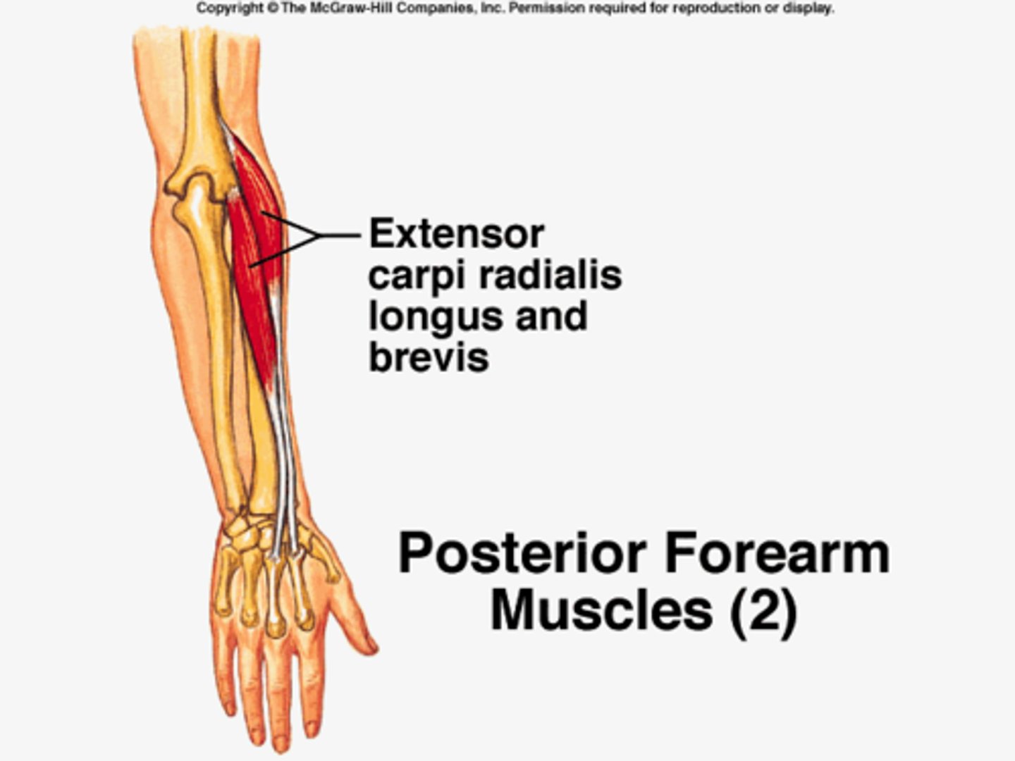
Extensor carpi radialis brevis
extensor carpi ulnaris
-Origin Common extensor tendon (lateral epicondyle), ulna
-Insertion 5th metacarpal
-Nerve Deep branch of the radial nerve (C7, C8)
-Actions extends and adducts the wrist
-Antagonist Flexor carpi radialis
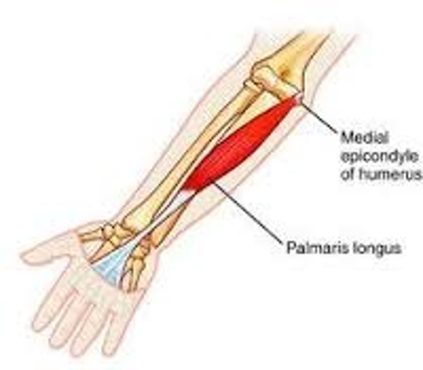
palmaris longus
-Origin medial epicondyle of humerus (common flexor tendon)
-Insertion palmar aponeurosis
-Nerve median nerve
-Actions wrist flexor
-Antagonist Extensor carpi radialis brevis, Extensor carpi radialis longus, Extensor carpi ulnaris
iliopsoas
-Origin Iliac fossa and lumbar spine
-Insertion Lesser trochanter of femur
-Nerve Branches from L1-L3
-Actions Flexion of hip
-Antagonist Gluteus maximus and the posterior compartment of thigh

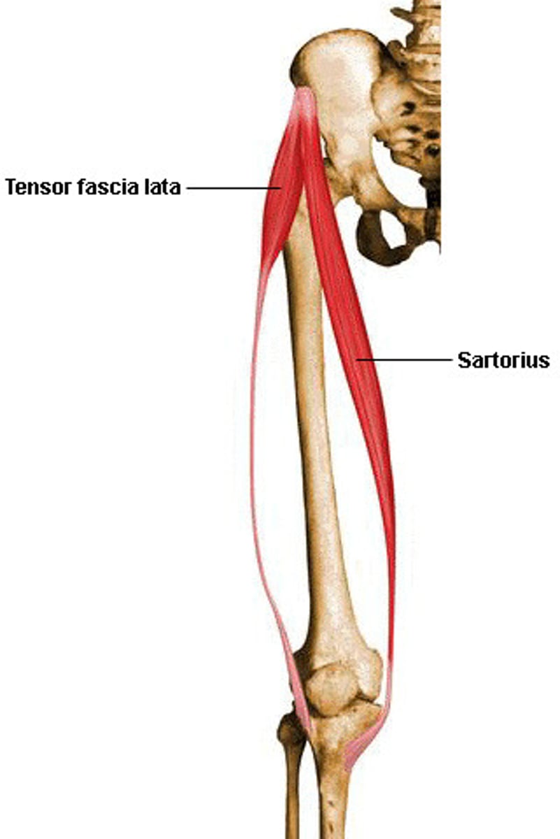
sartorius
-Origin Anterior superior iliac spine of the pelvic bone
-Insertion anteromedial surface of the upper tibia in the pes anserinus
-Nerve femoral nerve (sometimes from the intermediate cutaneous nerve of thigh)
-Actions Flexion, abduction, and lateral rotation of the hip, flexion of the knee
-tailor's muscle and longest.
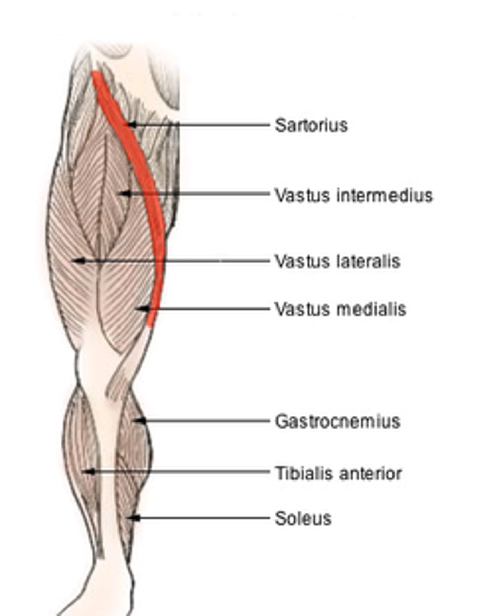
rectus femoris
-Origin anterior inferior iliac spine and the exterior surface of the bony ridge which forms the groove on the iliac portion of the acetabulum
-Insertion inserts into the patellar tendon as one of the four quadriceps muscles
-Nerve femoral nerve
-Actions knee extension; hip flexion
-Antagonist Hamstring

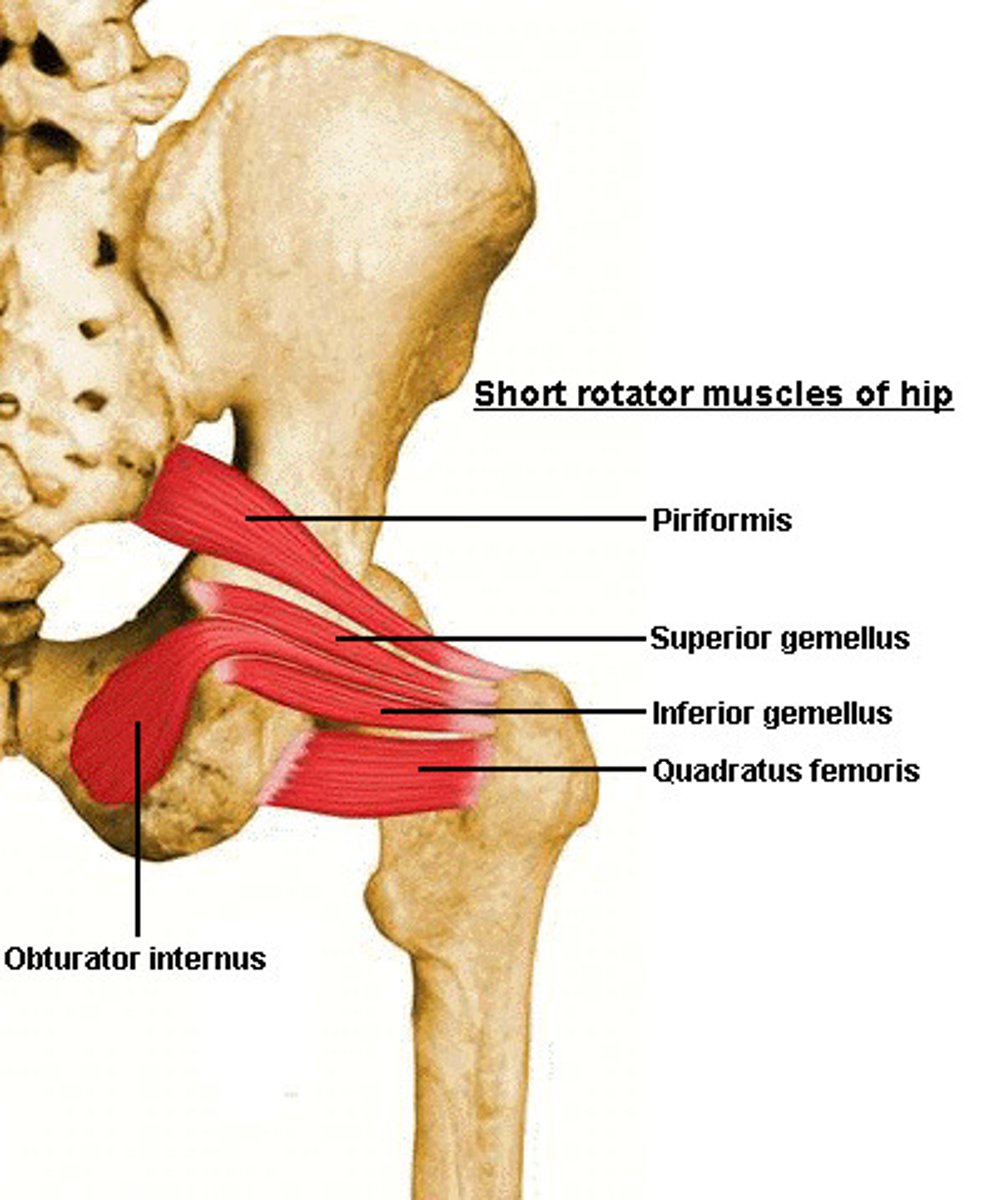
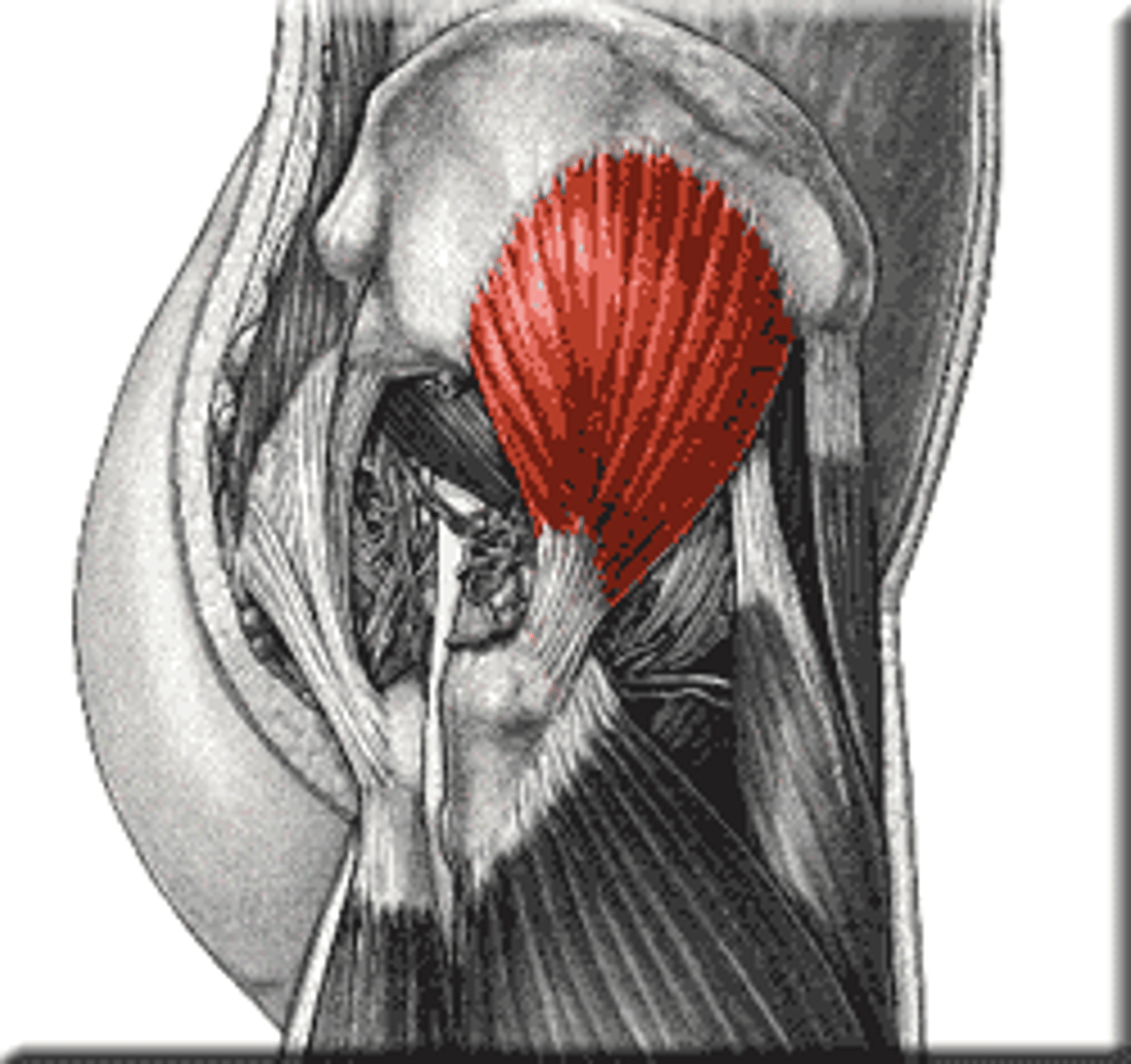
piriformis
-Origin Sacrum
-Insertion Greater trochanter
-Nerve Nerve to the piriformis (L5, S1, and S2)
-Actions External rotator of the thigh
gracilis
-Origin ischiopubic ramus
-Insertion tibia (pes anserinus)
-Nerve anterior branch of obturator nerve
-Actions flexes, medially rotates, and adducts the hip,
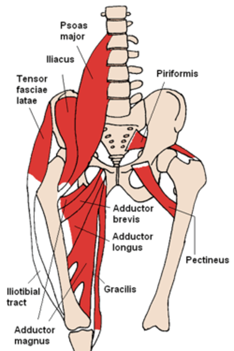
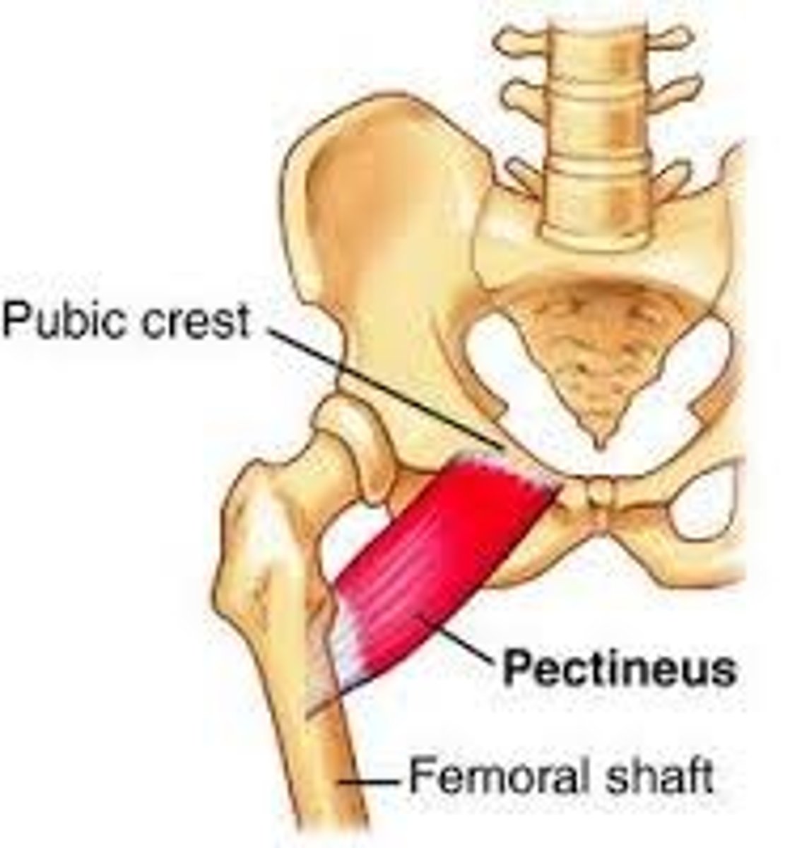
pectinus
-Origin Pectineal line of the pubic bone
-Insertion Pectineal line of the femur
-Nerve Femoral nerve, sometimes obturator nerve
-Actions flexion and adduction of thigh
tensor fascia latae
-Origin Iliac crest
-Insertion Iliotibial tract
-Nerve Superior gluteal nerve (L4, L5, S1)
-Actions
=Hip; flexion, medial rotation, abduction
*hamstring*
-Origin tuberosity of the ischium, linea aspera
-Insertion tibia, fibula
-Nerve: sciatic nerve (tibial nerve and common fibular nerve)
-Actions: flexion of knee, extension of hip
-Antagonist: Rectus femoris muscle

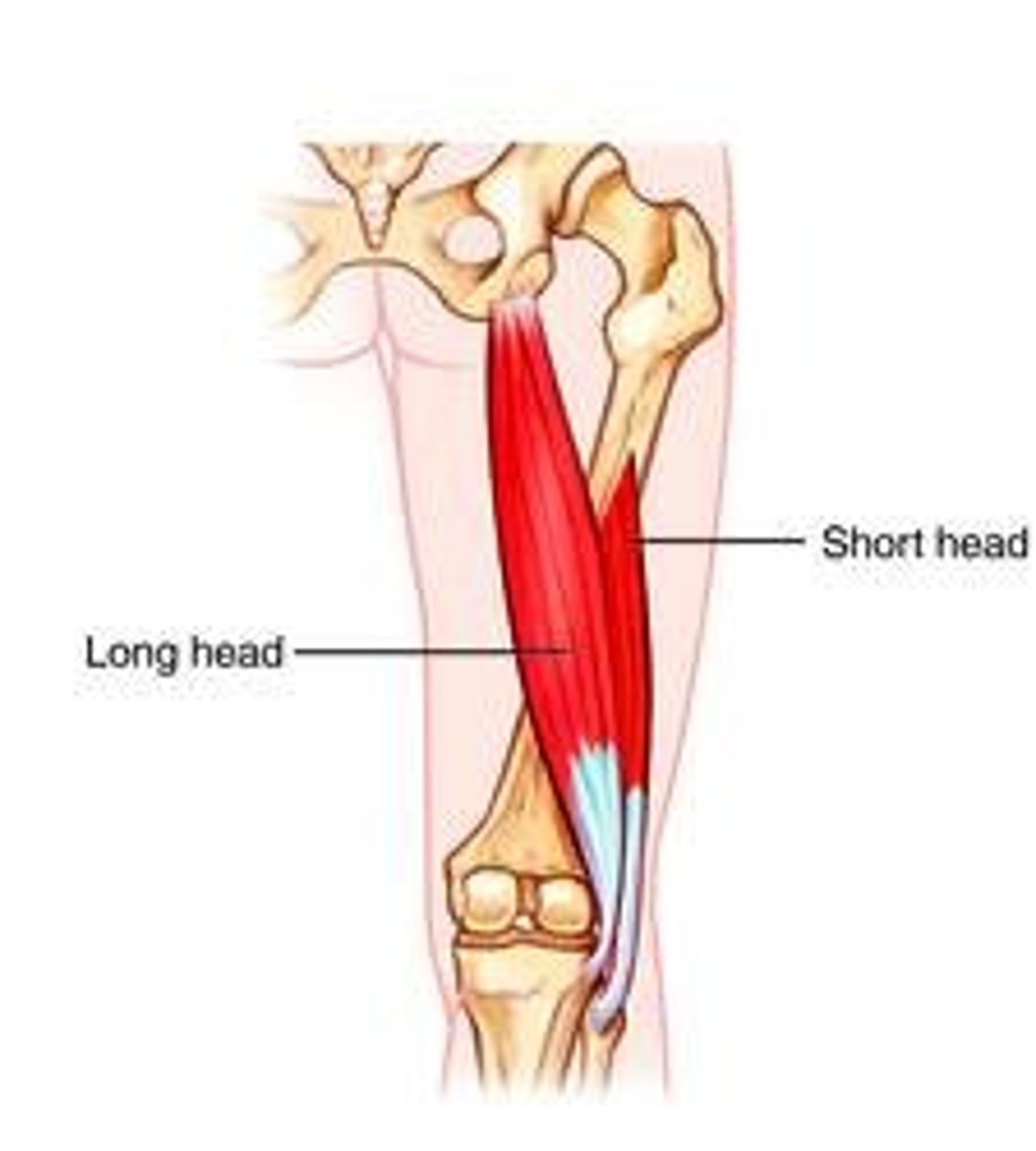
biceps femoris
-Origin tuberosity of the ischium, linea aspera, femur
-Insertion the head of the fibula which articulates with the back of the lateral tibial condyle
-Nerve
=long head: tibial nerve
=short head: common fibular nerve
-Actions
=flexes knee joint,
=laterally rotates knee joint (when knee is flexed),
=extends hip joint (long head only)
-Antagonist Quadriceps muscle
semitendinosus
-Origin Tuberosity of the ischium
-Insertion Pes anserinus (tibia)
-Nerve Sciatic (tibial, L5, S1, S2)
-Actions Flexion of knee, extension of the hip joint
-Antagonist Quadriceps muscle
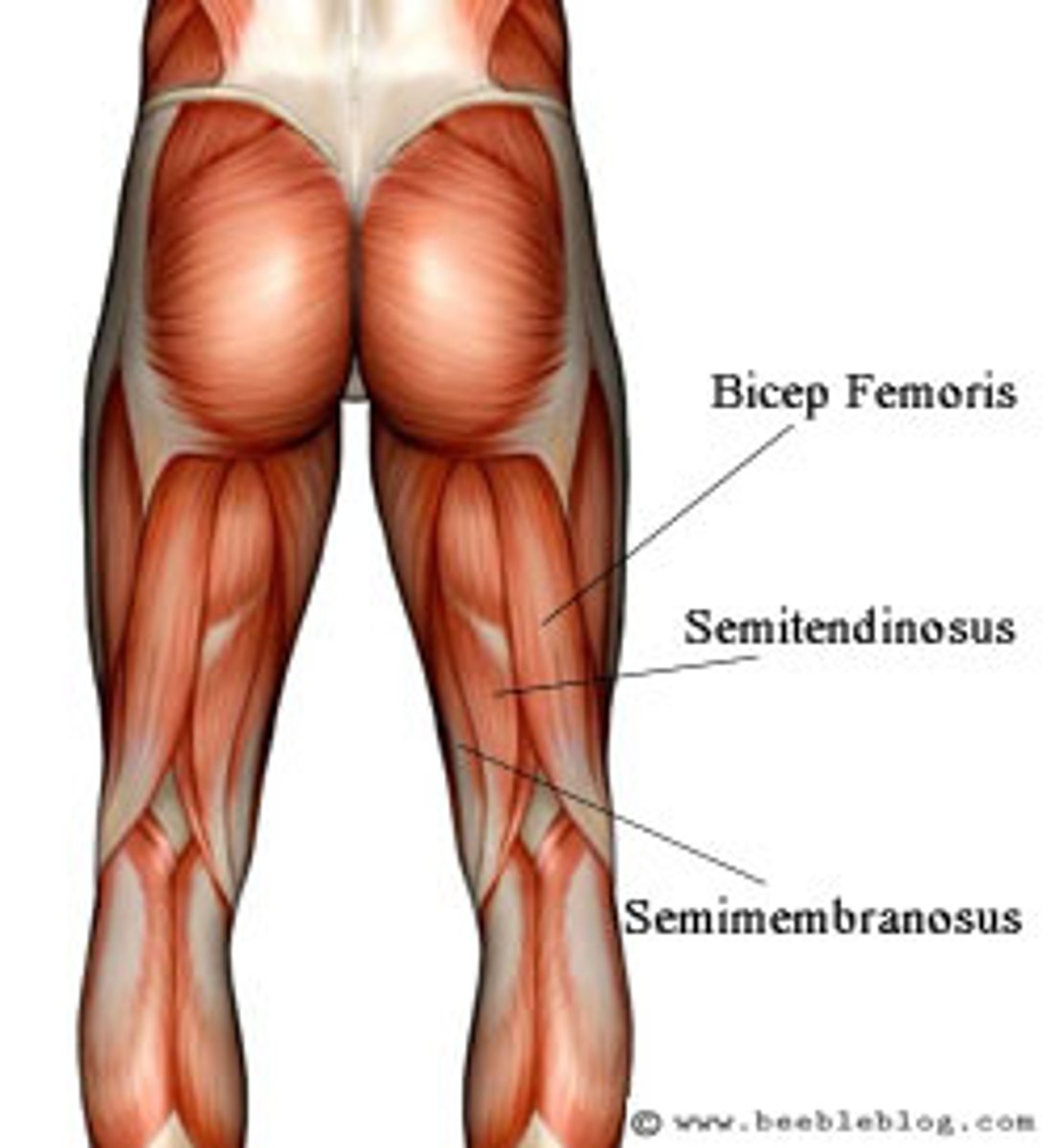
semimembranosus
-Origin Ischial tuberosity
-Insertion Medial condyle of tibia
-Nerve Tibial part of sciatic nerve
(L5, S1 and S2)
-Actions Extension of hip and flexion of knee
-Antagonist Quadriceps muscle and Tensor fasciae latae
*quadriceps*
-Origin Combined rectus: femoris and vastus muscles
-Insertion: Tibial tuberosity
-Nerve: Femoral nerve
-Actions
=Knee extension
=Hip flexion (Rectus femoris only)
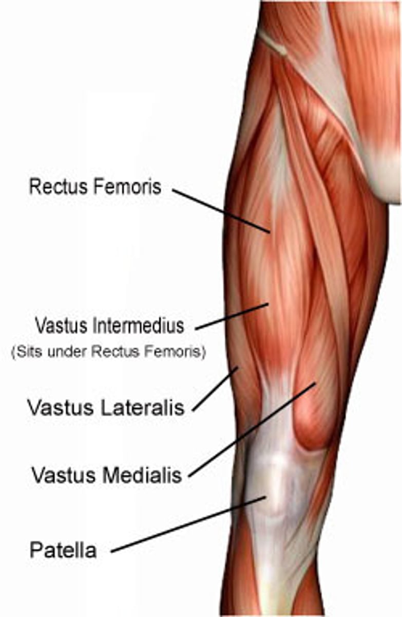
vastus lateralis
-Origin Greater trochanter, Intertrochanteric line, and Linea aspera of the Femur
-Insertion Patella via the Quadriceps tendon and Tibial tuberosity via the Patellar ligament
-Nerve femoral nerve
-Actions Extends and stabilizes knee
-Antagonist Hamstring

vastus intermedius
-Origin antero/ lateral femur
-Insertion Quadriceps tendon
-Nerve femoral nerve
-Actions Extension of knee joint
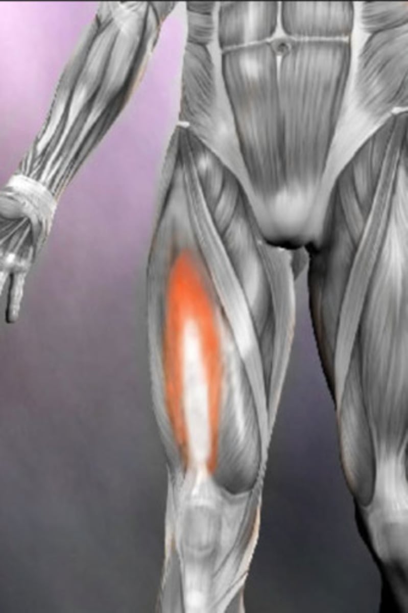
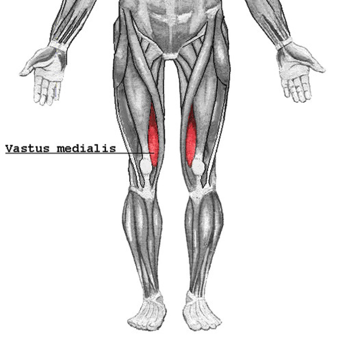
vastus medialis
-Origin Medial side of femur
-Insertion Quadriceps tendon
-Nerve Femoral nerve
-Actions Extends leg
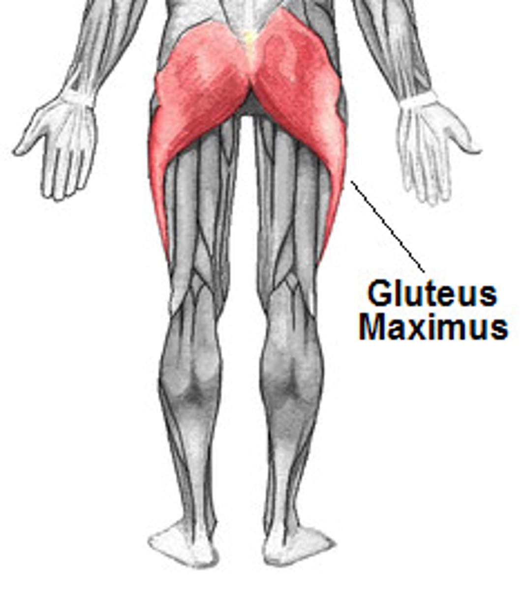
*gluteus maximus*
-Origin Gluteal surface of ilium, lumbar fascia, sacrum, sacrotuberous ligament
-Insertion Gluteal tuberosity of the femur and iliotibial tract
-Nerve Inferior gluteal nerve (L5, S1 and S2 nerve roots)
-Actions
=External rotation and extension of the hip joint
=chief antigravity muscle in sitting and abduction of the hip
-Antagonist iliopsoas
*gluteus medius*
-Origin Gluteal surface of ilium, under gluteus maximus
-Insertion Greater trochanter of the femur
-Nerve superior gluteal nerve (L4, L5, S1 nerve roots)
-Actions
=abduction of the hip
=Medial rotation of thigh.
-Antagonist adductors
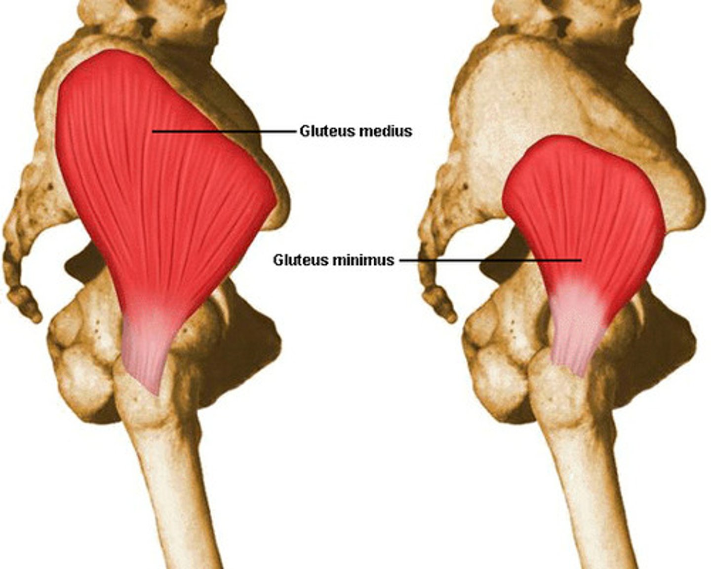
gluteus minimus
-Origin From area in between the anterior gluteal line and inferior gluteal line of Gluteal surface ilium, under gluteus medius.
-Insertion Greater trochanter of the femur
-Nerve superior gluteal nerve (L4, L5, S1 nerve roots)
-Actions
=Works in concert with gluteus medius: abduction of the hip
=Medial rotation of thigh.
-Antagonist lateral rotator group
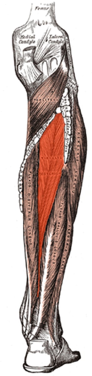
*tibialis posterior*
-Origin Tibia and fibula
-Insertion: Navicular and medial cuneiform bone
-Nerve Tibial nerve
-Actions
=Inversion of the foot.
-Antagonist Fibularis brevis
*GCM*
-Origin superior to articular surfaces of lateral condyle of femur and medial condyle of femur
-Insertion: tendon calcaneus (achilles tendon) into mid-posterior calcaneus
-Nerve tibial nerve from the sciatic, specifically, nerve roots S1-S2
-Actions:
= plantar flexes foot,
=flexes knee
-Antagonist Tibialis anterior muscle
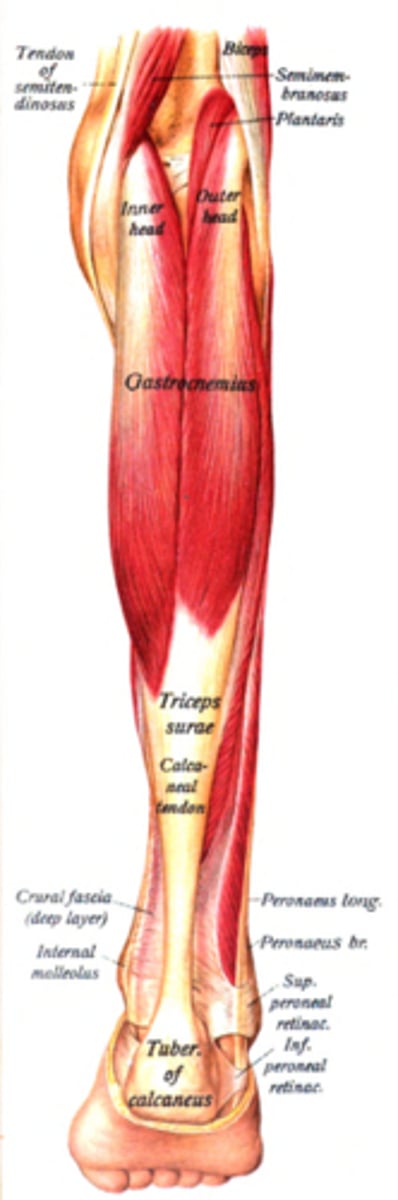
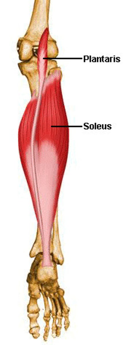
soleus
-Origin fibula, medial border of tibia (soleal line)
=Insertion tendo calcaneus
-Nerve tibial nerve, specifically, nerve roots L5-S2
-Actions plantarflexion
-Antagonist tibialis anterior
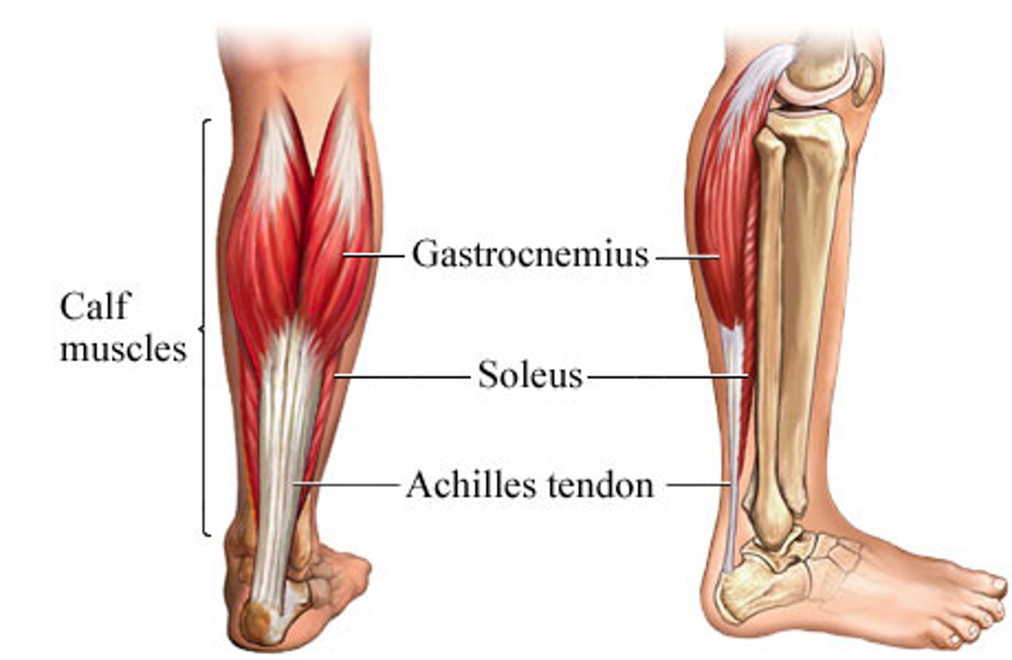
triceps surae
calf muscle:gcm, soleus, achilles tendon
peroneus longus
-Origin Upper lateral shaft of fibula
-Insertion first metatarsal, medial cuneiform
[same insertion as Tibialis Anterior]
-Nerve Superficial fibular (peroneal) nerve
-Actions plantarflexion, eversion,
-Antagonist Tibialis anterior muscle that does Inversion and Dorsiflexion
![<p>-Origin Upper lateral shaft of fibula</p><p>-Insertion first metatarsal, medial cuneiform</p><p>[same insertion as Tibialis Anterior]</p><p>-Nerve Superficial fibular (peroneal) nerve</p><p>-Actions plantarflexion, eversion,</p><p>-Antagonist Tibialis anterior muscle that does Inversion and Dorsiflexion</p>](https://knowt-user-attachments.s3.amazonaws.com/b5a97660-64af-4a22-a26b-495e7d325d9a.jpg)
peroneus brevis
-Origin Lower two-thirds of lateral fibula
-Insertion Fifth metatarsal
-Nerve Superficial fibular nerve
-Actions Plantarflexion, eversion
peroneus tertius
-Origin distal anterior surface of the fibula also the interosseous membrane
-Insertion dorsal surface of metatarsal 5
-Nerve deep fibular nerve
-Actions dorsiflexion and eversion of the foot

plantaris
-Origin: Lateral supracondylar ridge of femur above lateral head of gastrocnemius
-Insertion: Tendon calcaneus (medial side, deep to gastrocnemius tendon)
-Nerve tibial nerve from S1-S2
-Actions Plantar flexes foot and flexes knee
-Antagonist Tibialis anterior muscle
flexor hallucis
-Origin fibula, posterior aspect of middle 1/3
-Insertion Plantar surface; base of distal phalanx of hallux
-Nerve tibial nerve, S2 & S3 nerve roots
-Actions
=flexes all joints of the big toe,
=plantar flexion of the ankle joint
-Antagonist Extensor hallucis longus muscle
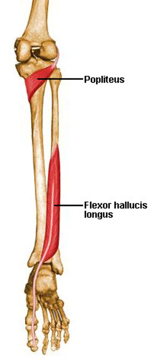
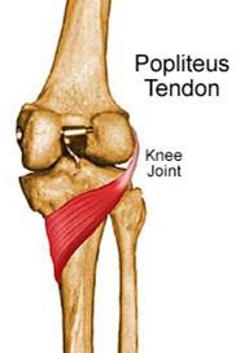
*popliteus*
-Origin lateral femoral epicondyle
-Insertion posterior surface of tibia proximal to soleus line
-Nerve Tibial nerve
-*Actions*
=Medially rotates tibia on the femur if the femur is fixed (sitting down) or
= laterally rotates femur on the tibia if tibia is fixed (standing up),
=unlocks the knee to allow flexion (bending)
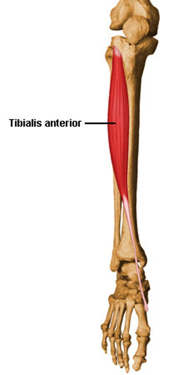
tibialis anterior
-Origin Upper 1/2 & Lateral Condyle of Tibia
-Insertion medial cuneiform and first metatarsal bones of the foot
-Nerve: Deep Fibular (peroneal) nerve (L5)
-Actions Dorsiflexion and Inversion of the foot
-Antagonist Fibularis longus, Gastrocnemius, Soleus, Plantaris, Tibialis posterior
extensor hallucis longus
-Origin Arises from the middle portion of the fibula on the anterior surface and the interosseous membrane
-Insertion Inserts on the dorsal side of the base of the distal phalanx of the big toe
-Nerve deep fibular nerve
deep peroneal nerve, L5 (L4-S1)
-Actions
=Extends the big toe and assists in dorsiflexion of the foot at the ankle.
-Antagonist Flexor hallucis longus, Flexor hallucis brevis

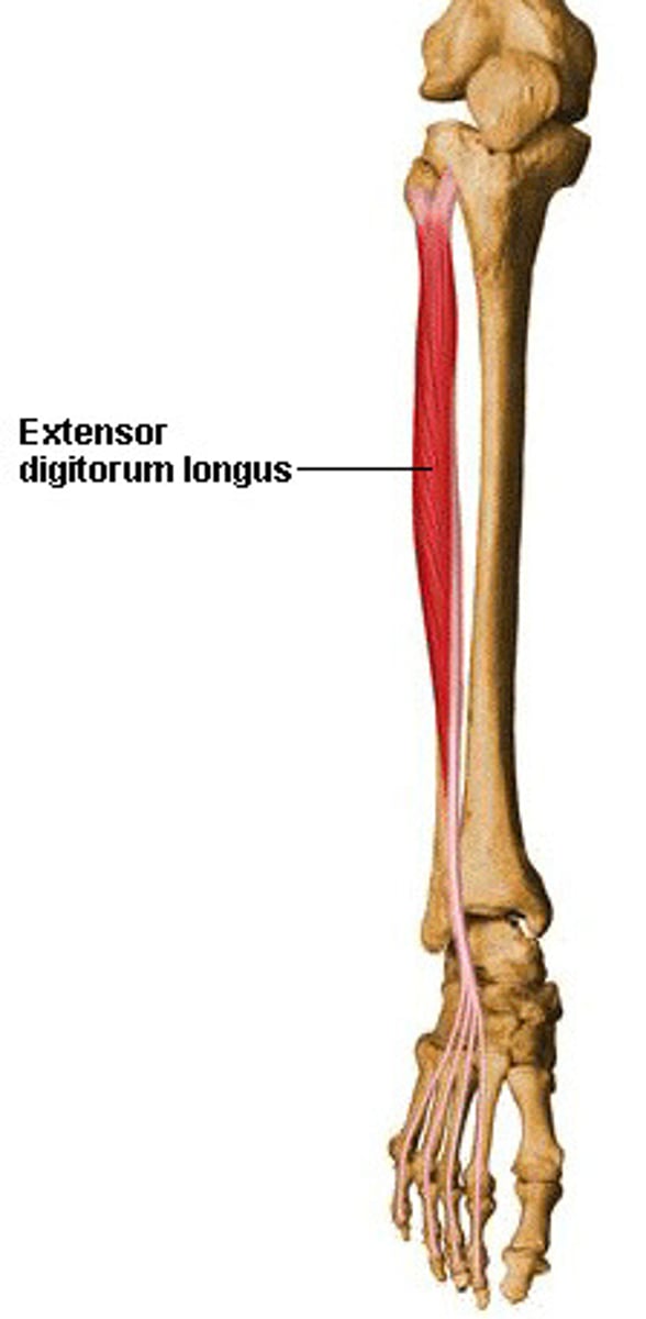
extensor digitorum longus
-Origin Anterior lateral condyle of tibia, anterior shaft of fibula and superior 3⁄4 of interosseous membrane
-Insertion: Dorsal surface; middle and distal phalanges of lateral four digits
-Nerve deep fibular nerve
-Actions
=extension of toes
=dorsiflexion of ankle
-Antagonist Flexor digitorum longus, Flexor digitorum brevis
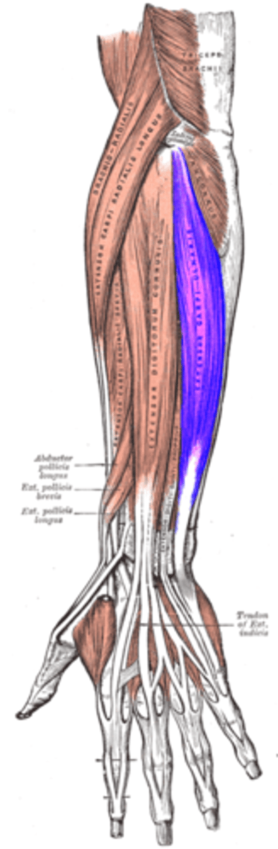
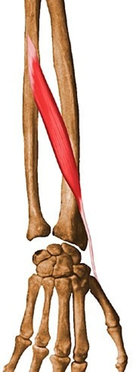
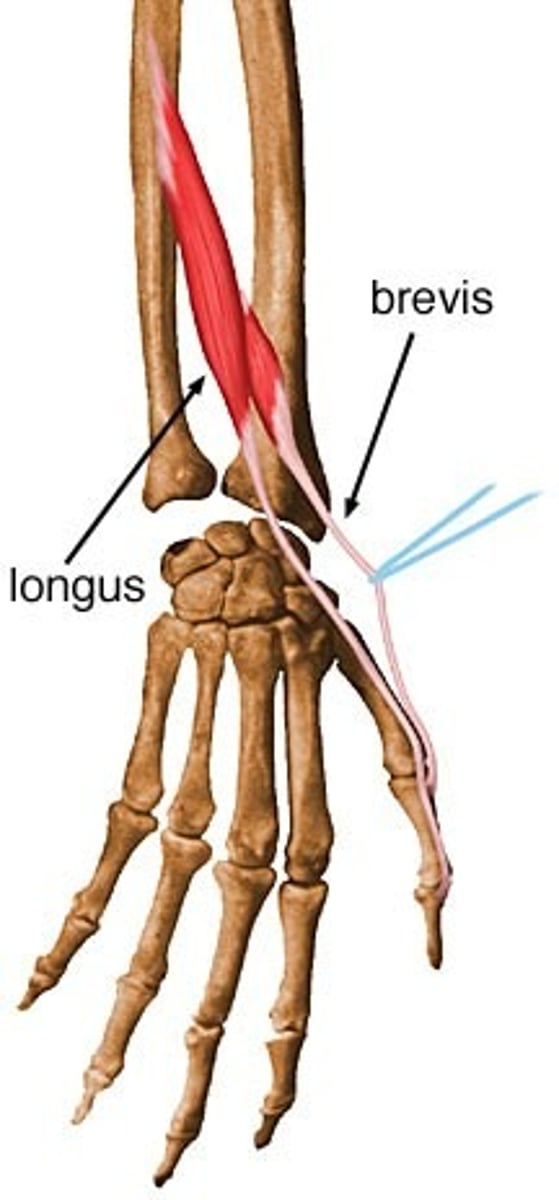
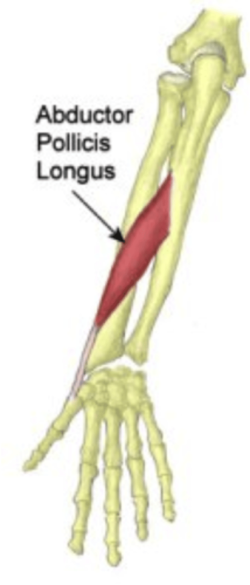
Abductor pollicis longus
Origin; ulna, radius, Interosseous membrane
Insertion; 1st metacarpal
Nerve; Posterior interosseous nerve
from deep branch of radial nerve
C7, C8
Actions; abduction, extension of thumb
Antagonist; Adductor pollicis muscle
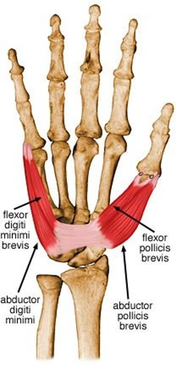
Abductor pollicis brevis
Origin Transverse carpal ligament, the scaphoid and trapezium
Insertion Radial base of proximal phalanx of thumb and the thumb extensors
Nerve Recurrent branch of the median nerve
Actions
=Abduction of the thumb by acting across the carpometacarpal joint and the metacarpophalangeal joint.
=It also assists in opposition and extension of the thumb.
Antagonist Adductor pollicis muscle

Extensor pollicis longus
Origin Middle third of posterior surface of ulna, interosseous membrane
Insertion thumb, distal phalanx
Nerve posterior interosseous nerve (branching from the radial nerve)
Actions extension of the thumb (metacarpophalangeal and interphalangeal)
Antagonist Flexor pollicis longus muscle, Flexor pollicis brevis muscle
Extensor pollicis brevis
Origin radius and the interosseous membrane
Insertion thumb, proximal phalanx
Nerve posterior interosseous nerve
Actions extension of thumb at metacarpophalangeal joint
Antagonist Flexor pollicis longus muscle, Flexor pollicis brevis muscle
Extensor digitorum muscle
Origin lateral epicondyle (common extensor tendon)
Insertion extensor expansion of middle and distal phalanges of the 2nd, 3rd, 4th, and 5th fingers[1]
Nerve radial nerve
Actions extension of hand, wrist and fingers
Antagonist Flexor digitorum superficialis muscle, Flexor digitorum profundus muscle
![<p>Origin lateral epicondyle (common extensor tendon)</p><p>Insertion extensor expansion of middle and distal phalanges of the 2nd, 3rd, 4th, and 5th fingers[1]</p><p>Nerve radial nerve</p><p>Actions extension of hand, wrist and fingers</p><p>Antagonist Flexor digitorum superficialis muscle, Flexor digitorum profundus muscle</p>](https://knowt-user-attachments.s3.amazonaws.com/b11f314f-8843-4c3d-9aee-7fead848c784.jpg)
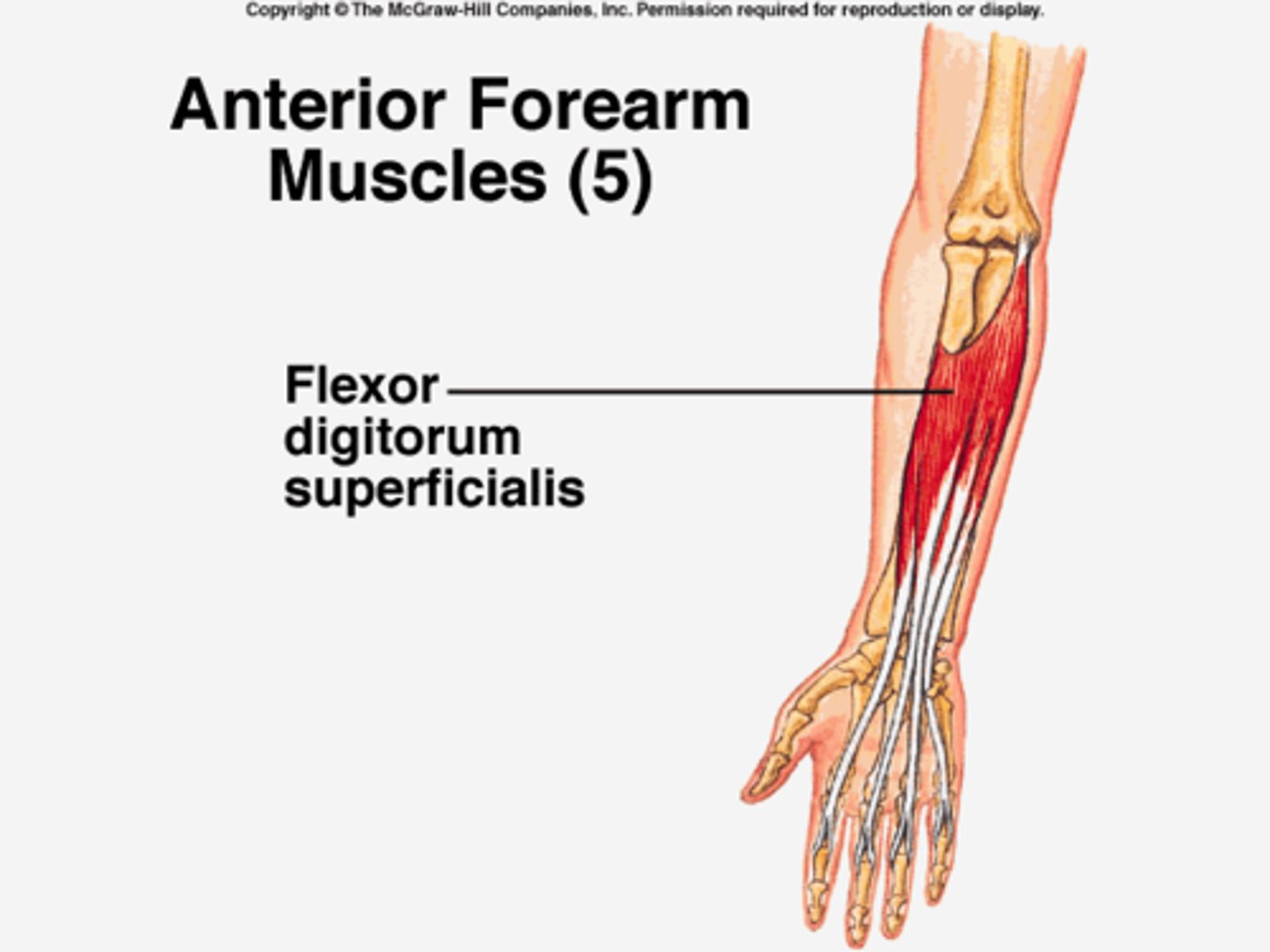
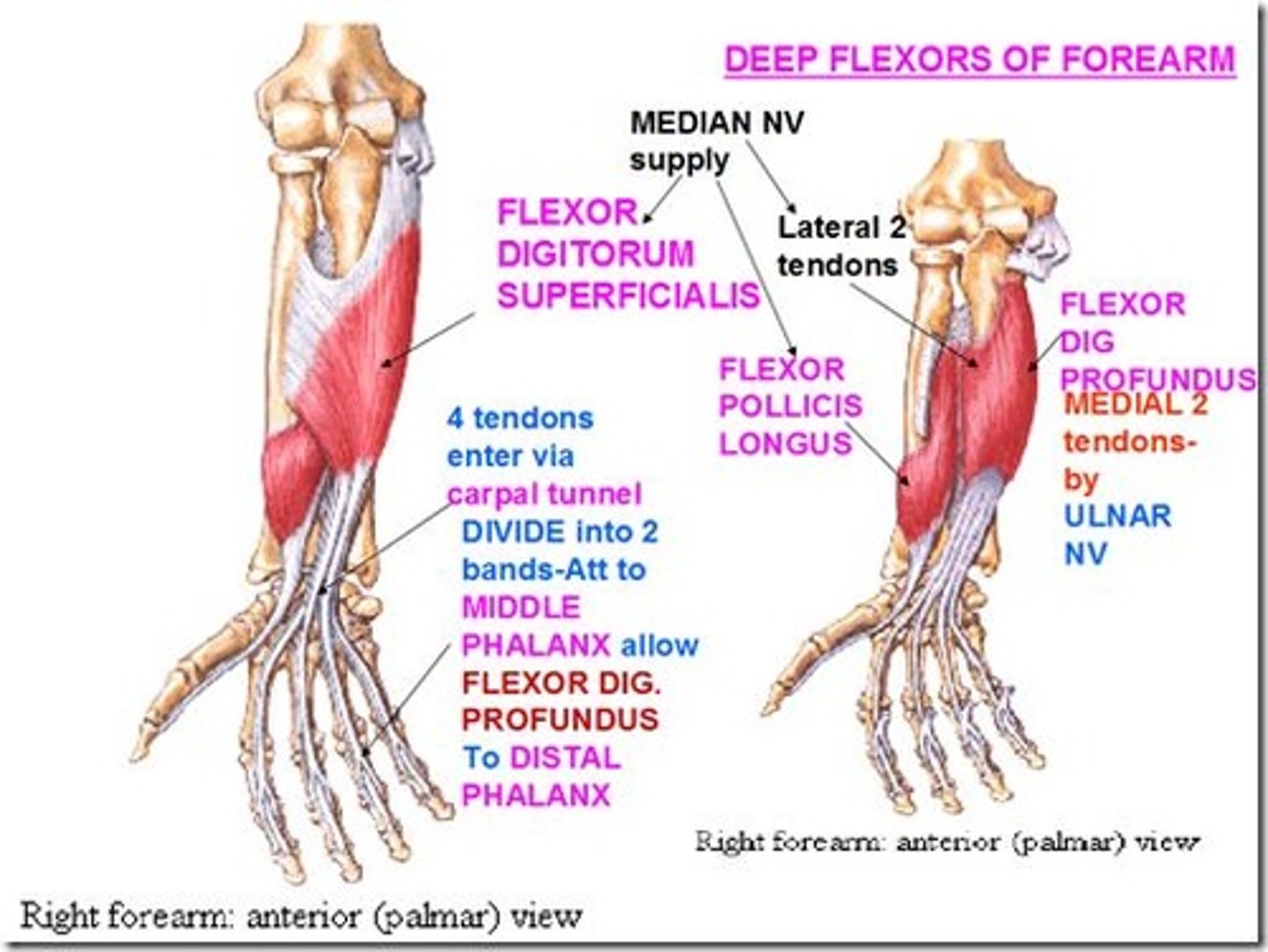
Flexor digitorum superficialis
Origin medial epicondyle of the humerus (common flexor tendon) as well as parts of the radius and ulna.
Insertion anterior margins on the bases of the middle phalanges of the four fingers
Nerve median nerve
Actions flexor of fingers (primarily at proximal interphalangeal joints)
Antagonist Extensor digitorum muscle
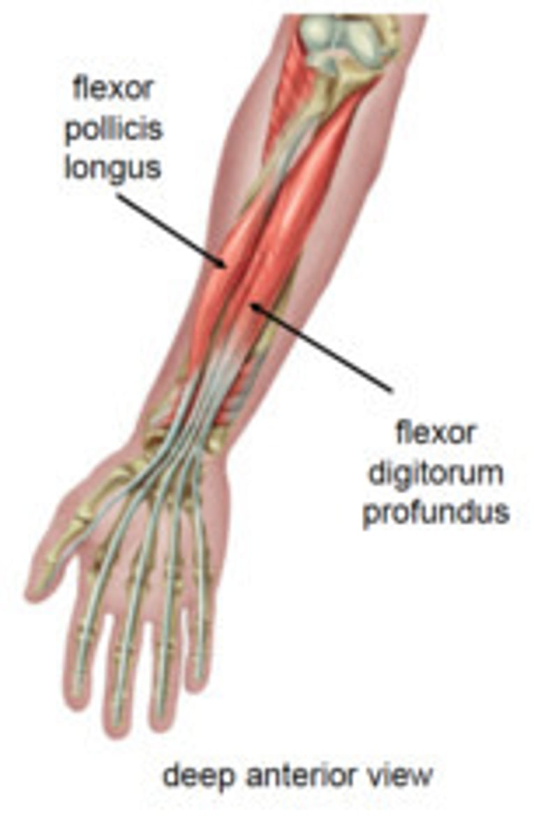
Flexor digitorum profundus
Origin upper 3/4 of the anterior and medial surfaces of the body of the ulna, interosseous membrane and deep fascia of the forearm
Insertion base of the distal phalanges of the fingers
Nerve median (anterior interosseous), muscular branches of ulnar
Actions flex hand and both interphalangeal joints
Antagonist Extensor digitorum muscle
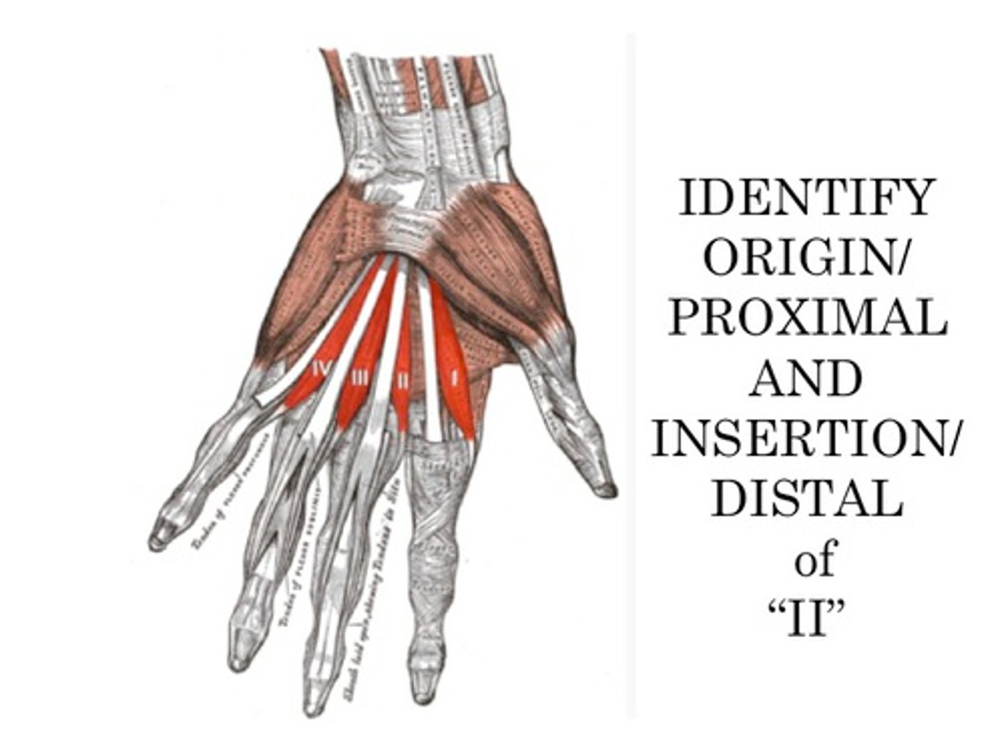
Lumbricals
Origin flexor digitorum profundus
Insertion extensor expansion
Nerve
= 3rd and 4th deep branch of ulnar nerve,
= 1st and 2nd median nerve
Actions
=flex metacarpophalangeal joints,
=extend interphalangeal joints
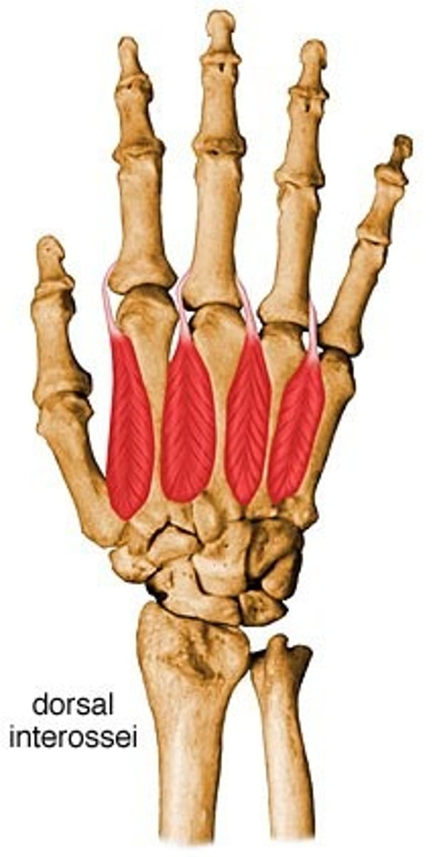
Dorsal interossei
Origin Metacarpals
Insertion Proximal phalanges and extensor expansions
Nerve Deep branch of ulnar nerve
Actions Abduct finger
Antagonist Palmar interossei
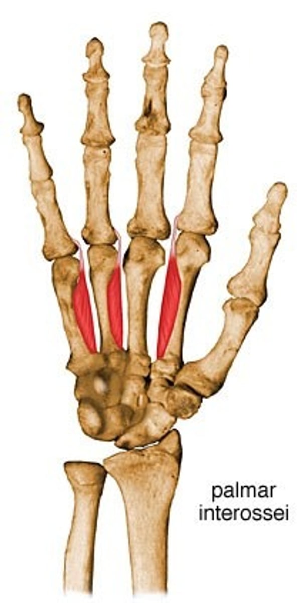
Palmar interossei
Origin Sides of metacarpals facing midline
Insertion Bases of proximal phalanges, extensor expansions
Nerve Deep branch of ulnar nerve
Actions Adduction, flexion and extension of fingers
Antagonist Dorsal interossei
Abductor pollicis longus
Origin ulna, radius, Interosseous membrane
Insertion 1st metacarpal
Nerve Posterior interosseous nerve
from deep branch of radial nerve
C7, C8
Actions abduction, extension of thumb
Antagonist Adductor pollicis muscle
Abductor pollicis brevis
Origin Transverse carpal ligament, the scaphoid and trapezium
Insertion Radial base of proximal phalanx of thumb and the thumb extensors
Nerve Recurrent branch of the median nerve
Actions
=Abduction of the thumb by acting across the carpometacarpal joint and the metacarpophalangeal joint.
= It also assists in opposition and extension of the thumb.
Antagonist Adductor pollicis muscle
Flexor pollicis brevis
Origin trapezium, flexor retinaculum
Insertion thumb, proximal phalanx
Nerve
=Recurrent branch of the median nerve, =deep branch of ulnar nerve (medial head)
Actions Flexes the thumb at the first metacarpophalangeal joint
Antagonist Extensor pollicis longus and brevis

Flexor pollicis longus
Origin The middle 2/4 of the anterior surface of the radius and the adjacent interosseus membrane.
Insertion The base of the distal phalanx of the thumb
Nerve Anterior interosseous nerve (branch of median nerve) (C8, T1)
Actions Flexion of the thumb.
Antagonist Extensor pollicis longus muscle, Extensor pollicis brevis muscle
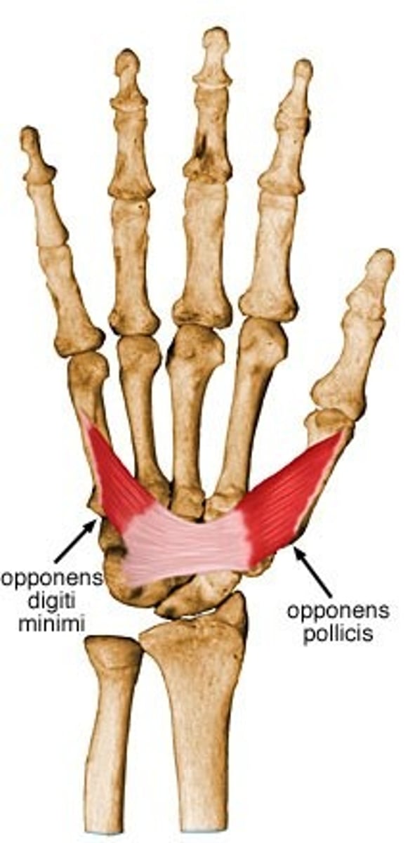
Opponens pollicis
Origin trapezium and transverse carpal ligament
Insertion metacarpal bone of the thumb on its radial side
Nerve Recurrent branch of the median nerve
Actions Flexion of the thumb's metacarpal at the first carpometacarpal joint, which aids in opposition of the thumb
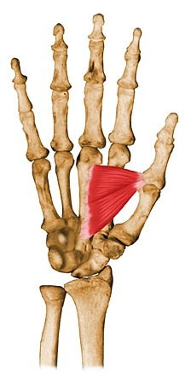
Adductor pollicis
Origin Transverse head: anterior body of the third metacarpal
Oblique head: bases of the second and the third metacarpals and the adjacent trapezoid and capitate bones
Insertion medial side of the base of the proximal phalanx of the thumb and the ulnar sesamoid
Nerve deep branch of the ulnar nerve
Actions adducts the thumb at the carpometacarpal joint
Antagonist Abductor pollicis longus muscle, Abductor pollicis brevis muscle