Oral pathology LO 8 pt. 2`
1/37
There's no tags or description
Looks like no tags are added yet.
Name | Mastery | Learn | Test | Matching | Spaced |
|---|
No study sessions yet.
38 Terms
Adenomas
Benign tumour of the salivary glands.
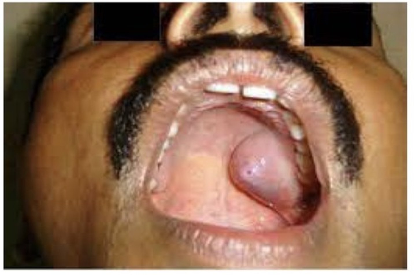
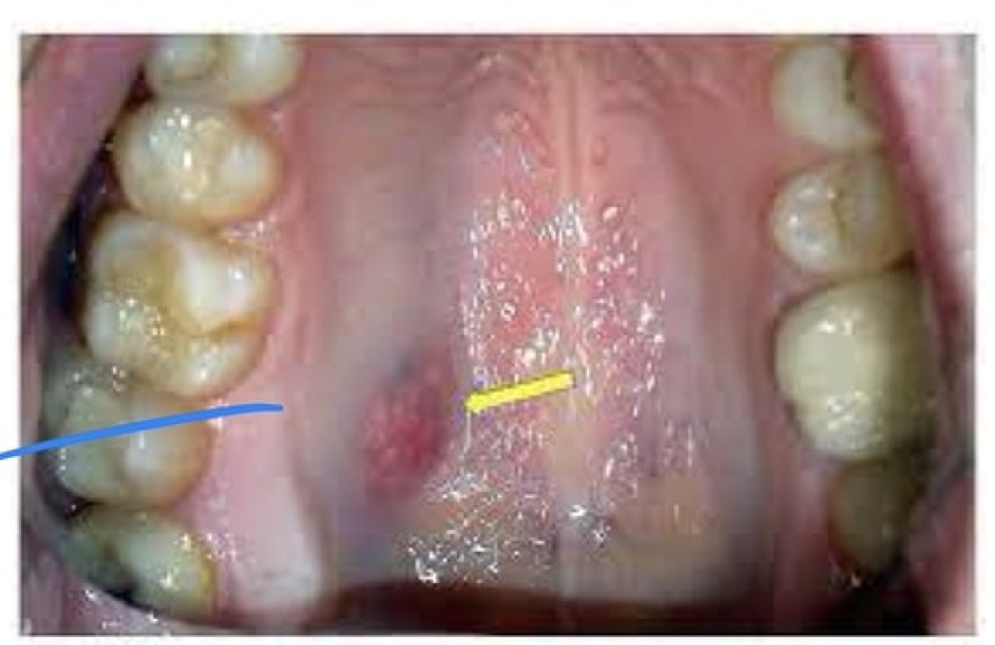
Pleomorphic adenoma (benign mixed tumour)
A benign salivary gland tumour. 90% of all salivary gland tumours. The most common extraoral location is the parotid gland; the most common intraoral location is the palate. Clinically appears as a slowly enlarging, non-ulcerated, painless, dome shaped mass. Tx: surgical excision, recurrence rates vary, they are related to the success of the initial surgical removal. Lesions are known to undergo malignant transformation.
Adenoid cystic carcinoma (cylindroma)
A slow growing malignant tumour of either major or minor salivary gland tissue. May be ulcerated or painful. Most common site: parotid gland. Microscopic: encapsulated, infiltrates surrounding tissue. Small, deeply stinging, uniform epithelial cells that resemble "Swiss cheese". Tx: surgical excision.
Mucoepidermoid carcinoma
A malignant salivary gland tumours. Unencapsulated, infiltrating tumour. Major gland tumours are most often found in the parotid gland, minor gland tumours on the palate. Appear clinically as slowly enlarging masses. May appear in the bone as wither a unilocular or multilocular radiolucency. May occur over a wide age range. Usually in adults but most common malignant salivary tumour in children, occurs most often in females.
Odontogenic tumours
Tumours derived from tooth-forming tissues. They may be composed of epithelium, mesenchyme, or a combination of both. Most are benign, but rare malignant forms exist.
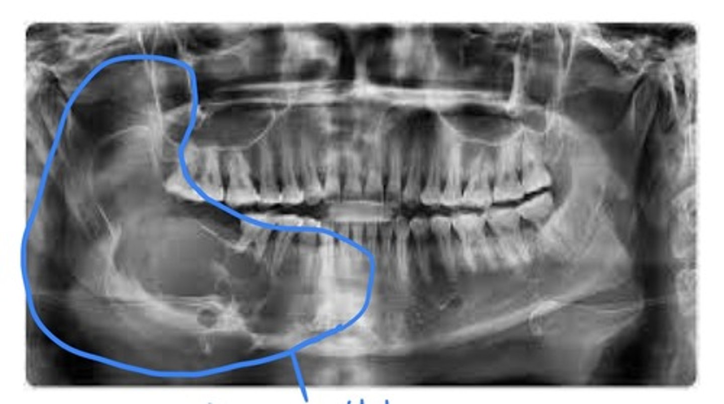
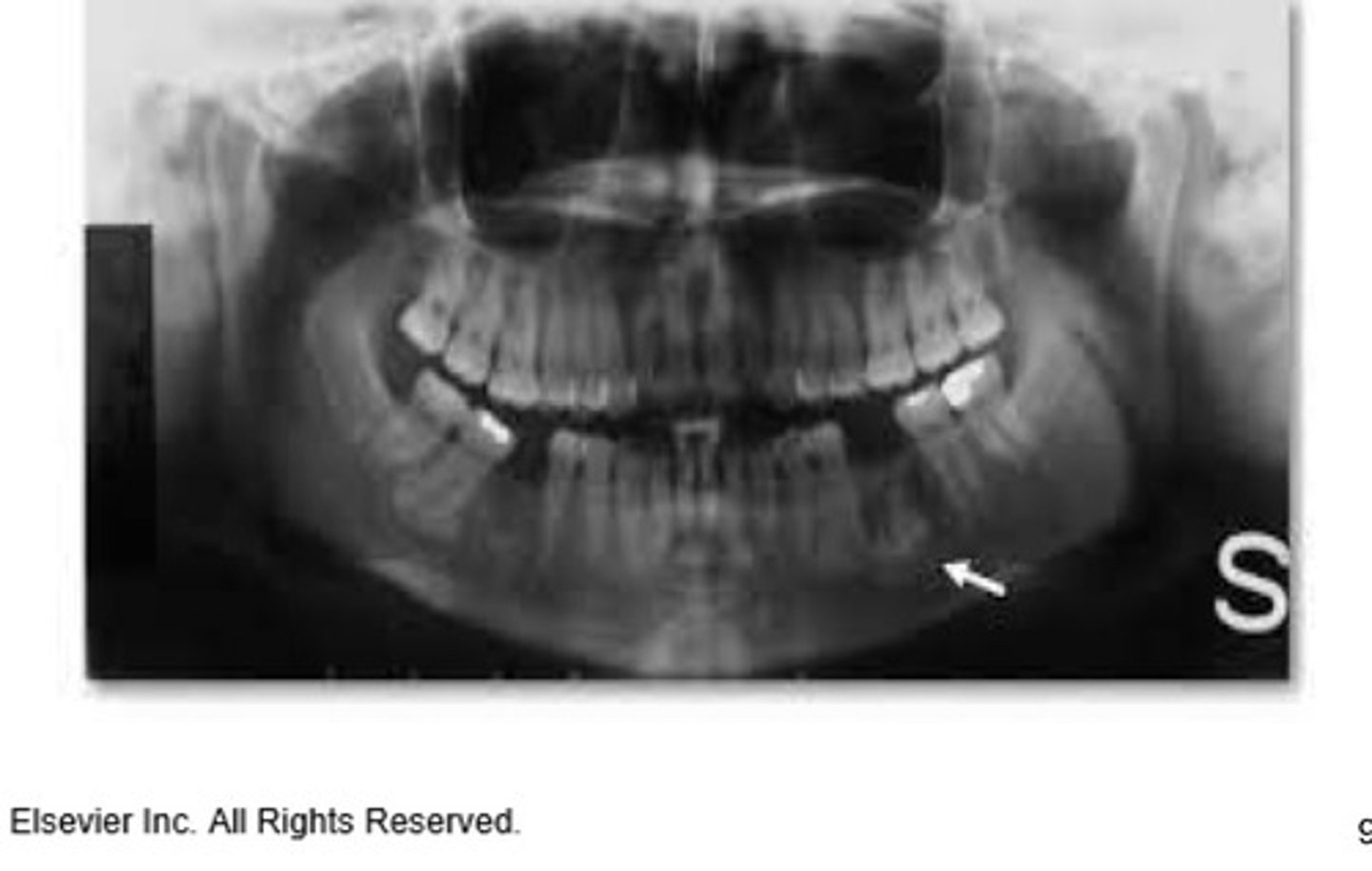
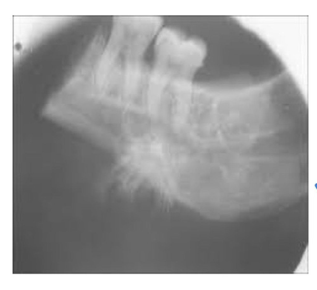
Ameloblastoma
A benign, slow-growing but locally aggressive epithelial odontogenic tumour. May occur in either mandible or maxilla, most often in the mandible in the molar or ramus area. Unencapsulated, infiltrates into surrounding tissue. Radiographic: a multilocular soap bubble-like or honeycombed radiolucency. May occur anywhere in jaws; can occur in association with a dentigerous cyst, may cause bone expansion. Recurrence is common.
Calcifying epithelial ondontogenic tumour
Radiographically: a unilocular or multilocular radiolucency. Occurs more often in the man than max, most often in the bicuspid and molar area.
Adenoid odontogenic tumour (AOT)
An encapsulated, benign epithelial odontogenic tumour. 70% occur in females less than the age of 20. 70% involve the anterior portion of the jaws. More common in MAX, many associated with impacted teeth. Related to puberty/hormonal changes and imbalances. Tumour is surrounded by a dense, fibrous connective tissue capsule. Consists of duct like structures, whorls, and large masses of cuboidal and spindle shaped epithelial cells.
Odontogenic myxoma
A type of mesenchymal odontogenic tumour. A benign nonencapsulated infiltrating tumour. Most often occurs in young ppl age 10-29, no sex predilection. Often brought on by puberty/hormonal changes. Radiographically: multilocular, honeycombed radiolucency w/ poorly defined margins, may become large and displace teeth. Most often occurs in mand.
Central cementifying and central ossifying fibromas
A type of mesenchymal odontogenic tumour. A benign well-circumscribed tumour. Usually occurs in adults in the 3rd and 4th decades. Female sex predilection, more often in mand. Contains connective tissue and calcifications.
Benign cementoblastoma
A type of mesenchymal odontogenic tumour. A cementum-producing lesion. Radiographically: a well defined radiopaque mass with a surrounding radiolucent halo. Tx: enucleation (removal) of the tumour and removal of the involved tooth, does not recur.
Ameloblastic fibroma
This is a type of mixed odontogenic tumour. A benign, nonencapsulated odontogenic tumour. Occurs in young children and young adults, more often in males. Most commonly in the mand bicuspid and molar region.
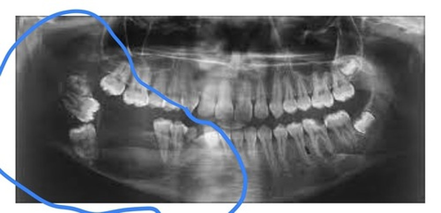
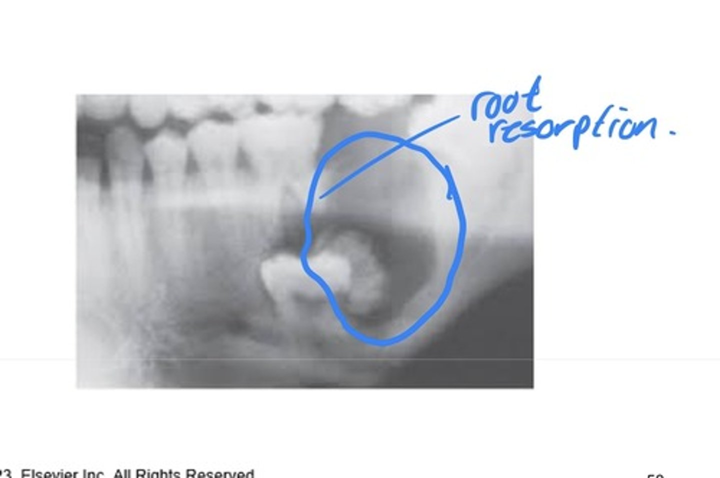
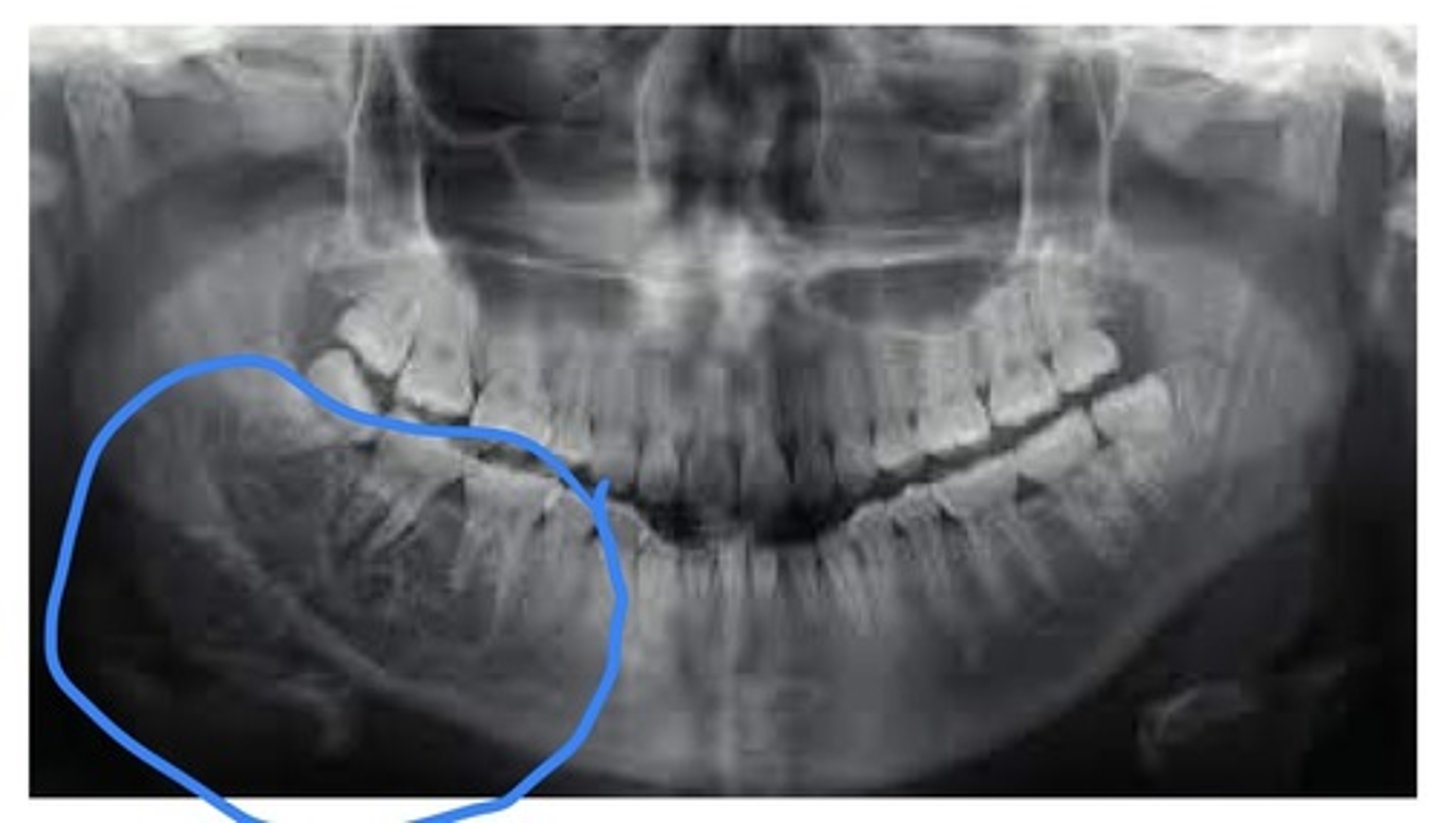
Compound odontoma
An odontoma that resembles teeth usually in the anterior maxilla.
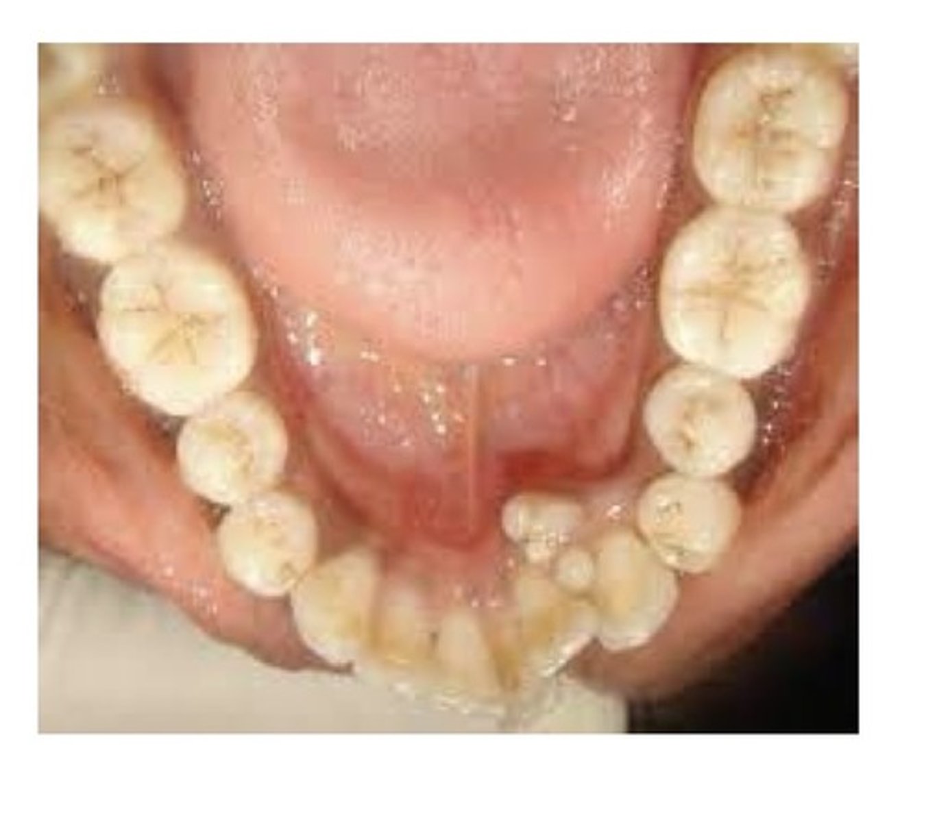
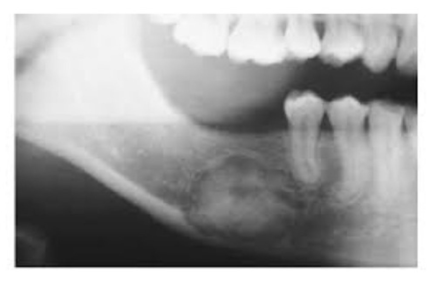
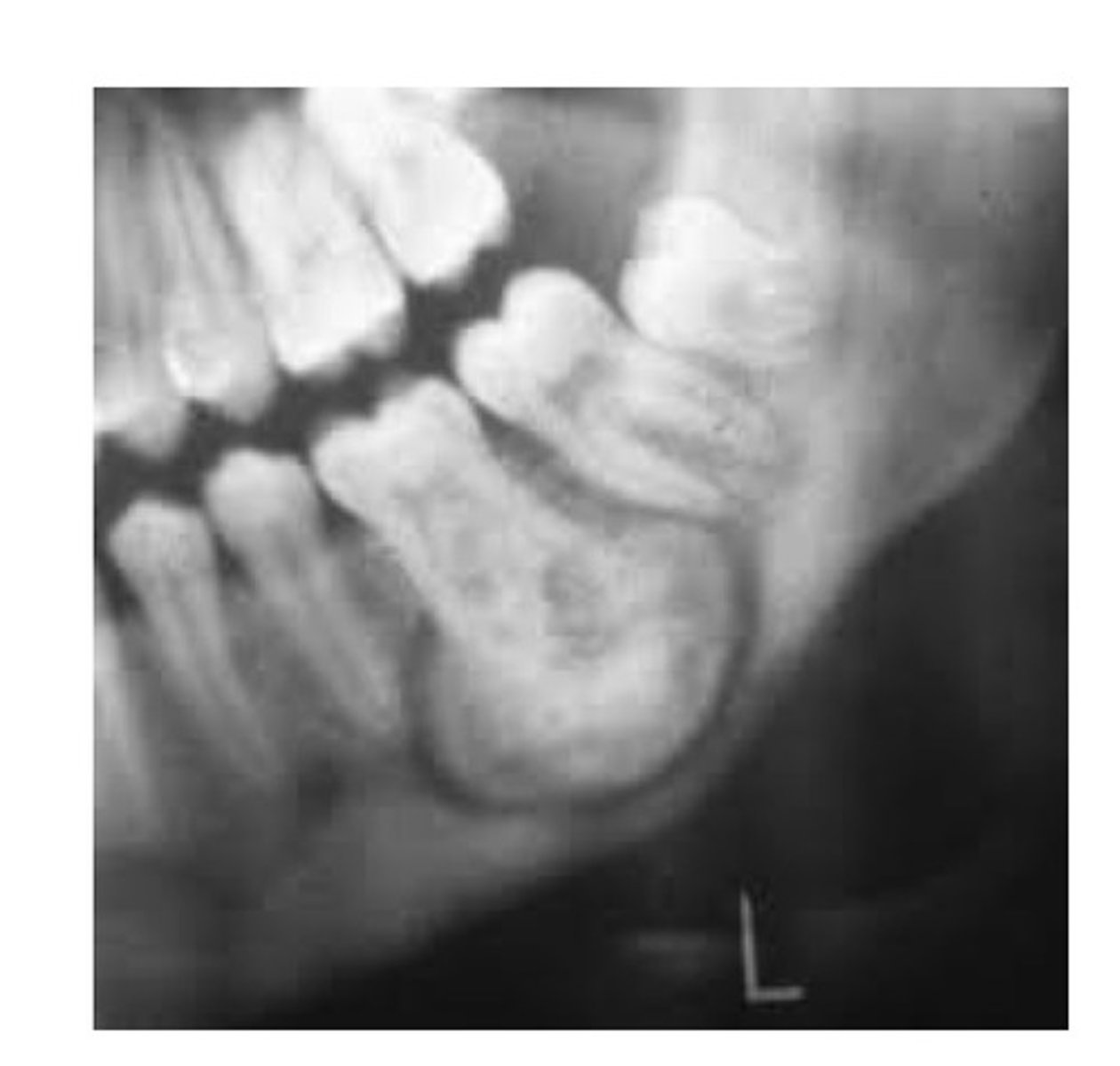
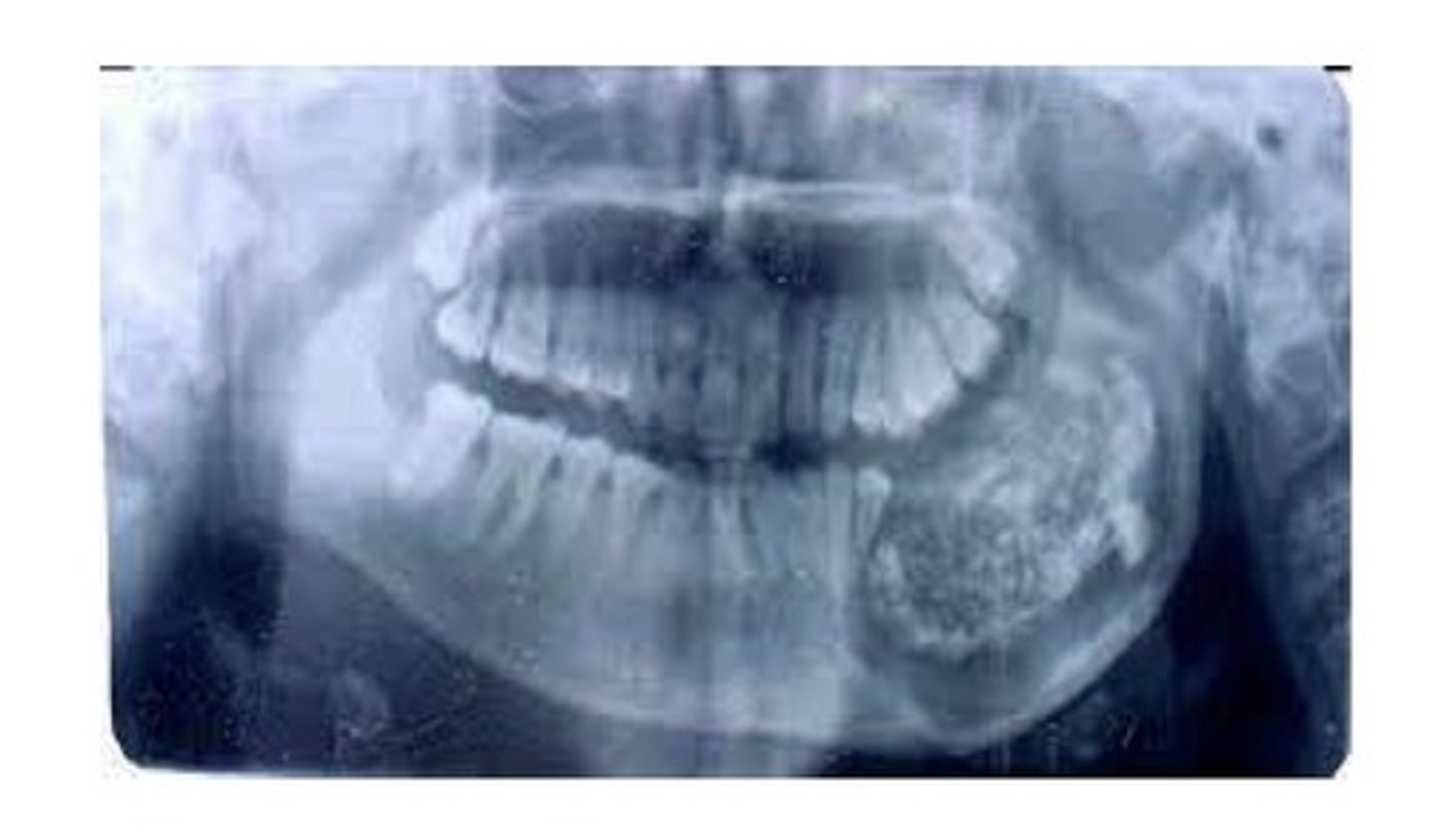
Complex odontoma
An odontoma that does not resemble teeth, usually located in the posterior mandible.
Failure of tooth to erupt
most common clinical manifestation of an ondontoma is:
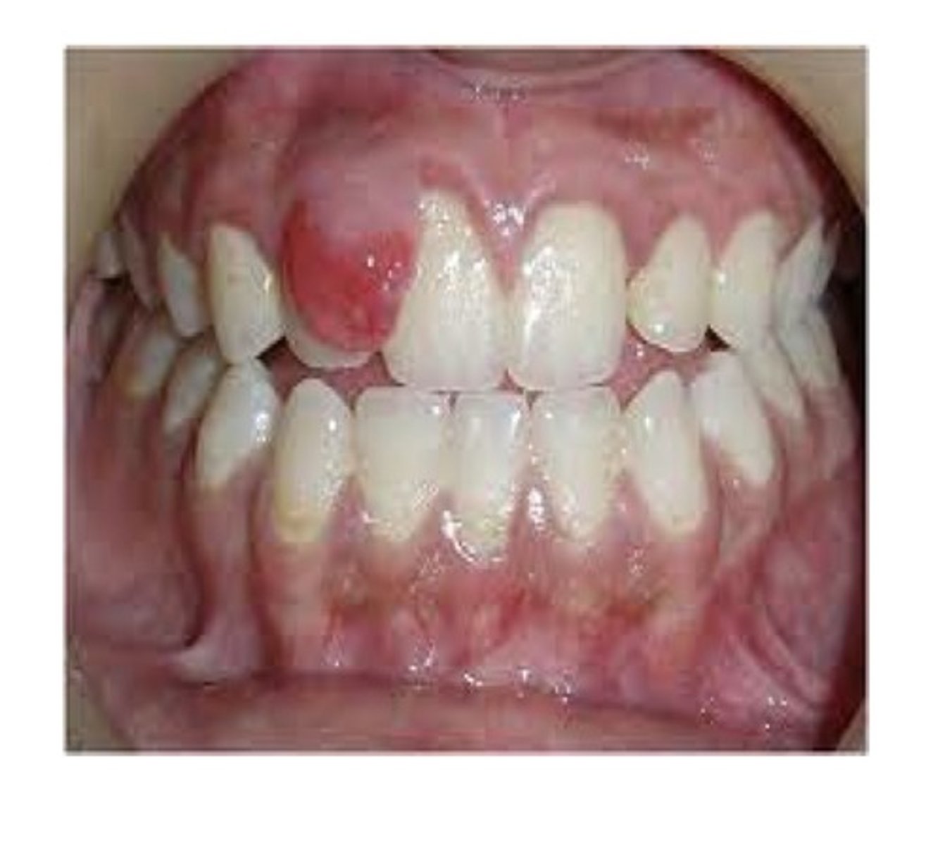
Peripheral ossifying fibroma
A well demarcated sessile or pedunculated lesion, most likely derived from cells of the periodontal ligament. More common in females. Often occurs in young people. Related to hormones and bacterial influence like calculus. Composed of cellular fibrous connective tissue interspersed with scattered bone and cementum-like calcifications. Looks like a pyogenic granuloma (pregnancy tumour)but is different bc it's calcifying.
Lipoma
A benign tumour of mature fat cells. Clinically: a yellowish mass surrounded by a thin layer of epithelium. Most commonly located on the buccal mucosa and vestibule. Most occur in individuals over 40. Microscopic: a well-delineated tumour with mature fat cells uniform in size and shape.
Neurofibroma and schwannoma
These 2 are benign tumours derived from Schwann cells in nerve tissue. The tongue is the most common intraoral location. May occur at any age, with no sex predilection.
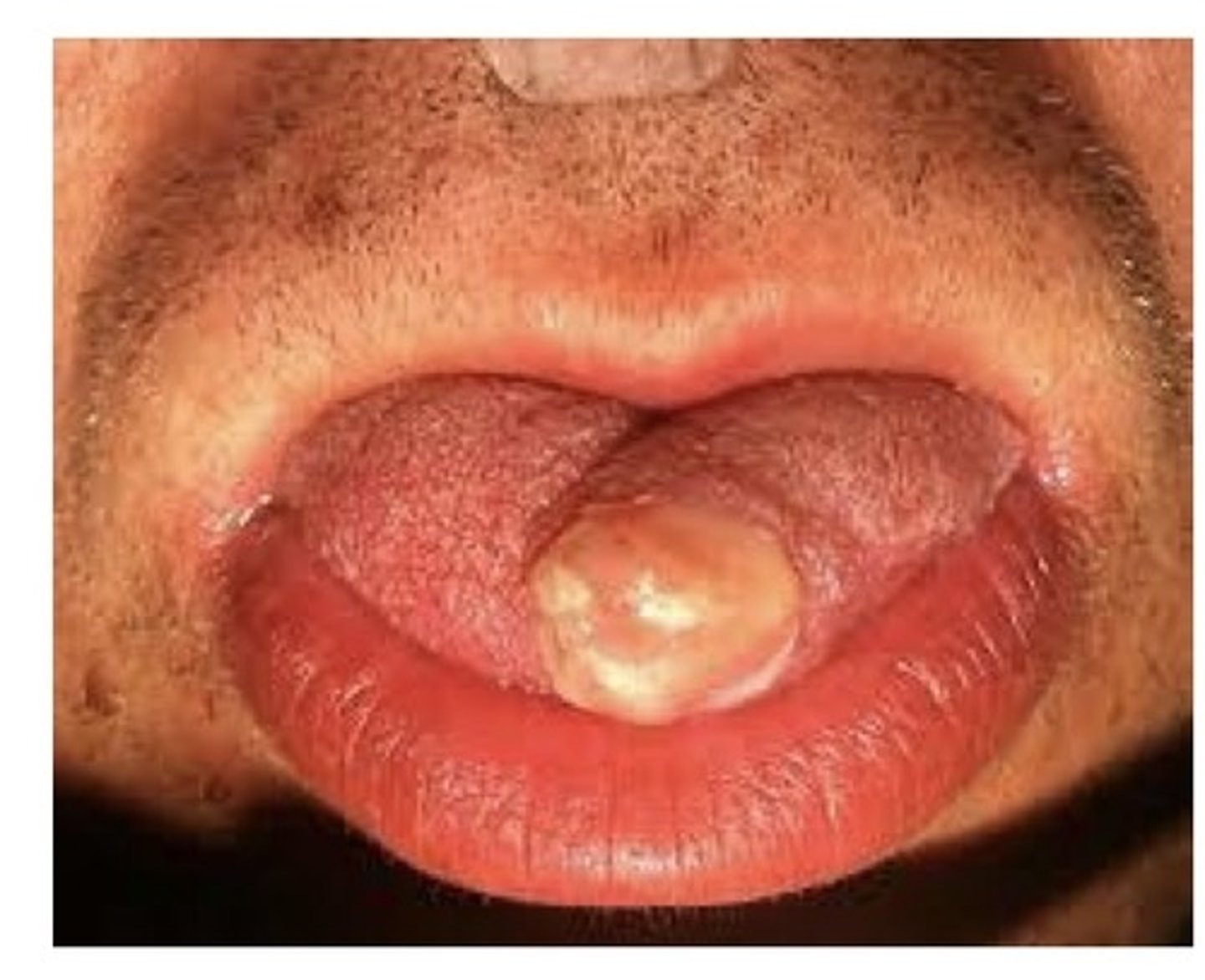
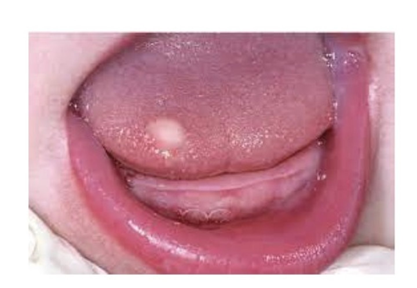
Granular cell tumour
A benign tumour composed of large cells with a granular cytoplasm. Most often occurs on the tongue, followed by the buccal mucosa. A painless, non ulcerated nodule. Most are found in adults, with a female sex predilection.
Rhabdomyoma
A benign tumour of striated muscle.
Leiomyoma
A benign tumour of smooth muscle.
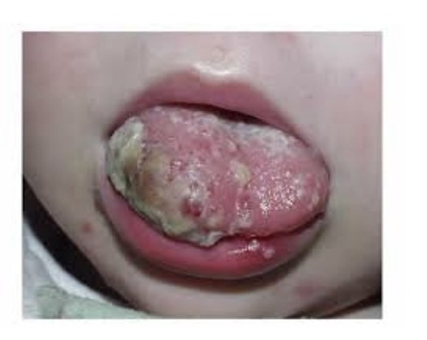
Rhabdomyosarcoma
A malignant tumour of striated muscle. Oral cancer in children is uncommon but out of all kids oral cancer, this is the most common. Typically occurs in children under 10, male sex predilection. Rapidly growing, destructive tumour.
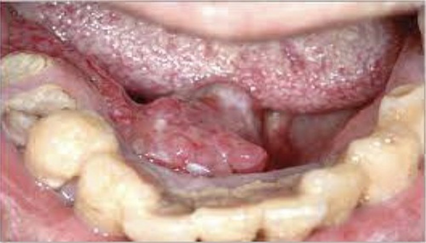
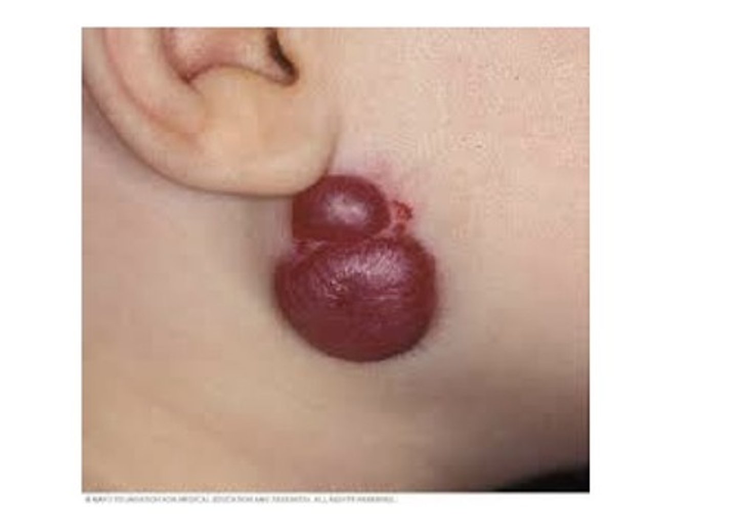
Hemangioma
Sometimes called a birthmark, raspberries, strawberries. A benign proliferation of capillaries. A capillary ———————: contains numerous small capillaries. A cavernous ———————: contains larger blood vessels. Most are present at birth or arise shortly after. Most commonly in the head and neck area, tongue is the most common intraoral location. More common in females, may occur in adults in response to trauma.
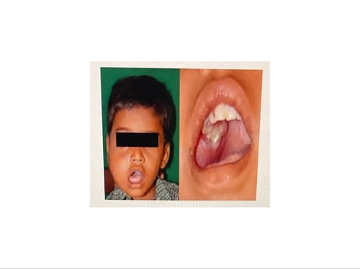
Lymphangioma
A benign tumour of lymphatic vessels. Most are present at birth, half arise in the head and neck area. Intraorally, the most common location is the tongue, where it is an ill-defined mass with a pebbly surface. A cystic —————————— in the neck is called a cystic hygroma.
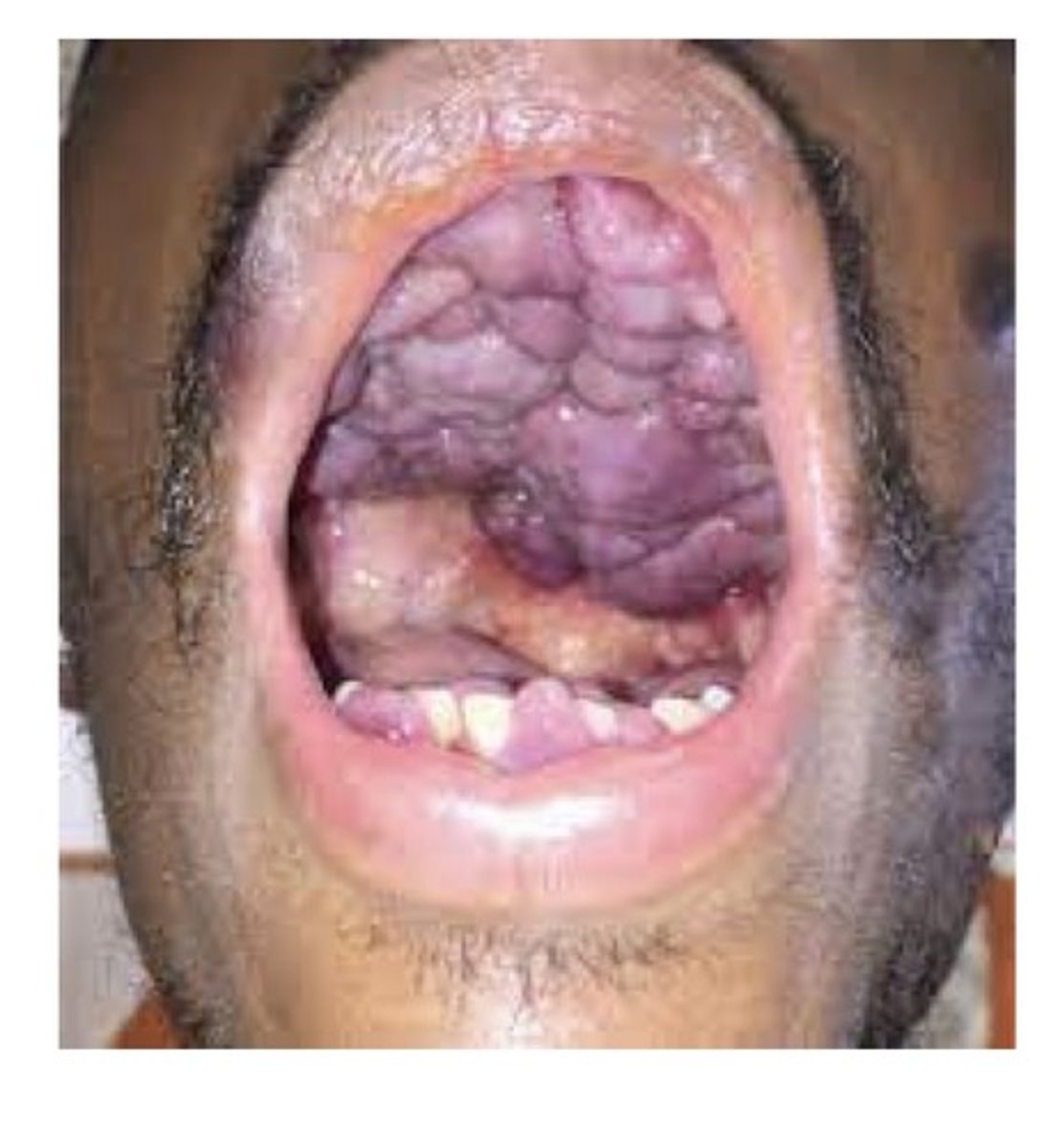
Kaposi sarcoma
These may arise in multiple sites including the skin and oral mucosa. Historically, seen in older men, more aggressive form has arisen with HIV. Lesions are often seen in the oral cavity as purple macules, plaques, or exophytic tumours. Most commonly on the hard palate and gingiva. May also occur in pts with immunodeficiency.
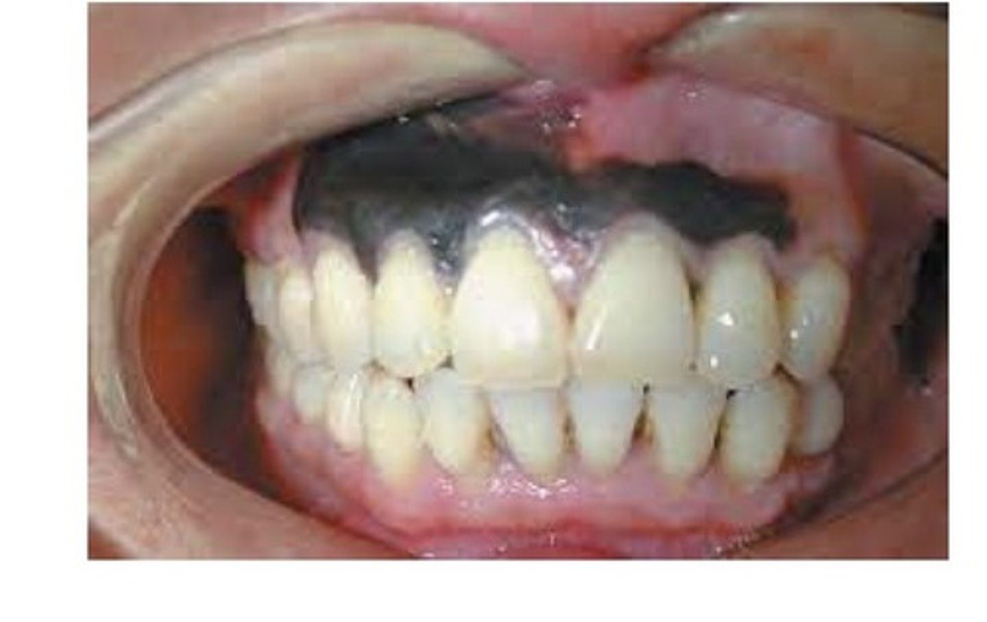
Melanocytic nevus
———— may refer to either a developmental tumour of melanocytes or a pigmented congenital lesion, also known commonly as a "mole". Can arise on the skin or oral mucosa. Intraoral tumours consist of tan-to-brown macules or paperless. Occur most often on the hard palate or buccal mucosa. Occur twice as often in woman as men.
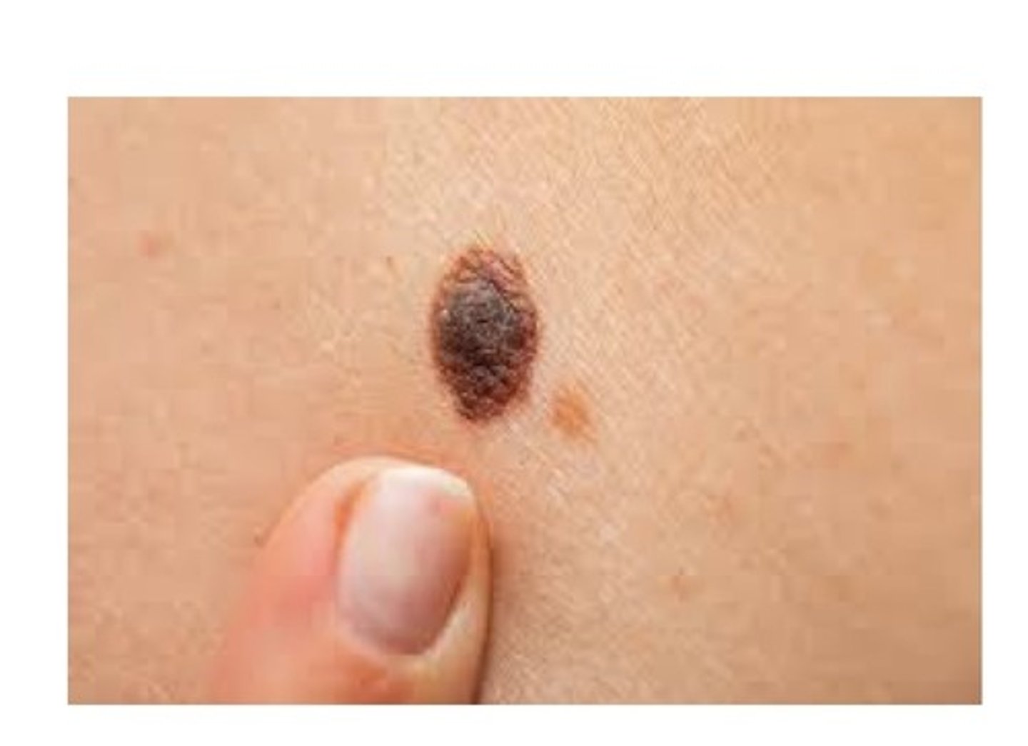
Raised, texture (scaly, flaky, ulcerated), colour, diffuse boarder, size change.
What are some signs that a mole has become malignant?
Malignant melanoma
A malignant tumour of melanocytes. Most arise on the skin in response to prolonged exposure to sunlight. Usually rapidly enlarging black-to-blue mass. And aggressive tumour with unpredictable behaviour and early metastasis. Most common intraoral locations are palate and max gingiva. Adults over 40.
Osteoma
An asymptomatic meningitis tumour composed of benign compact bone. Radiographically: appears as an either a sharply delineated radiopaque mass within a bone or attached to outer surfaces of bone.
Osteosarcoma (osteogenic sarcoma)
A malignant tumour of bone forming tissue. Most common primary malignant tumour of bone in patients less than 40 yrs of age. Occurs twice as frequently in mandible as maxilla. More common in males. Radiographically: asymmetric widening of the PDL space, and a "sunburst" pattern may be seen.
Chondroma
A benign tumour of cartilage is called a:
Chondrosarcoma
A malignant tumour of cartilage is called:
Leukemia
A broad group of disorders characterized by an overproduction of atypical white blood cells. The types of ——————— seen are classified according to the kinds of cells that are proliferating.
Acute leukemia
This type of leukemia is most common in children and young adults. Characterized by a proliferation of immature white blood cells.
Chronic leukemia
This type of leukemia most frequently occurs in middle aged adults. Characterized by excess proliferation of mature white blood cells.
Lymphoma (non-Hodgkin lymphoma)
A malignant tumour of lymphoid tissue. Clinical presentation: granule enlargement of lymph nodes. The most common intraoral location is the tonsils. Usually occurs in adults. More common in males than females.
Multiple myeloma
A systemic, malignant proliferation of plasma cells. Causes destructive lesions in bone. Most patients are older than 40 years, occurs most common in a pts 70s. Males are affected more than females. Pts usually experience bone pain and swelling. Mand more than max.
Metastatic tumours
——————— tumours from primary sites elsewhere in the body are rare. Most tumours arise from the thyroid, breast, lungs, prostate gland, and kidneys. Most frequent intraoral site for ——————— tumours in the mandible. Pts may experience pain, parasthesia or anesthesia of the lip, swelling, expansion of the affected bone, and loosening of the affected area.most pts are adults, males more often than females.