IGCSE Edexcel Biology: structure of heart + circulatory system
1/23
Earn XP
Description and Tags
Name | Mastery | Learn | Test | Matching | Spaced |
|---|
No study sessions yet.
24 Terms

Name structures 1,2,5
1 — aorta
2 — superior vena cava
5 — right atrium
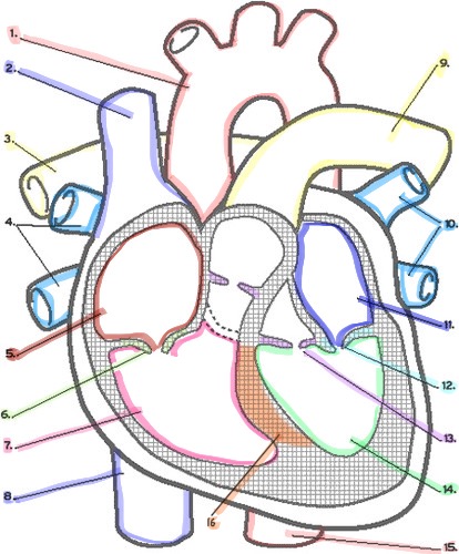
Name structures 6,7,8
6 — tricuspid valve
7 — right ventricle
8 — inferior vena cava (same part as superior vena cava)

Name structures 9,10,11
9 — pulmonary artery
10 — pulmonary vein
11 — left atrium

Name structures 12,13,14,16
12 — bicuspid valve
13 — semilunar valve
14 — left ventricle
16 — septum
Show the pathway of blood. El diagram de la miss Kitchen
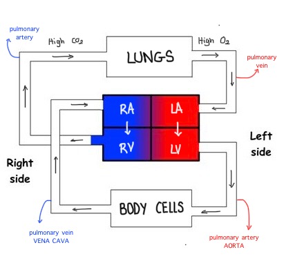
What is meant by the term double circulatory system? In which organisms?
In one complete cycle, blood is pumped into the heart twice (blood pumped from heart → gas exchange organ (lung) → back to heart → rest of body) It invokes 2 circulation:
Pulmonary (circulation between lung and heart. From deoxygenated to oxygenated)
Systemic (circulation between heart and body. From oxygenated to deoxygenated)
Humans and other mammals
What is meant by the single circulatory system? Which organisms?
A system where the blood is pumped from the heart to the gas exchange organ, and then directly to the rest of the body.
Fish
What type of circulatory system is more efficient?
Double circulatory system
From textbook: it is more efficient because heart pumps twice, so higher pressure maintained = blood travels quickly to organs (O2).
Not in textbook: It prevents oxygenated and deoxygenated blood from mixing which increases the efficiency of oxygen transport and energy production in the body. And helps to alter the pressure from the different chambers of the heart (both circulation systemic and pulmonary can be controlled independently)
The human circulatory system comprises
The heart - pump
Blood vessels - carry blood around body
Blood - the transport medium
Which side does oxygenated (high O2) blood enter? What is the destination? Why kind of circulation is this + path?
Left side of the heart → body cells
Systemic circulation:
Left side of the heart — oxygenated blood through atrium → ventricle → aorta → rest of the body cells → returns deoxygenated blood through vena cava to right side of heart
Which side does deoxygenated blood (high CO2) enter? What is the destination? Why kind of circulation is this + path?
Right side of heart → lungs
Pulmonary circulation
Right side of heart — deoxygenated blood through atrium → ventricle → pulmonary artery → lungs → oxygenated blood returns to left side heart through pulmonary vein.
How is the left ventricle different from the right? Why? (4)
The left ventricle has a thicker (1) muscle (1) wall than the right ventricle as it has to pump blood at high pressure (1) around the entire body (1).
The right ventricle is pumping blood at a lower pressure to the lungs prevents damage to capillaries in the lungs and lungs are closer.
How are the walls of the atria? What does this allow the atria to do?
The walls of the atria are thin. This allows them to be stretched to receive blood as it returns to the heart, but it can contract with enough force to push blood through the bicuspid and tricuspid valves into the ventricles
- Higher pressure
What is the septum + its function? What would happen if there was no septum?
It is a muscle wall that separates both sides of the heart so that oxygenated blood doesn't mix with deoxygenated blood.
Mixing of theses two will decrease the efficency of blood to carry oxygen and our organs won't be able to work properly.
Describe what would happen to the flow of blood in the left side of the heart if the bicuspid valve did not function effectively (2)
Flow of blood cannot pass to ventricle (goes backwards)
Less oxygenated blood pumped to body
Blood is pumped ____ the heart in veins and ____ from the heart in arteries.
towards
away (arteries away)
The coronary arteries supply the ____ of the heart with oxygenated blood. Why?
Cardiac muscle tissue.
As the heart is a muscle it needs constant supply of oxygen (and glucose) for aerobic respiration to release energy to allow continued muscle contraction. If not enough oxygen is supplied, it can damage or result in the death of the heart muscle.
How are the cardiac muscles different from other muscles in our body?
They can contract and relax continuously be without becoming fatigued
Aorta — where blood flows away from the heart TOWARDS the body cells.
Vena cava — where blood flows from body cells INTO the right atrium.
- Deoxygenated blood coming from the body flows through the vena cava and into the right atrium.
- The atrium contracts and the blood is forced through the tricuspid valve into the right ventricle.
- The ventricle contracts and the blood is pushed through the semilunar valve into the pulmonary artery.
- The blood travels to the lungs and moves through the capillaries past the alveoli where gas exchange takes place
- Low pressure blood flow on this side of the heart prevents damage to the capillaries in the lungs.
- Oxygenated blood returns via the pulmonary vein to the left atrium.
- The atrium contracts and forces the blood through the bicuspid valve into the left ventricle.
- The ventricle contracts and the blood is forced through the semilunar valve and out through the aorta.
- Thicker muscle walls of the left ventricle produce a high enough pressure for the blood to travel around the whole body.