Chapter 1
1/40
Earn XP
Description and Tags
Name | Mastery | Learn | Test | Matching | Spaced |
|---|
No study sessions yet.
41 Terms
how are microorganisms grown and studied?
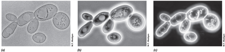
Microscopy
Culture: cells grown in/on nutrient medium
Very specific needs
Medium: liquid/solid mixture containing all required nutrients
Growth analysis
Assays
Spectrophotometer
Etc.
Bacterial colonies
Shapes, growth, color, etc.
Molecular, biochemical, genetic analysis
what are some key features of microbial cells?
Create energy
metabolic/anabolic reactions
Harness energy
Must replicate
Mitosis or binary fission
Evolution
Goes faster than most species
Horizontal gene transfer
Can share once alive, not vertically like we do (birth)
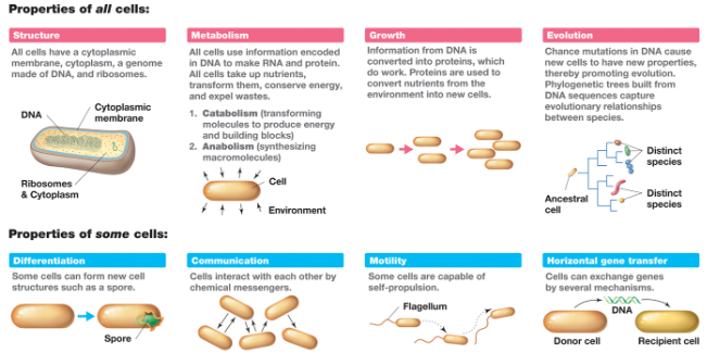
what are some additional features microbial cells may have?
Can become different types of cells
Endospores
Typically triggered by a crappy environment (too cold, hot, treated incorrectly, etc.)
Tough sugary coat and some ribosomes, really tough seed, goes dormant
Better place with better nutrients = new home and can grow
Autoclave is the only way we can destroy it
Can talk to each other
Corum sensing
Motility
Some move
Single flagella, some have multiple
Same movement as sperm

cell size of prokaryotes
0.2 to 600 um in diameter
cell size of eukaryotes
5-100 um in length
why is cell size important for microorganisms?
They are small because they are simple, not very complex
Uses diffusion to get rid of waste products and get nutrients
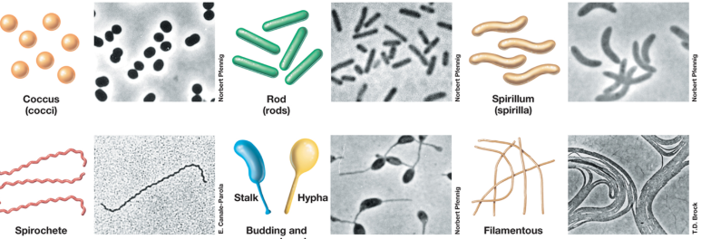
what are some bacteria shapes?
Coccus (circle)
Rod, bacillus, bacilli (rod)
Spirillum (spirula) (squiggle)
Spirochete (squiggle)
Much more rigid in shape than spirilla
Budding and appendaged (balloon shaped)
Filamentous (looks like spaghetti)
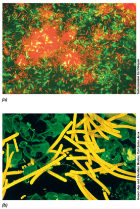
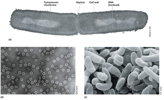
bacteria
Prokaryotes
Usually undifferentiated single cells 0.5-10 um
80+ phylogenetic lineages
archaea
Prokaryotes
Historically associated with extreme environments but not all of them are extremophiles
Thermal vents
Glaciers
Lack known parasites or pathogens of plants and animals
12+ phyla
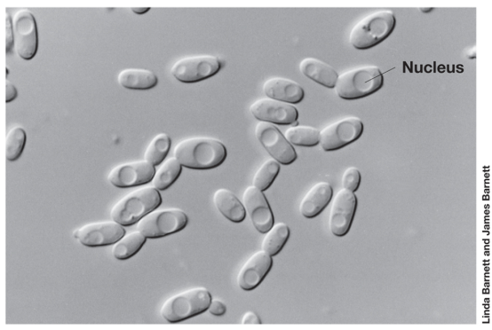
eukarya
Plants, animals, fungi
First were unicellular, may have appeared 2 billion years ago
At least 6 kingdoms
Varies dramatically in size, shape, physiology
Can change shapes depending on environment
viruses
Obligate parasites that only replicate within host cell
Not cells
Doesn’t carry out metabolism; take over infected cells to replicate
Have small genomes of double-stranded or single-stranded DNA or RNA
Classified based on structure, genome composition, and host specificity
Naming is confusing
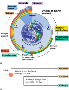
origin of cell domains
The origin of earth
Bacteria, archaea, and eukarya distinguished by 4 BYA
Descended from last universal common ancestor (LUCA)
The sun then let them go through photosynthesis (anoxic photosynthesis first, then eventually produces oxygen aka anaerobic (photosynthesis)
what are the good and the bad of microorganisms?
Bad
Can cause food spoilage and foodborne disease
Fermentation process
Glycolysis into fermentation paths
Good
Improve food safety
Preservation
Yummy foods and drinks
what types of cells are hypothermophiles?
archaea because they like extremely high temperatures
what types of cells are psychrophiles?
bacteria because they like extremely low temperatures
what types of cells are acidophiles?
archaea because their pH are extremely low
what types of cells are alkaliphiles?
archaea because their pH are extremely high
what types of cells are barophiles?
bacteria because they like extremely high pressure
what types of cells are halophile?
archaea because they like extremely high concentrations of salt
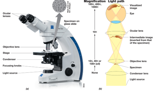
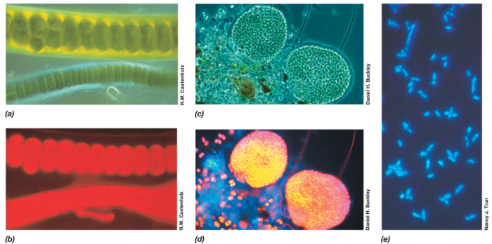
light microscope
Ocular lens = 10x magnification
Low power = 4x
10x x 4x = 40x magnification
10x x 10x = 100x magnification
Oil immersion = 100x
10x x 100x = 100x magnification
DON’T USE UNLESS YOU HAVE OIL
Condenser to control how much light you have in the microscope
When light moves through different medium, it can show very differently due to how fast/slow the light is
phase contrast microscope
converts phase shifts in light passing through a transparent specimen to brightness to change the image
fluorescence microscopy
optical microscope that uses fluorescent light instead of scattering light
differential interference contrast microscopy
uses polarized light and specialized prisms to enhance contrast in unstained samples, which reveals a detailed surface
confocal scanning laser microscopy
uses a laser beam to scan a sample point-by-point, and a pinhole to block out-of-focus light
electron microscope
Uses electrons instead of light to image cells and structures
Electromagnents function as lenses
Operates in a vacuum
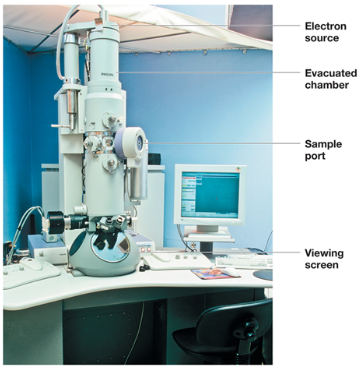
transmission electron microscope (TEM)
uses a beam of high-energy electrons that can pass through a thin sample to produce a highly magnified image

scanning electron microscope (SEM)
scans the surface with a focused beam of electrons
magnification
ability to enlarge an image
resolution
the ability to distinguish 2 adjacent objects as distinct and separate
who was the first to describe microbes
Robert Hooke
who was the first to see bacteria?
Antoni van Leeuwenhoek
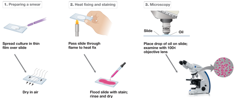
specimen preparation
No cover slips because it creates too wide of a slide so the oil immersion can’t see anything
Take glass slide and apply culture containing bacteria in center of the slide
Use an inoculating loop to transfer it
Spread very thin
We need it to dry completely
If we move too quickly, we will boil the bacteria if we heat fix
When you boil them, it will burst open and distort what the bacteria looks like
Stain to create contrast so we can visualize microbes when we get to the light microscope
stains to create contrast (positive charge; basic dyes)
methylene blue
crystal violet
safranin
stains to create contrast (negative charge; acidic dyes)
nigrosin
India ink
what do basic dyes adhere to?
negatively charged proteins
what do acidic dyes adhere to?
sticks to the background of the proteins, repels cells
gram positive bacteria
very thick layer of peptidoglycan
gram negative bacteria
very thin layer of peptidoglycan
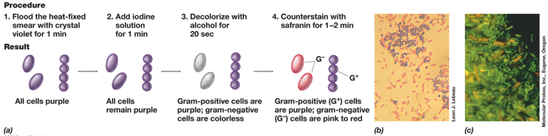
procedure for staining gram stains
Primary stain in gram stains is crystal violet
Sticks to all cells
All the cells then are purple
Rinse with water, then come in with iodine
Iodine creates complex with crystal violet, makes it though cell wall
Add 95% ethanol
Decolorizing step
Dehydrates some components of peptidoglycan layer
Creates tougher cell
Crystal violet and iodine can now sneak out, which makes it colorless
Gram positive cells are purple
Gram negative cells are colorless
We then apply safranin to cells, nothing will happen to the gram-positive cells, but the gram-negative cells will become pink in color
Koch’s postulates
When we have an animal with a certain disorder, if we take sample and grow it in culture, we have to identify the pathogen
Create a plate, use inoculating loop to collect sample and streak it across agar plate
Incinerate loop with flame
Drag initial swabbing
A lot of colonies on first swipe to just a little on second swipe
Single colony forms
Arisen from one single cell
Put into broth tube
Transfer suspected pathogen to healthy animal
Has to succumb to a disease as first animal
Repeat step 2, see if it is the same
If it is the same, we can say that the disease is capable of spreading and causing diseases
how can you use molecular biology to study microorganisms
Pasteur and Spontaneous Generation
Louis Pasteur: chemist and microscopist
Discovered alcoholic fermentation was a biological process, not just a chemical process
Developed vaccines for anthrax, fowl cholera, and rabies
Pasteur flask: swan-necked flask
Pasteur disproved the theory of spontaneous generation
Life arose spontaneously from nonliving matter