3.2.1 Cell Structure
1/62
There's no tags or description
Looks like no tags are added yet.
Name | Mastery | Learn | Test | Matching | Spaced | Call with Kai |
|---|
No analytics yet
Send a link to your students to track their progress
63 Terms
State two structural features that distinguish eukaryotic cells from prokaryotic cells. (2 marks)
- Contain membrane-bound organelles.
- DNA is enclosed within a nucleus.
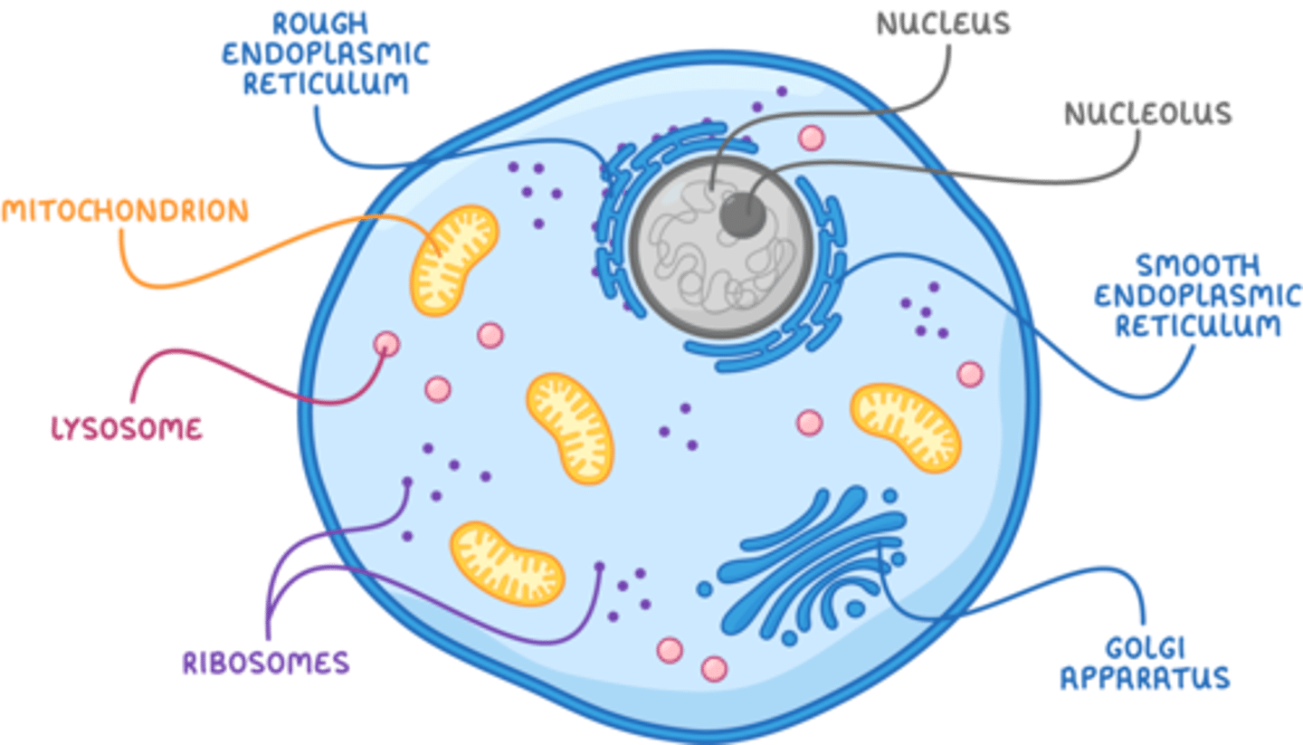
Draw a labelled diagram of the structure of a typical animal cell. (8 marks)
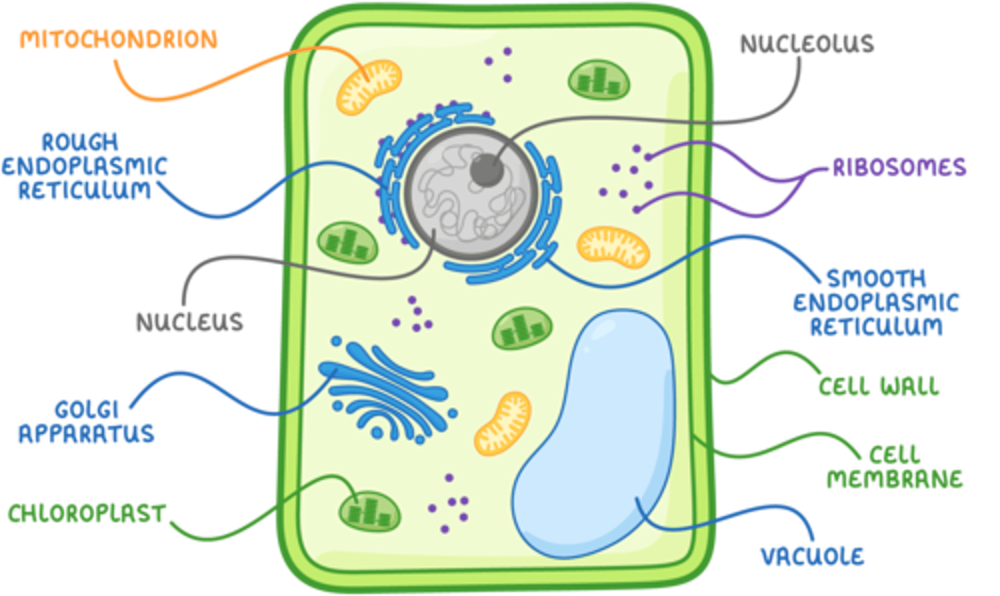
Draw a labelled diagram of the structure of a typical plant cell. (11 marks)
State the main differences between fungal and plant cells. (2 marks)
- Fungal cell walls are made of chitin, instead of cellulose.
- Fungal cells don't have chloroplasts.
State the three organelles that are only present in plant cells. (3 marks)
- Cellulose cell wall
- Chloroplasts
- Vacuole
Explain the arrangement of molecules in the cell-surface membrane. (2 marks)
- Phospholipids form a bilayer, with hydrophilic phosphate heads facing water and hydrophobic fatty acid tails facing inwards.
- Proteins are embedded within or attached to the bilayer.
State two functions of the cell-surface membrane. (2 marks)
- Controls movement of substances into and out of the cell (selectively permeable).
- Contains molecules for cell recognition and signalling.
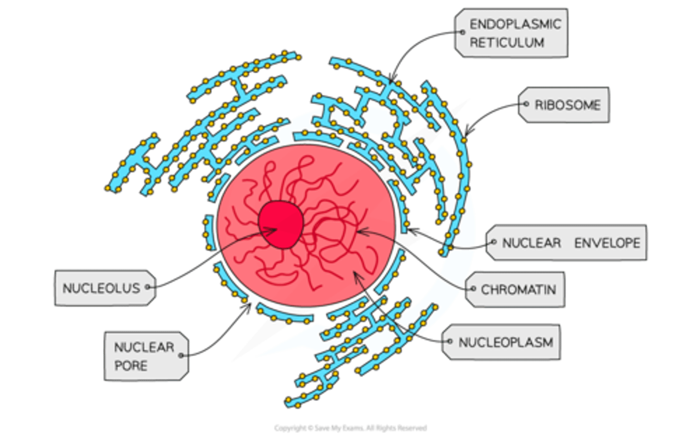
Draw and describe the structure of the nucleus. (5 marks)
- Enclosed by a double membrane called the nuclear envelope, containing pores.
- Contains nucleoplasm and a dense nucleolus.
- DNA is linear and bound to histone proteins, arranged as chromatin or chromosomes.
State the two functions of the nucleus. (2 marks)
- Stores genetic information for coding proteins.
- Site of DNA replication and transcription.
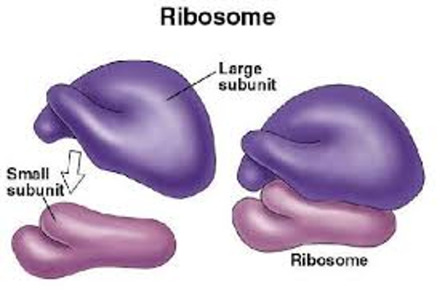
Describe the structure and function of a ribosome. (4 marks)
- Composed of ribosomal RNA and proteins, forming two subunits.
- Lacks a surrounding membrane.
- Site of protein synthesis (translation).
Draw and describe the structure of the rough and smooth endoplasmic reticulum. (4 marks)
- rER consists of membranes with ribosomes attached.
- sER is a similar membrane system but without ribosomes.
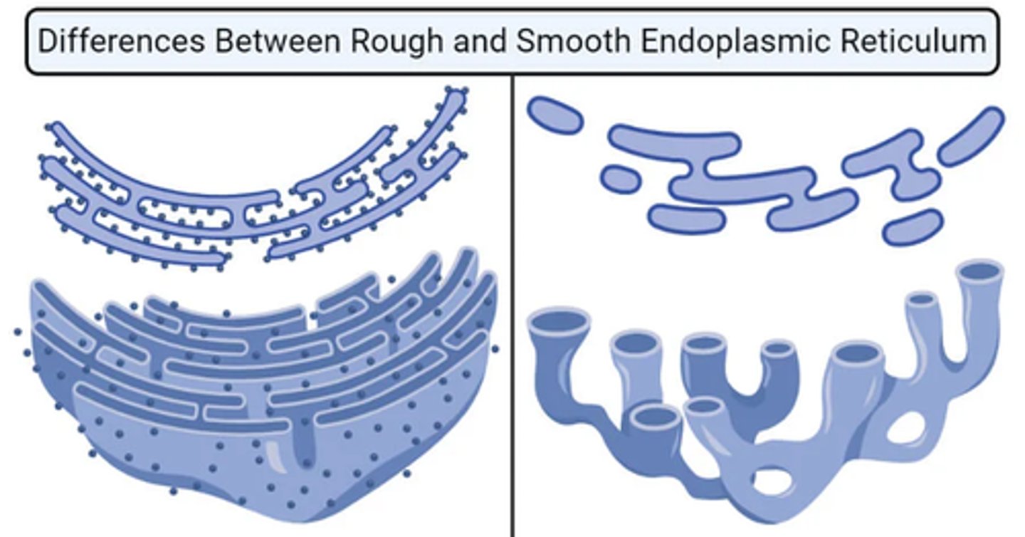
Describe the main functions of the rough and the smooth ER. (2 marks)
- rER synthesises, folds, and transports proteins, often packaging them into vesicles.
- sER synthesises and processes lipids and carbohydrates, including steroid hormones.
Draw a labelled diagram of the structure of the golgi apparatus. (4 marks)
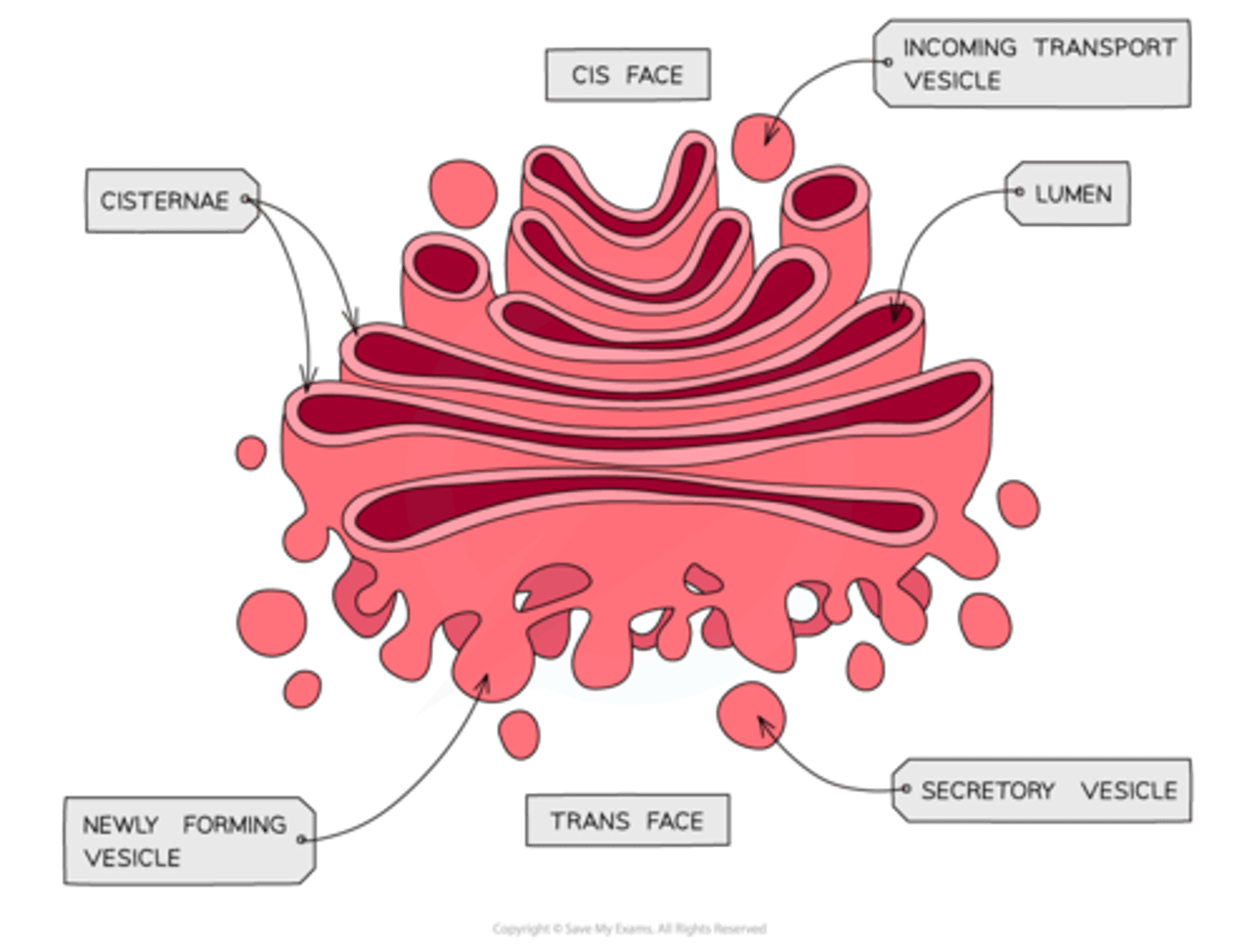
Describe the structure of the Golgi apparatus and Golgi vesicles. (2 marks)
- Golgi vesicles are small membrane-bound sacs.
- The Golgi apparatus consists of those flattened membrane sacs.
Explain the main functions of the Golgi apparatus. (2 marks)
- Modifies proteins and lipids, for example by adding carbohydrates to form glycoproteins or glycolipids.
- Packages proteins and lipids into vesicles, including the production of lysosomes.
Describe the main role of the Golgi vesicles. (2 marks)
- Transport proteins and lipids to their destination.
- Often fusing with the cell-surface membrane.
Describe the structure of a lysosome and state the function of the lysosomes. (2 marks)
- A membrane-bound sac containing hydrolytic enzymes.
- Release enzymes such as lysozymes to break down pathogens or worn-out cell components.
Draw a labelled diagram of the structure of a mitochondrion. (5 marks)
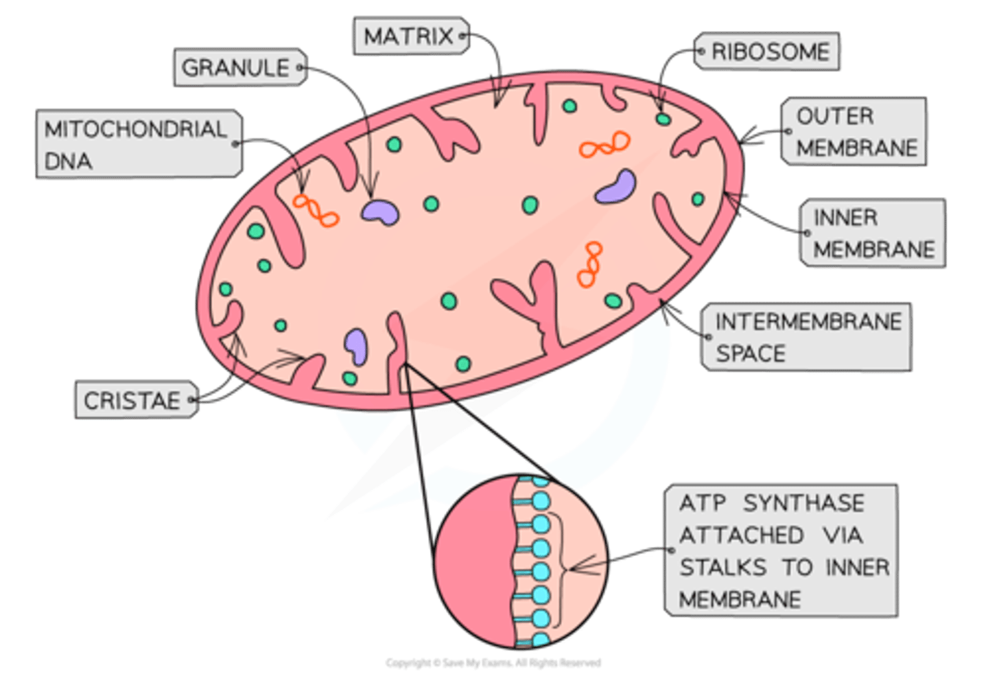
Describe the structure and function of mitochondria. (2 marks)
- Surrounded by an outer membrane and an inner membrane folded into cristae.
- The matrix contains 70S ribosomes and circular DNA.
- It is the site of aerobic respiration to produce ATP for energy-requiring processes.
Draw a labelled diagram of the structure of a chloroplast. (5 marks)
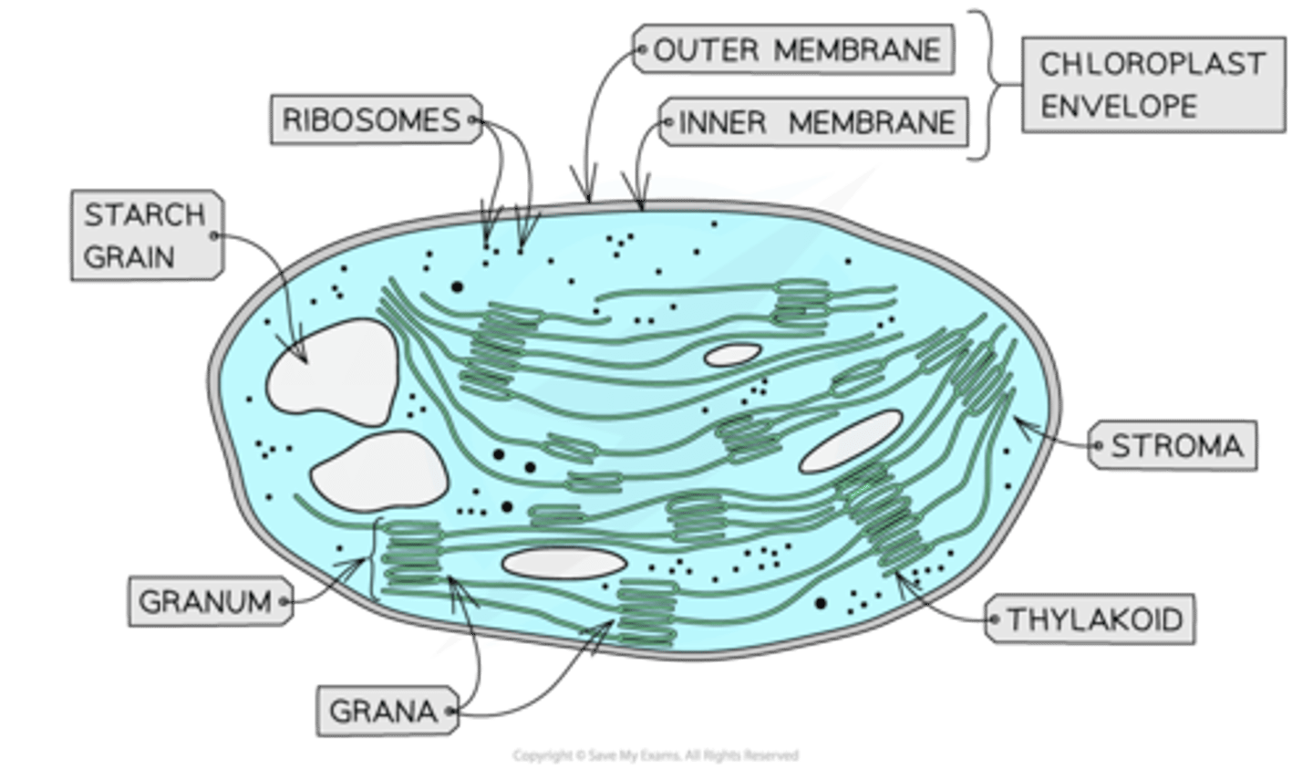
Describe the structure of a chloroplast. (3 marks)
- Enclosed by a double membrane with internal stacks of thylakoids (grana) connected by lamellae.
- The stroma contains circular DNA, 70S ribosomes, starch granules, and lipid droplets.
Explain the main function of chloroplasts. (2 marks)
- Contain pigments such as chlorophyll.
- That absorb light for photosynthesis, producing carbohydrates and lipids.
Describe the structure of the cell wall in plants, algae, and fungi. (2 marks)
- In plants and algae, made of cellulose.
- In fungi, made of chitin.
State one function of the cell wall. (2 marks)
- Provides structural strength to the plant cell.
- Prevents the cell from bursting under osmotic pressure.
Draw and describe the structure of the plant cell vacuole. (3 marks)
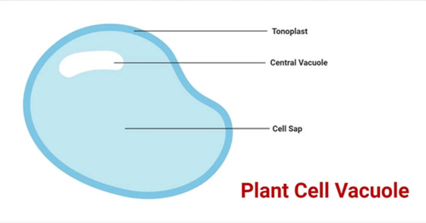
State the main function of the plant vacuole. (2 marks)
- Maintains turgor pressure in the plant cell.
- Stores substances such as sugars, amino acids, pigments, and waste products.
Explain how eukaryotic cells are organised in multicellular organisms. (3 marks)
- Specialised cells with a similar structure and origin work together to form tissues that perform a specific function.
- Different tissues combine to form organs, each carrying out particular roles.
- Groups of organs work together in organ systems to carry out complex processes.
State an example of how a cell's organelle composition is adapted to its function. (2 marks)
- A plasma cell contains many ribosomes.
- To produce large amounts of antibodies through protein synthesis.
Explain why some cells may contain lots of mitochondria and state an an example. (2 marks)
- To provide energy to cells that require a large amount of ATP for movement or contraction.
- For example, muscle cells.
Describe the roles of the organelles that are involved in the production, transport and release of proteins from eukaryotic cells. (5 marks)
1. DNA in the nucleus codes for these proteins
2. Ribosomes in the RER produce these proteins via protein synthesis
3. Mitochondria produces the ATP that is required for protein synthesis
4. Golgi apparatus packages and modifies these proteins into vesicles
5. Vesicles fuse with the cell membrane and release proteins outside of cell
State the main structural features that distinguishes prokaryotic cells from eukaryotic cells. (2 marks)
- Prokaryotic cells lack membrane-bound organelles.
- Their DNA is not enclosed within a nucleus.
Draw and describe the general structure of a prokaryotic cell. (6 marks)
- Surrounded by a cell-surface membrane and a cell wall containing murein.
- Contains cytoplasm with small (70S) ribosomes.
- Has circular DNA free in the cytoplasm, sometimes with additional plasmids.
- May have a capsule and flagella for protection and movement.
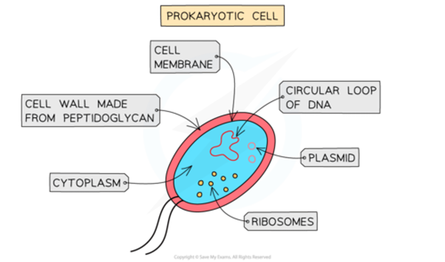
State the structures present in all prokaryotic cells. (5 marks)
- Cell wall (made of murein - a glycoprotein).
- Cell surface membrane.
- Free circular DNA molecule in cytoplasm.
- Ribosomes (70s ribosomes)
Cytoplasm.
State the structures present in some prokaryotic cells. (5 marks)
- Capsule surrounding the cell wall.
- One or more plasmids.
- One or more flagella.
Describe the structure and organisation of DNA of prokaryotic cells. (4 marks)
- No nucleus is present.
- DNA is free in the cytoplasm.
- It contains circular DNA and sometimes additional plasmids.
- DNA is not attached to any histone proteins.
Describe the main properties of plasmids found in prokaryotic cells. (3 marks)
- They contain antibiotic resistance genes. (amongst others)
- Plasmids are able to be passed between prokaryotes.
- Some prokaryotic cells have several plasmids, others have no plasmids.
Compare and contrast eukaryotic and prokaryotic cells. (4 marks)
- Eukaryotes have membrane-bound organelles; prokaryotes do not.
- Eukaryotes have a nucleus with linear DNA; prokaryotes have circular DNA free in the cytoplasm.
- Eukaryotic ribosomes are larger (80S) than prokaryotic ribosomes (70S).
- Prokaryotic cell walls contain murein, whereas eukaryotic cell walls (if present) contain cellulose or chitin.
State what two properties viruses are often described as. (2 marks)
- Non-living.
- Acellular.
Explain why viruses are described as acellular. (2 marks)
- They are not made of cells.
- They have no cell membrane, cytoplasm, or organelles.
Explain why viruses are described as non-living. (2 marks)
- They have no metabolism.
- They therefore, cannot move, respire, replicate, or excrete without a host cell.
Draw a labelled diagram of the structure of a typical viral particle. (3 marks)
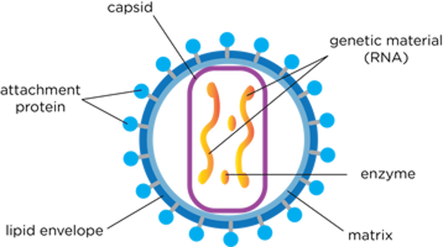
Describe the general structure of a virus particle. (4 marks)
- Contains nucleic acid (DNA or RNA) enclosed within a protein coat called a capsid.
- Has attachment proteins on its surface for binding to receptors on host cells.
- Lacks cytoplasm, ribosomes, and other cell structures.
- Some viruses have an additional lipid envelope surrounding the capsid.
Define the terms magnification and resolution. (2 marks)
- Magnification is how many times larger an image is compared to the actual object.
- Resolution is the minimum distance at which two objects can be seen as separate.
State one principle and one limitation of an optical microscope. (2 marks)
- Uses light focused by glass lenses to view specimens.
- Has low resolution due to the long wavelength of light.
Explain why a specimen must be thin in optical microscopy. (2 marks)
- So that light can pass through the specimen.
- So that a single layer of cells is visible.
State why specimens must be stained in optical microscopes and state the two main stains that we use during microscopy. (3 marks)
- So that the cell structures are visible.
- Eosin is used to highlight the cytoplasm,
- Iodine contained in potassium iodide solution highlights starch grains.
State the five structures that cannot be identified using an optical microscope. (5 marks)
- Mitochondrion.
- Ribosome.
- Endoplasmic Reticulum.
- Lysosome.
- Cell-surface membrane.
Describe how a transmission electron microscope produces a micrograph. (4 marks)
1. Specimen is stained with electron dense substances e.g. heavy metal salts.
2. Beam of electrons are transmitted through the specimen.
3. Staining substances deflect the electrons in the beam.
4. The pattern that the remaining electrons produce as they pass through specimen is converted into an image.
Compare optical and electron microscopes. (6 marks)
- Electron microscopes use a beam of electrons, while optical microscopes use a beam of light.
- Electron microscopes have much greater resolution as electrons have a shorter wavelength, while optical microscopes have relatively low resolution.
- Electron microscopes are focused using magnets, while optical microscopes are focused using glass lenses.
- Electron microscopes can view smaller structures, while optical microscopes cannot see smaller structures.
- Electron microscopes require the specimen to be dead, while optical microscopes can view living specimens.
- Electron microscopes produce images not in colour, while optical microscopes produce images in colour.
State one principle and one limitation of a transmission electron microscope. (2 marks)
- Passes electrons through the specimen to create a 2D image of internal structures.
- Requires very thin specimens and can only be used with dead material.
Describe how a scanning electron microscope produces a micrograph. (3 marks)
1. Specimen is coated with a thin film of heavy metal e.g. gold.
2. Electron beam is scanned to and across the specimen.
3. Electrons that are reflected from the surface are collected and produce an image on a viewing screen.
State one principle and one limitation of a scanning electron microscope. (2 marks)
- Scans electrons over the surface to produce a 3D image.
- Cannot view internal structures and can only be used with dead specimens.
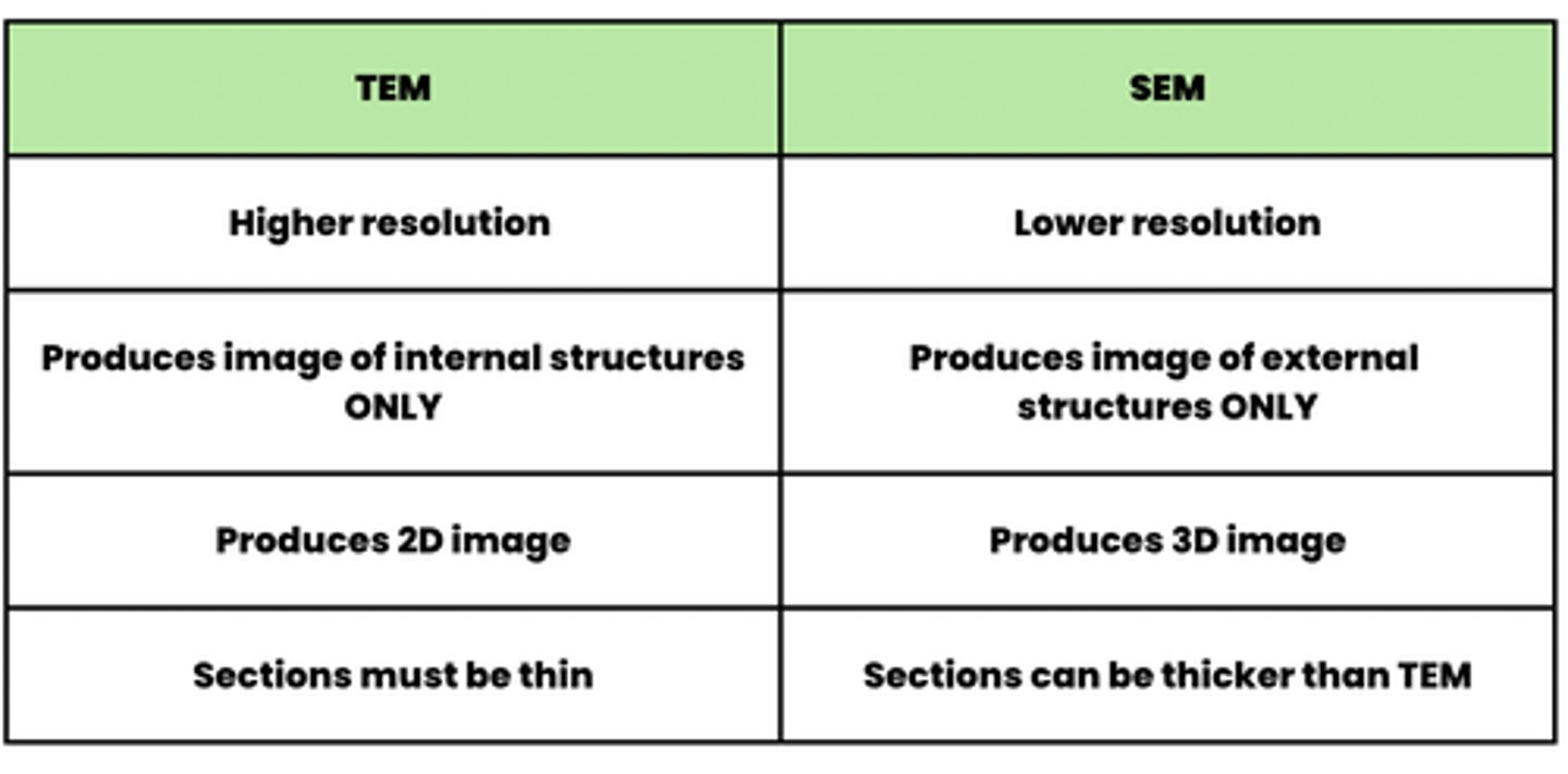
Compare transmission electron microscopes and scanning electron microscopes. (4 marks)
Explain how scientists distinguish between artefacts and actual cell structures. (2 marks)
- They prepare specimens using different techniques.
- If a feature appears with one method but not another, it is more likely to be an artefact.
Describe how you would calculate magnification, image size, or real size. (3 marks)
1. Write down and rearrange the formula (I = A × M).
2. Convert all measurements to the same unit.
3. Calculate the answer and check if the correct units are used.
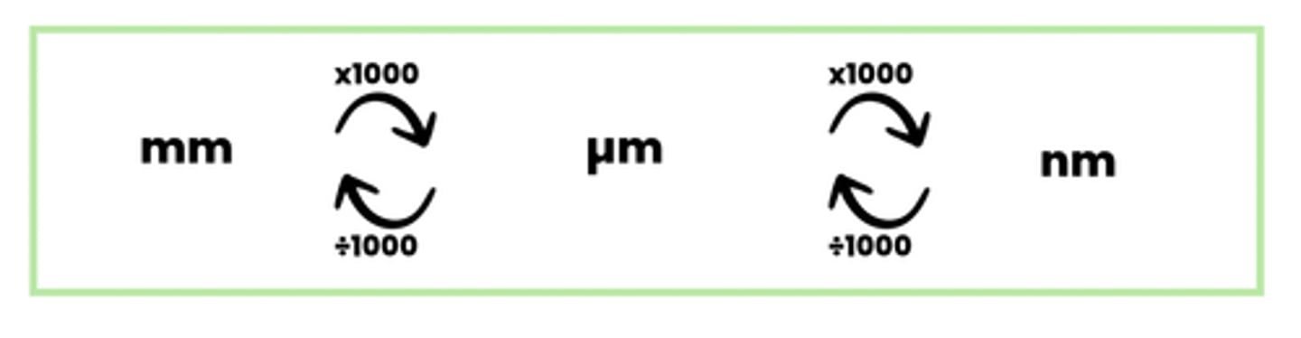
Draw a diagram to show the unit conversions that are needed in magnification. (3 marks)
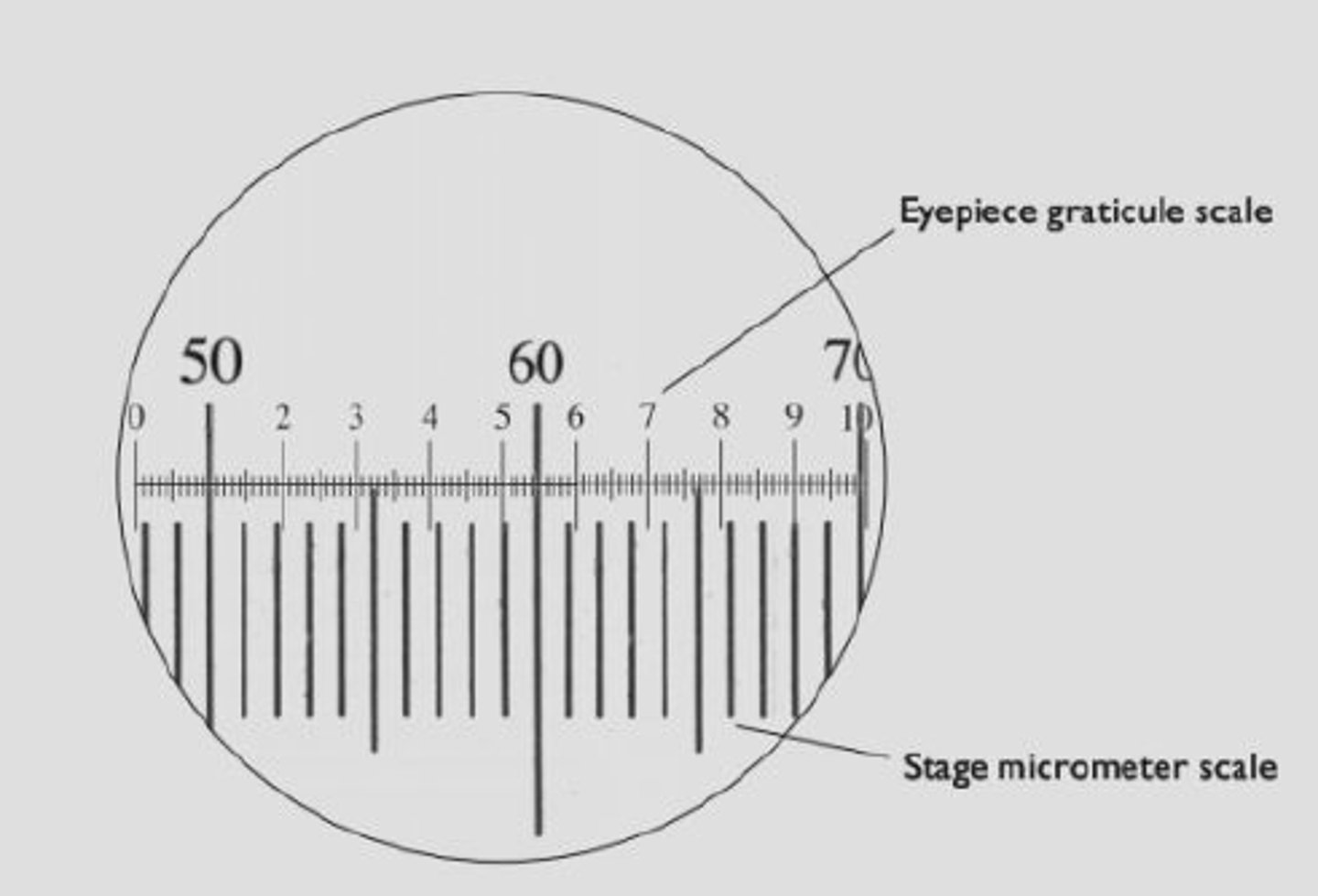
Describe the method for measuring the size of an object under an optical microscope. (6 marks)
1. Align the eyepiece graticule scale with the stage micrometer scale.
2. Calibrate the eyepiece graticule using the stage micrometer.
3. Remove the micrometer and measure the specimen using the graticule.
4. Multiply the number of graticule divisions by the size of one division.
5. Recalibrate if changing magnification.
Describe the process of cell fractionation. (4 marks)
1. Homogenise tissue using a blender.
2. Place your sample in an ice-cold, isotonic, buffered solution.
3. Filter homogenate.
4. Perform ultracentrifugation to separate organelles in order of density.
State the purpose of homogenising tissue in cell fractionation. (2 marks)
- Breaks open the cell membrane to release organelles.
- Ensures cellular contents are freed for separation.
Explain why the homogenate is placed in an ice-cold, isotonic, buffered solution. (3 marks)
- Ice-cold reduces enzyme activity so organelles are not damaged.
- Isotonic prevents water movement into or out of organelles, avoiding bursting or shrinking.
- Buffer maintains constant pH so enzymes do not denature.
State the purpose of filtering the homogenate. (2 marks)
- Removes large debris.
- Such as whole cells or connective tissue.
Describe the process of ultracentrifugation. (3 marks)
1. Spin the homogenate at low speed to pellet the heaviest organelles.
2. Remove the pellet and respin the remaining liquid at a higher speed.
3. Continue at increasing speeds to separate lighter organelles.
State the order in which cell organelles are isolated during ultracentrifugation. (5 marks)
1. Nuclei
2. Chloroplasts (if its a plant cell)
3. Mitochondria
4. Endoplasmic reticulum (SER and RER)
5. Ribosomes