Ch. 11 - Functional Organization of Nervous Tissue
1/58
There's no tags or description
Looks like no tags are added yet.
Name | Mastery | Learn | Test | Matching | Spaced | Call with Kai |
|---|
No analytics yet
Send a link to your students to track their progress
59 Terms
functions of the nervous system
sensory input
integration
control of muscles and glands
homeostasis
mental activities
central nervous system
processes, integrates, stores, responds to information from PNS
brain & spinal cord
encased in bone
peripheral nervous system
detects stimuli, transmits info to and receives info from CNS
sensory receptors & nerves
nervous tissue outside of CNS
2 divisions: sensory & motor
sensory division (PNS)
transmits action potentials from sensory receptors to the CNS
motor division (PNS)
carries action potentials away from the CNS in cranial or spinal nerves
2 subdivisions: somatic & autonomic
somatic nervous system (m)
innervates skeletal muscle
autonomic nervous system (m)
innervates cardiac muscle, smooth muscle, and glands
3 subdivisions: sympathetic, parasympathetic, enteric
sympathetic division (ANS)
“fight-or-flight”; most active during physical activity
parasympathetic division (AMS)
“rest-and-digest”; regulates resting functions
enteric nervous system (ANS)
controls the digestive system
neurons
excitable cells that transmit electrical signals
3 components: soma, dendrites, axon
nuclei
clusters of cell bodies in the CNS
ganglia
clusters of cell bodies in the PNS
soma (cell body)
nissl substance: primary site of protein synthesis
rough ER and free ribosomes
contains nucleus, nucleolus, Golgi apparatus, mitochondria
axons (nerve fibers)
slender processes: uniform diameter, length varies
trigger zone: where axon originates and action potential is generated
presynaptic terminal
branched terminus of an axon
synapse
junction between a nerve cell and another cell
multipolar neurons
have several dendrites and a single axon
interneurons & motor neurons
bipolar neurons
have a single axon and dendrite
components of sensory organs
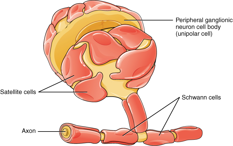
unipolar neurons
have a single axon
most sensory neurons
glial cells
cells that surround neurons, accounting for over half of the brain’s weight
supportive scaffolding: “neuroglia”
segregate and insulate neurons
guide young neurons to proper connections
promote health and growth
glial cells of the CNS
astrocytes
microglial cells
ependymal cells
oligodendrocytes
glial cells of the PNS
satellite cells
Schwann cells
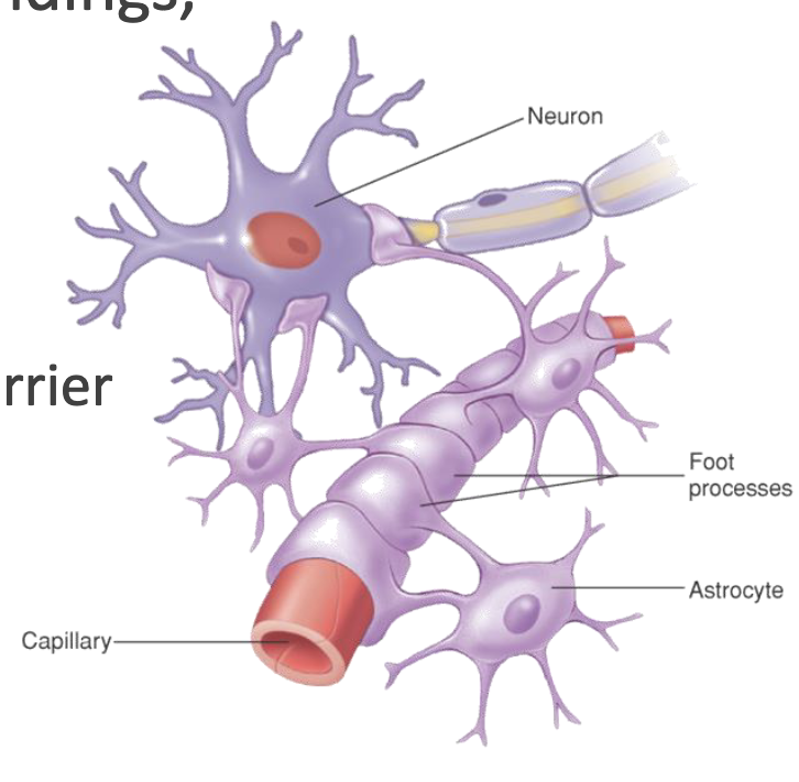
astrocytes
most abundant, branched, and versatile; cling to neurons/synapses and cover capillaries
support neurons & blood vessels
anchor neurons to nutrient supplies
influence function of blood-brain barrier
produce secondary energy to neurons
process substances: clean up K ions & recycle neurotransmitters
isolate damaged tissue & limit spread of infection
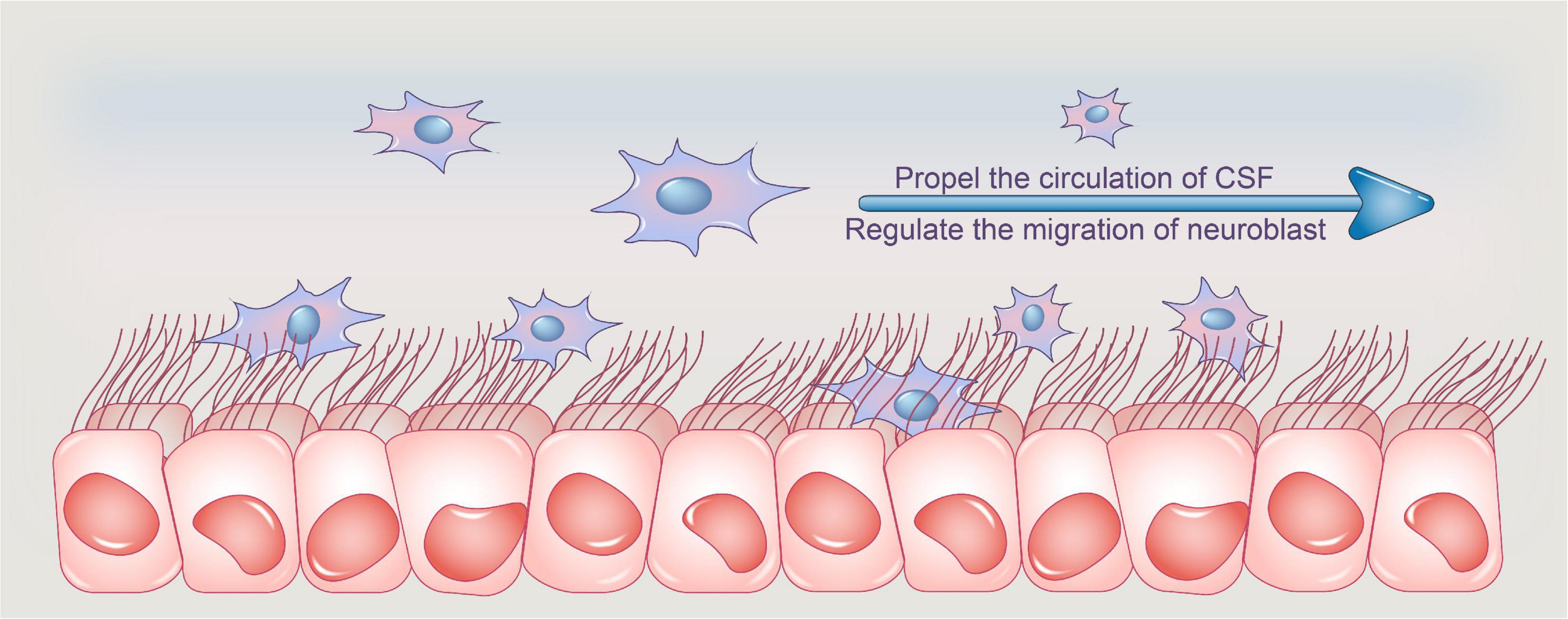
ependymal cells
range in shape and are often ciliated; line ventricles of brain and central canal of spinal cord
produce cerebrospinal fluid (CSF)
help to circulate CSF using cilia
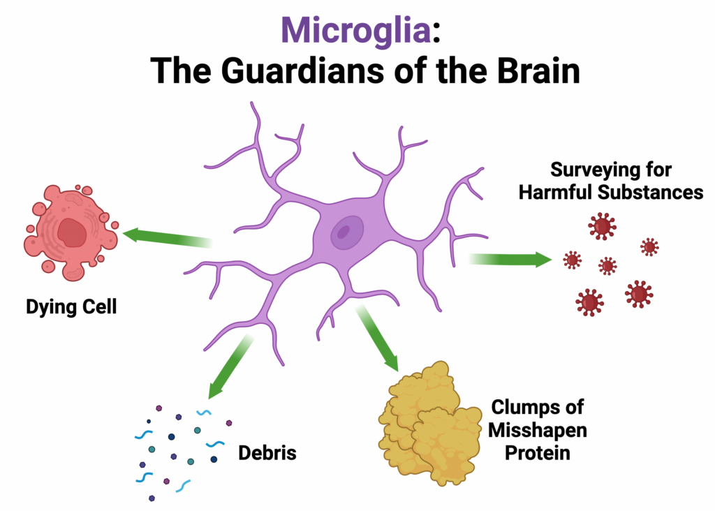
microglia
small and ovoid with tiny processes; migrate to areas of inflammation/damage/death
macrophages (immune cells)
phagocytes, monitor the health of neurons
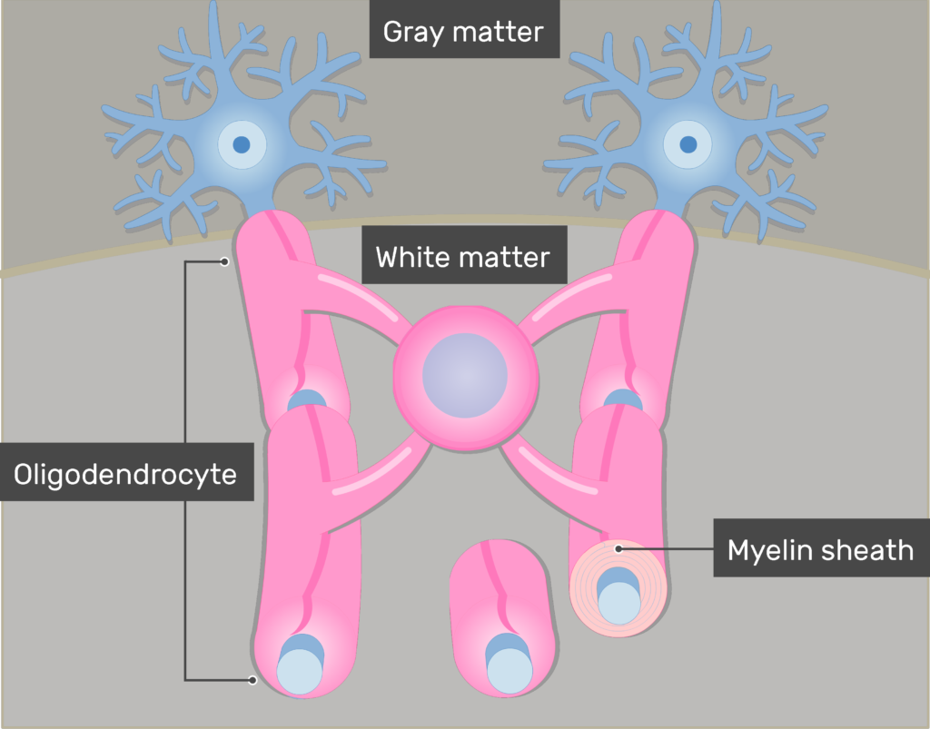
oligodendrocytes
form myelin sheaths around the axons of several CNS neurons
‘octopus’
MS: attacks, breaks down myelin sheaths
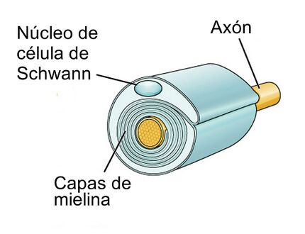
Schwann cells (PNS)
form myelin sheath around part of one axon of a PNS neuron
satellite cells (PNS)
support/nourish cell bodies within ganglia
protect cell from heavy metals
myelin
whitish, fatty segmented sheath around most long axons
protects axon
electrically insulates fibers from one another
increases speed of nerve impulse transmission
nodes of Ranvier
gaps in myelin sheath
unmyelinated axons
rest in invaginations of Schwann cells (PNS) or oligodendrocytes (CNS)
conduct action potentials slowly
still technically myelinated, but much less so
white matter
consists of myelinated axons
propagates action potentials
CNS: forms nerve tracts
PNS: forms nerves
gray matter
collections of neuron cell bodies or unmyelinated axons
CNS: forms cortex & nuclei
PNS: forms ganglia
resting membrane potential
charge difference across the plasma membrane when the cell is not being stimulated
inside of the cell is more negatively charged
K+ diffuses out of cell
action potentials
electrical signals produced by cells
occur when a graded potential causes depolarization of the the PM to a level called threshold
all-or-none fashion, of the same magnitude no matter the stimulus strength
results of action potentials
sensations
complex mental activities
contraction of muscles
secretion of certain glands
electrical properties of cells result from:
permeability characteristics and ionic concentration differences across the plasma membrane
channels within cell body
50-70 channels per micrometer
channels within trigger zone
~350 channels per micrometer
concentration differences across PM
Sodium, Calcium, and Chloride ions are in much greater concentration outside the cell
Potassium ions and negatively charged molecules (e.g. proteins) are in much greater concentration inside the cell
negatively charged proteins are synthesized inside the cell, cannot diffuse out of it
sodium-potassium pump
moves ions by active transport
K+ ions moved into the cell
Na+ ions moved out of the cell
leak channels
non-gated (always open)
K+ channels more numerous
plasma membrane is more permeable to K+ when at rest
gated ion channels
include ligand-gated channels, voltage-gated channels, etc.
ligand-gated ion channels
open/close with the binding of a specific ligand (neurotransmitter)
common in nervous & muscle tissue, glands
voltage-gated ion channels
open/close in response to small voltage changes across the plasma membrane
common in nervous & muscle tissues
depolarization
inside of the cell becomes more positive
Na+ diffuses into the cell through voltage-gated ion channels
repolarization phase
return of the membrane potential to the resting membrane potential
voltage-gated Na+ channels close
voltage-gated K+ channels open, K+ diffuses out of the cell
afterpotential
brief period of hyperpolarization following repolarization
graded potentials
magnitude varies from small to large depending on stimulus strength or frequency
can by hyperpolarizing or depolarizing
can summate (add on to each other)
decrease in magnitude as they spread across membrane
can cause generation of APs
propagation of action potentials
unmyelinated: immediately adjacent to previous APs
myelinated: at successive Nodes of Ranvier
synapse
junction between two cells where communication takes place
presynaptic cell: transmits signal
postsynaptic cell: receives the signal
electrical synapses
gap junctions in which tubular proteins called connexons allow ionic currents to move between cells
APs are conducted rapidly between cells, synchronized activity
common in cardiac muscle and many types of smooth muscle where coordinated contractions are essential
chemical synapses
have three anatomical components:
presynaptic terminals
postsynaptic membranes
synaptic cleft
presynaptic terminals
enlarged ends of the axon containing synaptic membranes
postsynaptic membranes
contain receptors for the neurotransmitter
chemical synapse activity
APs arriving at presynaptic terminal cause voltage-gated Ca channels to open
Ca ions diffuse into the cell, synaptic vesicles release neurotransmitters
neurotransmitters diffuse from presynaptic terminal across synaptic cleft
neurotransmitters combine with their receptor sites and cause ligand-gated channels to open. ions diffuse in/out of cell, membrane potential changes
excitatory post-synaptic potential
a depolarizing graded potential of the postsynaptic membrane
increases neurotransmitter release
inhibitory postsynaptic potential
a hyperpolarizing graded potential of the postsynaptic membrane
decreases neurotransmitter release
decreases likelihood of action potential by moving membrane potential farther from threshold