9.6 Detection of Light
1/26
There's no tags or description
Looks like no tags are added yet.
Name | Mastery | Learn | Test | Matching | Spaced | Call with Kai |
|---|
No analytics yet
Send a link to your students to track their progress
27 Terms
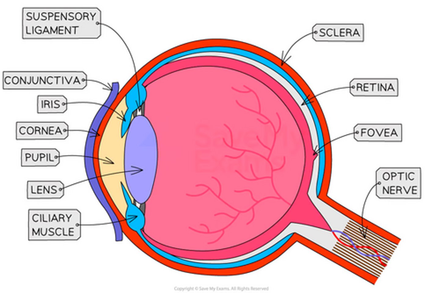
How would you draw and label a diagram showing a cross-section of the mammalian eye? (5)
- Draw the three main layers of the eye: the sclera, choroid, and retina.
- At the front of the eye, draw and label the cornea, iris, pupil, and lens.
- At the back of the eye, draw and label the retina, fovea, and the optic nerve leading from the optic disc.
- Label the ciliary body which is attached to the lens.
- Label the main chambers, including the anterior chamber and the vitreous body.
What are the functions of the retina, choroid, and sclera? (3)
- The retina is a layer of light-sensitive cells, known as rods and cones, and the neurones that lead from them to the optic nerve.
- The choroid is composed of pigmented cells that prevent internal reflection, and its vascular layer nourishes the retina.
- The sclera is a thick, protective connective tissue membrane that surrounds the eye.
What is the structure of the lens and its supporting components? (3)
- The lens is a transparent structure responsible for the fine focussing of light onto the retina.
- It is held in position by the suspensory ligament, the fibres of which are attached to the ciliary body.
- The ciliary body contains a complex set of smooth muscles that are controlled by the autonomic nervous system.
How is the shape of the lens altered for focusing? (2)
- When the ciliary muscles relax, the suspensory ligaments pull the lens, causing it to flatten to focus on distant objects.
- When the ciliary muscles contract, the tension on the lens is released, and it returns to a more spherical shape to focus on closer objects.
What are the iris and the cornea? (3)
- The cornea is a transparent outer layer that protects the eye and is responsible for the majority of light refraction.
- The iris is the coloured part of the eye that contains circular and radial muscles.
- The differential contraction of these muscles varies the size of the aperture, or pupil, controlling how much light enters the eye.
What are the fovea, the blind spot, and the optic nerve? (3)
- The fovea is an area of the retina containing only cone cells and is the region of highest visual acuity.
- The blind spot is the point where the optic nerve leaves the eye and is so-named because it contains no photoreceptor cells.
- The optic nerve is a bundle of nerve fibres that carries impulses from the retina to the brain for processing.
What are the function and location of cone cells? (3)
- Cone cells are photoreceptors that are concentrated in the fovea area of the retina.
- They function to perceive the environment in conditions of high light intensity.
- They are capable of colour perception and provide high visual acuity.
Why do cone cells have high visual acuity? (2)
- Each individual cone cell synapses with a single individual sensory neurone.
- This one-to-one connection means the brain receives very precise information, resulting in high visual acuity.
What are the function and location of rod cells? (2)
- Rod cells are photoreceptors that are located outside the fovea in the peripheral parts of the retina.
- They provide greyscale vision and are used for seeing in low light intensities or at night, as they contain the highly sensitive pigment rhodopsin.
What is retinal convergence? (3)
- Rod cells show retinal convergence, a phenomenon where several rod cells synapse with the same sensory neurone.
- This arrangement makes them highly sensitive to low light levels and movement in the visual field.
- Several small generator potentials from different rods can summate to trigger an action potential in the sensory neurone.
What is the path light takes to reach the photoreceptors? (3)
- The path of light passes through the pupil and is focused by the lens onto the retina.
- The neurones of the retina are located at the interior edge of the cell layer.
- Therefore, light must pass through the synaptic layers before reaching the photosensitive part of the rods and cones.
What is the composition of rhodopsin? (3)
- Rod cells contain the visual pigment rhodopsin, which is also known as visual purple.
- It is formed from two components: a lipoprotein called opsin and a light-absorbing molecule called retinal.
- Retinal is a derivative of vitamin A and exists as two different isomers, cis-retinal and trans-retinal.
How does the process of bleaching occur in a rod cell? (3)
- In the dark, the retinal component of rhodopsin exists in the cis-form.
- When a molecule of rhodopsin is struck by a photon of light, it converts the cis-retinal into the trans-form.
- This change in the shape of the retinal causes the rhodopsin molecule to break down into its constituent parts of opsin and retinal.
How is a generator potential formed in a rod cell? (3)
- When rhodopsin is bleached by light, a cascade of chemical reactions is triggered.
- This cascade causes the sodium ion channels in the cell membrane to become less permeable to sodium ions.
- The interior of the cell therefore becomes more negative relative to the outside, and this state of hyperpolarisation is known as a generator potential.
How is a signal passed from a photoreceptor to a sensory neurone? (3)
- The extent of hyperpolarisation in the photoreceptor depends on the intensity of the light stimulus.
- A neurotransmitter is released into the synapse with a bipolar cell, which connects the photoreceptor to the sensory neurone.
- If the stimulus is sufficient, an action potential is set up in the bipolar cell, which then passes onto the sensory neurone.
Why can a rod not be stimulated twice in quick succession? (2)
- Once the rhodopsin in a rod cell has been bleached by light, it becomes unresponsive.
- The rod cannot be stimulated again until the rhodopsin pigment has been resynthesised.
What is the process of rhodopsin resynthesis? (3)
- The resynthesis of rhodopsin from opsin and retinal is an endergonic process requiring energy.
- ATP for this process is provided by the mitochondria located in the inner segment of the rod cell.
- This energy is used to convert the trans-retinal back to cis-retinal, which can then re-associate with opsin to reform rhodopsin.
What is dark adaptation? (3)
- In bright daylight, the rods are entirely bleached and can no longer respond to dim light.
- The process of the rhodopsin reforming in low light or darkness is known as dark adaptation.
- When the rhodopsin is fully reformed, the eye is then highly sensitive to dim light and is said to be dark-adapted.
What are two adaptations of a rod cell? (2)
- The rod cell has numerous mitochondria in its inner segment to provide ATP for the resynthesis of rhodopsin.
- It has lamellae in its outer segment which increase the surface area for holding the pigment molecules.
What is iodopsin? (3)
- The visual pigment found in cone cells is known as iodopsin.
- There are three different types of iodopsin, with each being sensitive to one of the primary colours of light: red, green, or blue.
- Iodopsin requires more light energy to break down than rhodopsin, and therefore it is not sensitive to low intensities of light.
How do cones provide colour vision? (2)
- The brain interprets the numbers of different types of cones that are stimulated as different colours, which provides colour vision.
- This function complements the low light vision and sensitivity to movement that is provided by the rods.
What is the basis of the trichromatic theory? (2)
- The trichromatic theory depends on the principle of colour mixing.
- It states that all colours can be produced by mixing the primary colours of light, which are red, blue, and green, in various proportions.
How is a specific colour perceived? (2)
- A particular colour is perceived when its specific wavelengths stimulate one, two, or all three types of cone cells to a varying degree.
- The final colour that is determined is a result of the relative excitation of these three receptor types.
How would you explain the different sensitivities of rod and cone cells to light of varying wavelengths, as shown in the graph? (5)
- The graph shows three different types of cone cells, each showing peak sensitivity to a different wavelength of light corresponding to blue, green, or red, which provides the basis for colour vision.
- Rod cells show a single peak sensitivity at a wavelength of approximately 500 nm, meaning they are unable to distinguish between different colours, resulting in monochromatic vision.
- Rod cells are more sensitive to light intensity than cone cells and can therefore detect light at lower levels, making them responsible for vision in low light conditions.
- The higher sensitivity of rod cells is due to spatial summation, where signals from multiple rods converge on a single bipolar neurone, amplifying the stimulus.
- The different absorption spectra of the visual pigments in these photoreceptors, rhodopsin in rods and iodopsin in cones, account for these different sensitivities.
What is the role of rod cells in initiating action potentials to the brain of the mouse? (5)
1. When light is absorbed by a rod cell, the visual pigment rhodopsin is bleached, breaking down into opsin and retinal.
2. This causes sodium ion channels on the rod cell membrane to close, making the membrane less permeable to sodium ions.
3. As the sodium pump continues to remove sodium ions, the inside of the cell becomes more negative, resulting in hyperpolarisation.
4. The hyperpolarisation of the rod cell stops the release of the inhibitory neurotransmitter glutamate at the synapse.
5. Without the inhibitory neurotransmitter, the bipolar cell depolarises, triggering an action potential in the ganglion cell which is transmitted to the brain.
What are the results of the light detection investigation, explain using the information from the graphs and table? (5)
- The table shows that no light is detected at 15 degrees from the fovea, which corresponds to the blind spot shown in Graph 1, an area that lacks any photoreceptor cells.
- Bright red light (670 nm) is only detected near the fovea because this is where cone cells are highly concentrated, and Graph 2 shows that only red-sensitive cones can respond to this wavelength.
- Dim red light is never detected because rod cells, which are responsible for dim light vision, are not sensitive to the 670 nm wavelength, as shown in Graph 2.
- Bright green light (525 nm) is detected across the retina (except the blind spot) as both the cones at the fovea and the rods in the periphery are sensitive to this wavelength.
- Dim green light is only detected in the periphery (at angles greater than 10 degrees) because this is where the highly sensitive rod cells are located, and they are responsible for detecting low-intensity light.
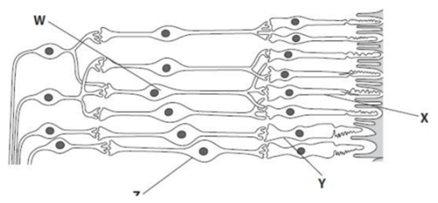
Why is a higher light intensity required to produce an action potential in cell Z than in cell W? (3)
- Many rod cells (X) connect to a single bipolar cell (W), which is known as retinal convergence, whereas only one cone cell (Y) connects to bipolar cell (Z).
- This convergence allows for spatial summation in the rod pathway, where small generator potentials from multiple rod cells are combined at the bipolar cell (W).
- As a result, the threshold potential is reached more easily in cell W even in low light, while cell Z requires a higher light intensity to generate a potential large enough to reach the threshold on its own.