Bio 225 Umich Exam 2
0.0(0)
Card Sorting
1/273
Earn XP
Description and Tags
Last updated 12:13 AM on 10/18/22
Name | Mastery | Learn | Test | Matching | Spaced | Call with Kai |
|---|
No analytics yet
Send a link to your students to track their progress
274 Terms
1
New cards
multipolar neuron
many processes from cell body -- many dendrites and one axon
2
New cards
bipolar neuron
- two main processes from cell body -- one dendrite and one axon
- usually unmylinated and very short
- in highly specialized places
- some sensory neurons
- usually unmylinated and very short
- in highly specialized places
- some sensory neurons
3
New cards
unipolar neuron
- one main process coming from cell body
- integration position no longer called axon hillock
- signal moves past cell body towards axon terminal
- integration position no longer called axon hillock
- signal moves past cell body towards axon terminal
4
New cards
motor neuron
- efferent neuron
- carries signal out (from CNS to some effector neuron)
- transmission to muscle or glands
- multipolar
- carries signal out (from CNS to some effector neuron)
- transmission to muscle or glands
- multipolar
5
New cards
sensory neuron
- afferent neuron
- carries signal in (from periphery to central)
- unipolar (somatosensory); some bipolar
- carries signal in (from periphery to central)
- unipolar (somatosensory); some bipolar
6
New cards
interneuron
- functions between two other neurons (connects neurons to other neurons)
- both pre and post synaptic
- input: NT from other neuron
- output: NT signaling to post synaptic neuron
- multipolar
- both pre and post synaptic
- input: NT from other neuron
- output: NT signaling to post synaptic neuron
- multipolar
7
New cards
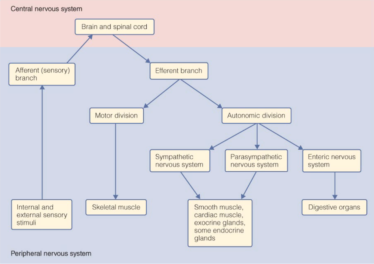
general organization of nervous system
incoming stimulus sensed by sensory receptors (either accessory structures or receptors on the end of the neuron, eg. mechanoreceptors, photoreceptors) -> triggers change in membrane potential, which sends signal up through afferent neuron -> signal reaches integrating center in CNS (eg. brain), and is transmitted through an interneuron and the cell body of the efferent neuron -> signal transmitted through the efferent neuron to the effector organs (eg. muscles, glands) -> output is achieved
beginning - reaching CNS = sensors
duration of time in CNS = integrating centers
leaving CNS - output = output pathways
beginning - reaching CNS = sensors
duration of time in CNS = integrating centers
leaving CNS - output = output pathways
8
New cards
nuclei
groups of neuronal cell bodies within the CNS
9
New cards
ganglia
groups of neuronal cell bodies outside of the CNS
10
New cards
tracts
bundles of many axons within the CNS
11
New cards
nerves
bundles of many axons outside of the CNS
12
New cards
major divisions of the vertebrate nervous system
13
New cards
parts of the central nervous system
- brain - integrating center made up of clusters of nuclei and tracts
- spinal cord - mediates information flow between brain and body (and vice versa), spinal cord has many nerves
- spinal cord - mediates information flow between brain and body (and vice versa), spinal cord has many nerves
14
New cards
peripheral nervous system
- the section of the nervous system lying outside the brain and spinal cord
15
New cards
nerves
- consist of myelinated and unmyelinated axons enclosed in connective tissue
- may contain sensory or motor or both
- cranial vs spinal
- associated cell bodies are located in (motor, nuclei) or near (sensory, ganglia) the CNS
- may contain sensory or motor or both
- cranial vs spinal
- associated cell bodies are located in (motor, nuclei) or near (sensory, ganglia) the CNS
16
New cards
grey matter
tissue that contains dendrites, cell bodies, unmyelinated axons
- in brain, mostly on outside with clusters on inside; in spinal cord, mostly inside
- in brain, mostly on outside with clusters on inside; in spinal cord, mostly inside
17
New cards
white matter
tissue that contains myelin sheaths
- in brain, mostly on inside; in spinal cord, mostly outside
- in brain, mostly on inside; in spinal cord, mostly outside
18
New cards
functions of spinal cord
- mediates spinal reflexes
- pathway for impulses to and from the brain
- not every stimulus elicits a reflex (some bring info to spine -> brain -> higher order processing -> decision to elicit a response -> transmitted down spinal cord (not a reflex)
- pathway for impulses to and from the brain
- not every stimulus elicits a reflex (some bring info to spine -> brain -> higher order processing -> decision to elicit a response -> transmitted down spinal cord (not a reflex)
19
New cards
dorsal horn
part of grey matter in spinal cord, closer to the back
20
New cards
ventral horn
part of grey matter in spinal cord, closer to the front
21
New cards
dorsal root
- part of grey matter in spinal cord, connected to dorsal horn
- contains sensory, afferent neurons (coming into the CNS)
- passes from dorsal root ganglion
- contains sensory, afferent neurons (coming into the CNS)
- passes from dorsal root ganglion
22
New cards
dorsal root ganglion
contains the cell bodies of afferent neurons (they are unipolar and sensory)
23
New cards
ventral root
- part of grey matter in spinal cord, connected to ventral horn
- contains efferent neuron cell body (signal coming from the CNS)(primarily motor effectors, cause some type of contraction as a response to the initial stimulus)
- leads to effector
- contains efferent neuron cell body (signal coming from the CNS)(primarily motor effectors, cause some type of contraction as a response to the initial stimulus)
- leads to effector
24
New cards
spinal nerve
joining of efferent and afferent nerves coming from the dorsal and ventral roots of the spinal cord (mixed nerve in this case)
25
New cards
reflex arcs
- do not require input from the brain
- no direct connection to the brain
- most commonly mediated by more than 2 neurons
- receptor -> sensory (afferent) neuron -> past cell body of unipolar sensory neuron into the dorsal root -> connects to interneuron (the synapse varies significantly) -> connects to the motor (efferent) neuron in the ventral root -> impulse is sent to the effector (muscle or gland)
26
New cards
simple reflex arc
involves only two neurons -- sensory, afferent neuron connected to the motor, sensory neuron in the spinal cord
27
New cards
reflex arc divergence
- allows for much greater coordination of response
- can layer onto interneuron many different connections -- interneuron has many axon terminals (have to coordinate many muscles)
- can layer onto interneuron many different connections -- interneuron has many axon terminals (have to coordinate many muscles)
28
New cards
descending tracts
- carry efferent neurons eliciting some sort of motor response
- myelinated (white matter)
- ipsilateral or contralateral
- myelinated (white matter)
- ipsilateral or contralateral
29
New cards
ipsilateral
tracts travel in a straight path and end up on the same side
30
New cards
contralateral
tracts cross over and end up on the opposite
-most signals from the motor cortex
-most signals from the motor cortex
31
New cards
ascending tracts
- carry afferent signals
- myelinated (white matter)
- carry information up
- contralateral
- myelinated (white matter)
- carry information up
- contralateral
32
New cards
three fundamental divisions of vertebrate brain
- hindbrain, midbrain, forebrain
- found in most vertebrates
- found in most vertebrates
33
New cards
hindbrain
- between spinal cord and rest of brain
- supports basic functions
- contains medula oblongata, pons, cerebellum
- supports basic functions
- contains medula oblongata, pons, cerebellum
34
New cards
midbrain
- coordinating/routing
-small in mammals
-small in mammals
35
New cards
forebrain
- larger portion of brain
- wrinkles increase surface area of brain
- higher order processing
- wrinkles increase surface area of brain
- higher order processing
36
New cards
medulla oblongata
- regulates many basic functions, including heart rate, breathing, and blood pressure
- first interface between spinal cord and brain
- first interface between spinal cord and brain
37
New cards
pons
- communicates between medulla, cerebellum, forebrain
- between spinal cord and forebrain
- between spinal cord and forebrain
38
New cards
cerebellum
- coordinates motor behaviors (subconscious - conscious is motor cortex)
- coordinates between different circuits
- coordinates between different circuits
39
New cards
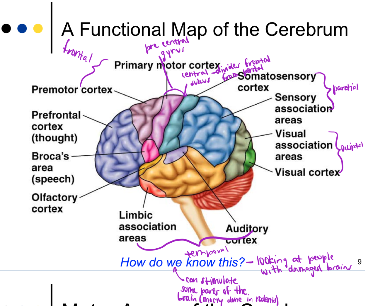
cortex
- outer part of the brain
- consists of lobes -- frontal, occipital, parietal, temporal (8 total, one on each side with separate functions)
- areas are devoted to different body parts (motor and sensory) -- larger areas designated for parts that require more motion/sensation, not parts with more body area
- consists of lobes -- frontal, occipital, parietal, temporal (8 total, one on each side with separate functions)
- areas are devoted to different body parts (motor and sensory) -- larger areas designated for parts that require more motion/sensation, not parts with more body area
40
New cards
cerebrum
- major part of the brain
- divided into 2 hemispheres
- connected via corpus callosum
- divided into 2 hemispheres
- connected via corpus callosum
41
New cards
gyrus
a convex fold or elevation in the surface of the brain
42
New cards
sulcus
any of the narrow grooves on the surface of the brain
43
New cards
primary motor cortex
- located in the frontal lobe
- controls movements of skeletal muscles
- stimulation results in specific movements (based on region)
- if damaged: paralysis of voluntary movements, reflexes remain intact -- ability to grasp cup
- controls movements of skeletal muscles
- stimulation results in specific movements (based on region)
- if damaged: paralysis of voluntary movements, reflexes remain intact -- ability to grasp cup
44
New cards
premotor cortex
- located in frontal cortex
- coordinates movements of groups of muscles
- if damaged: loss of skill, can be relearned (still have primary motor cortex, just have to rewire) -- ability to hold cup and bring to mouth
- coordinates movements of groups of muscles
- if damaged: loss of skill, can be relearned (still have primary motor cortex, just have to rewire) -- ability to hold cup and bring to mouth
45
New cards
primary somatosensory cortex
- located in parietal lobe, post central gyrus
- primary input from sensory receptors in skin and muscle
- if damaged: loss of sensation -- cannot feel coin in pocket
- primary input from sensory receptors in skin and muscle
- if damaged: loss of sensation -- cannot feel coin in pocket
46
New cards
somatosenses
senses distributed throughout the body (touch, pressure, pain, thermosensation)
47
New cards
somatosensory association area
- located in parietal lobe
- interpretation of sensation
- integration with memory
- if damaged: loss of identification -- cannot tell that object in pocket is a coin
- interpretation of sensation
- integration with memory
- if damaged: loss of identification -- cannot tell that object in pocket is a coin
48
New cards
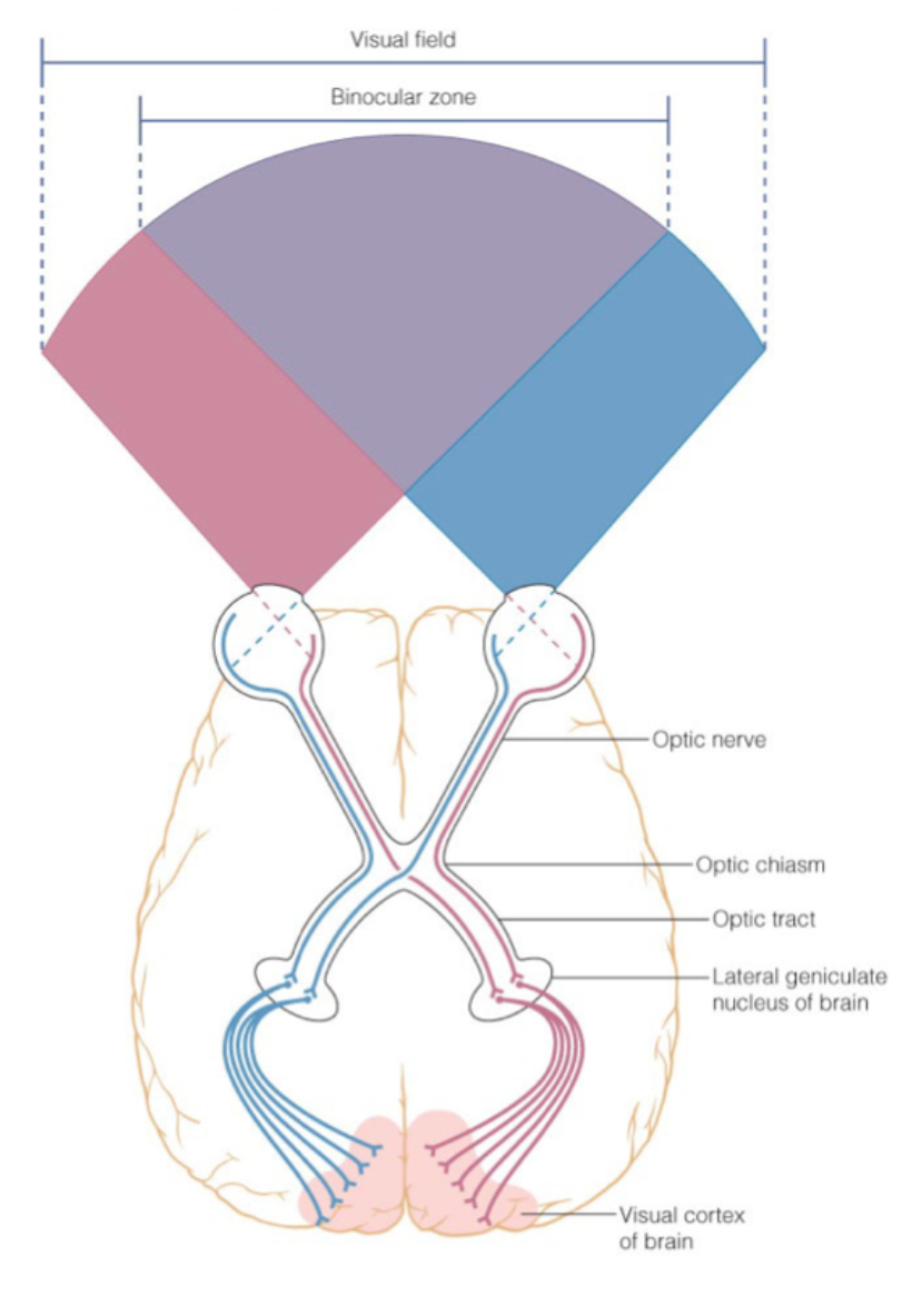
primary visual cortex
- located in occipital lobe
- receives light stimuli
- receives light stimuli
49
New cards
visual association areas
- located in occipital lobe
- puts together an interpretation (could recognize light forms rectangle)
- puts together an interpretation (could recognize light forms rectangle)
50
New cards
primary input from optic tracts
- significant ipsilateral signals
- if contralateral, cross at optic chiasm
- if contralateral, cross at optic chiasm
51
New cards
prefrontal cortex
- frontal lobe
- responsible for thought and personality
- responsible for thought and personality
52
New cards
broca's area
- interface between temporal lobe and motor cortex, left hemisphere
- association of signals to generate speech
- association of signals to generate speech
53
New cards
wernickie's area
- interface between sensory cortex and temporal lobe, right hemisphere
- connects the perception of speech
- connects the perception of speech
54
New cards
auditory cortex
receives signals about hearing
55
New cards
limbic system
situated below the cerebral cortex, behavioral and emotional responses
56
New cards
thalamus
- relay station, routes information
- sensory signals go through to get routed to the sensory cortex
- filters and sorts sensory inputs
- information from somatosensory regions synapsed here -> interneurons integrate signal -> decide whether it can pass through and where it goes
- sensory signals go through to get routed to the sensory cortex
- filters and sorts sensory inputs
- information from somatosensory regions synapsed here -> interneurons integrate signal -> decide whether it can pass through and where it goes
57
New cards
hippocampus
- learning and memory
- important for memory formation in mammals (especially spatial memory formation)
- important for memory formation in mammals (especially spatial memory formation)
58
New cards
amygdala
emotions, fear, and aggression (fear memory formation and innate fear)
59
New cards
hypothalamus
- homeostasis
- diverse set of homeostatic functions
- regulates pituitary gland
- regulated body temperature
- regulates food intake
- involved in stress response (form connections with pituitary gland, pituitary connected to adrenal gland (HPA axis))
- helps maintain ion and water balance
- regulates circadian rhythms
60
New cards
olfactory bulb
- sense of smell
- receives info from nose
- receives info from nose
61
New cards
cingulate gyrus
62
New cards
fornix
63
New cards
diencephalon
the posterior division of the forebrain; connects the cerebral hemispheres with the midbrain
64
New cards
telencephalon
the anterior division of the forebrain; the cerebrum and related parts of the hypothalamus and thalamus
65
New cards
long term potentiation
- form of synaptic plasticity
- causes long lasting increases in signal transmission between neurons
- thought to underlie learning and memory
- occurs at dendritic spin synapses of hippocampal neurons
- mechanism for tracking repetitive activity (salient (stand out) stimuli)
- stimulation of spine synapse -> leads to changes -> way of tracking repetitive activity
- provides a mechanism in which repetitive activity of a particular neuronal pathway can leave a record of itself even after the activity has stopped
- occurs after high frequency stimulation
66
New cards
high frequency stimulation triggers several key synaptic changes
- high frequency changes depend on spine density/number, spine morphology, and spine function
- in low-frequency, one sodium channel (with AMPA receptor), calcium channel (with NMDA channel) blocked by magnesium
- in high-frequency, increased depolarization displaces magnesium block, calcuim enters via NMDA receptors and activates CAMKIII and protein kinase C, causes phosphorylation of AMPA receptors, increases number at plasma membrane and response to glutamate because of increased conductance of sodium
67
New cards
sympathetic nervous system
- antagonistic to parasympathetic nervous system, homeostasis requires balance between the two
- most active during stress or physical activity
- fight or flight (or freeze in some organisms) in response to imminent threat
- increases heart rate and breathing, directs blood to working muscles (toward extremities, away from visceral organs)
- allows organism to respond quickly to danger
- initial cell body is in spinal cord -> connects to cluster of cell bodies outside of CNS (ganglion)
68
New cards
parasympathetic nervous system
- most active during periods of rest
- resting and digesting
- redirects energy toward maintenance activities, such as digestion (blood directed to digestive tract, increased mobility through digestive tract, lower heart rate)
- initial cell body is in spinal cord -> connects to cluster of cell bodies outside of CNS (ganglion)
- resting and digesting
- redirects energy toward maintenance activities, such as digestion (blood directed to digestive tract, increased mobility through digestive tract, lower heart rate)
- initial cell body is in spinal cord -> connects to cluster of cell bodies outside of CNS (ganglion)
69
New cards
dual innervation
- both sympathetic and parasympathetic have connections to most vital organs
- sympathetic collateral ganglion located very close to CNS, parasympathetic collateral ganglion located further from the CNS with shorter axons for secondary neurons
- some exceptions, including arterioles, adrenal medulla, and kidney that use basal tone for regulation
- sympathetic collateral ganglion located very close to CNS, parasympathetic collateral ganglion located further from the CNS with shorter axons for secondary neurons
- some exceptions, including arterioles, adrenal medulla, and kidney that use basal tone for regulation
70
New cards
effect of sympathetic and parasympathetic on pupil
sympathetic: dilates
parasympathetic: constricts
adrenergic receptor: alpha
parasympathetic: constricts
adrenergic receptor: alpha
71
New cards
effect of sympathetic and parasympathetic on heart
sympathetic: increases rate and force
parasympathetic: slows heart rate
adrenergic receptor: beta 1
parasympathetic: slows heart rate
adrenergic receptor: beta 1
72
New cards
effect of sympathetic and parasympathetic on bronchioles of lungs
sympathetic: dilates
parasympathetic: constricts
adrenergic receptor: beta 2
parasympathetic: constricts
adrenergic receptor: beta 2
73
New cards
effect of sympathetic and parasympathetic on digestive tract
sympathetic: decreases motility and secretion (less important in face of danger)
parasympathetic: increases motility and secretion
adrenergic receptor: alpha, beta 2
parasympathetic: increases motility and secretion
adrenergic receptor: alpha, beta 2
74
New cards
path of sympathetic nervous system
- starts in cell body within CNS (shorter than para, so ganglion are closer to CNS)
- NT acetylcholine is released and binds to nicotinic cholinergic receptor in and causes excitatory response in cell body of postganglionic neuron
- NT norepinephrine/noradrenaline is released and binds to adrenergic receptor in effector organ -- different sub types of adrenergic in different parts of the body, helps mediate different cell responses to stimulants
75
New cards
path of parasympathetic nervous system
- starts in cell body within CNS (longer than symp., so ganglion are further from CNS)
- NT acetylcholine is released and binds to nicotinic cholinergic receptor in and causes excitatory response in cell body of postganglionic neuron
- NT acetylcholine is released and binds to muscarinic cholinergic receptor in effector organ
- NT acetylcholine is released and binds to nicotinic cholinergic receptor in and causes excitatory response in cell body of postganglionic neuron
- NT acetylcholine is released and binds to muscarinic cholinergic receptor in effector organ
76
New cards
different subtypes of adrenergic receptors
- respond to both direct sympathetic nervous system response and metabotropic adrenergic receptors
- alpha 1, alpha 2, beta 1, beta 2
- differences in locations allow sympathetic nervous system to finely regulate where the blood is going
- differences in receptor subtypes among effector organs explain the diverse effects of sympathetic and parasympathetic stimulation in various tissues -- signal is the same, different receptors cause different responses
77
New cards
norepinephrine/noradrenaline
- behaves like neurotransmitter
- faster and more transient
- faster and more transient
78
New cards
epinephrine/adrenaline
- behaves like hormone because it is released into the blood and circulates before binding to post synaptic cell
- slower and longer lasting
- slower and longer lasting
79
New cards
alpha 1
location: smooth muscle of the blood vessels of skin, gut, kidneys, salivary glands
effect: vasoconstriction
second messenger system: G proteinactivates phospholipase C (PLC)
sensitivity: NE > E
effect: vasoconstriction
second messenger system: G proteinactivates phospholipase C (PLC)
sensitivity: NE > E
80
New cards
alpha 2
location: membrane of adrenergic axon terminals
effect: inhibits release of NE
second messenger system: G protein inactivates adenylate cyclase, inhibits cAMP production
sensitivity: NE > E
effect: inhibits release of NE
second messenger system: G protein inactivates adenylate cyclase, inhibits cAMP production
sensitivity: NE > E
81
New cards
beta 1
location: heart
effect: increases strength of cardiac contraction and increases heart rate
second messenger system: G protein activates adenylate cyclase, activates cAMP production
sensitivity: NE =E
effect: increases strength of cardiac contraction and increases heart rate
second messenger system: G protein activates adenylate cyclase, activates cAMP production
sensitivity: NE =E
82
New cards
beta 2
location: lungs, smooth muscles of blood vessels leading to skeletal and cardiac muscles
effect: dilates bronchial passages, vasodilation
second messenger system: G protein activates adenylate cyclase, cAMP production
sensitivity: E > NE
effect: dilates bronchial passages, vasodilation
second messenger system: G protein activates adenylate cyclase, cAMP production
sensitivity: E > NE
83
New cards
basal tone
- allows for one branch to control an effector organ bidirectionally
- arterioles in skin/gut: basal sympathetic signaling (some sympathetic signaling) = normal vascular tone, decreased sympathetic signaling = vasodilation, increased sympathetic signaling = vasoconstriction
- changes in AP frequency alters amount of sympathetic input -> normal vessels = moderate signal rate, blood vessels dilate = decreased signal rate, blood vessels constrict = increased signal rate
84
New cards
adrenal medulla
middle section of the adrenal gland, secretes epinepherine
85
New cards
adrenal cortex
exterior of adrenal gland
86
New cards
adrenal medulla
- innervated by only the sympathetic nervous system, no connections to parasympathetic
- preganglionic sympathetic neuron in CNS releases acetylcholine through axon varicosities (and sometimes en passant synapses) into the adrenal medulla -- allows all receptors to respond to acetylcholine by releasing epinephrine into circulatory system
- acetylcholine binds to nicotinic cholinergic receptors on chromaffin cell (looks a lot like postganglionic post synaptic neurons, like modified sympathetic ganglion with no neurons)
- epinephrine is released into circulatory system and travels through blood stream until it binds to adrenergic receptor on effector organ
- one way that nervous system and endocrine system work together
87
New cards
somatic motor neurons
- innervate skeletal muscle for voluntary movements
- cell bodies in CNS (voluntary muscle movements initiated in frontal lobe)
- release acetylcholine that binds to nicotinic receptor attached to skeletal muscle (neuromuscular junction/NMJ)
- located in frontal lobe
- cell bodies in CNS (voluntary muscle movements initiated in frontal lobe)
- release acetylcholine that binds to nicotinic receptor attached to skeletal muscle (neuromuscular junction/NMJ)
- located in frontal lobe
88
New cards
generator potential
- specialized form of graded potential directly detected by the neuron
- sensory stimulus binds to receptor protein in dendrite of afferent neuron
- causes change in MP
- sensory stimulus binds to receptor protein in dendrite of afferent neuron
- causes change in MP
89
New cards
receptor potential
- specialized form of graded potential detected by receptor in accessory cell that transmits signal to the neuron
- sensory stimulus detected by receptor protein in accessory cell -> change in MP of the cell -> calcium enters and NT is released -> binds to receptor on dendrite of afferent neuron
- sensory stimulus detected by receptor protein in accessory cell -> change in MP of the cell -> calcium enters and NT is released -> binds to receptor on dendrite of afferent neuron
90
New cards
sensory receptors
- convert incoming stimulus (type does not matter) into changes in membrane potential
- chemical stimulus: binds to receptor protein (chemoreceptors) -> signal transduction pathway opens ion channel -> change in MP -> signal to integrating center
- pressure stimulus: triggers pressure receptor (mechanoreceptors) -> signal transduction pathway opens ion channel -> change in MP -> signal to integrating center
- light stimulus: triggers light receptor (photoreceptors) -> signal transduction pathway opens ion channel -> change in MP -> signal to integrating center
91
New cards
types of receptor stimulus modalities
- chemoreceptors detect the presence of chemicals in the environment
- mechanoreceptors detect pressure and movement, including proprioception (internal reception within muscles)
- photoreceptors detect light
- thermoreceptors detect temperature
- electroreceptors detect electric fields (not in humans)
- magnetoreceptors detect magnetic fields (not in humans)
- mechanoreceptors detect pressure and movement, including proprioception (internal reception within muscles)
- photoreceptors detect light
- thermoreceptors detect temperature
- electroreceptors detect electric fields (not in humans)
- magnetoreceptors detect magnetic fields (not in humans)
92
New cards
adequate stimulus
preferred (most sensitive) stimulus modality (do not have to be strong)
93
New cards
multiple modalities
- many receptors can be excited by other stimuli, if sufficiently strong -- if it's not the adequate stimulus, it have to be very strong to excite the receptor
- receptors on eye are photoreceptors, but if enough pressure is sensed, we can perceive light
- receptors on eye are photoreceptors, but if enough pressure is sensed, we can perceive light
94
New cards
polymodal receptors
- sensitive to more than one stimulus modality
- nociceptors detect various strong, potentially damaging stimuli (pain)
- polymodal nociceptors transduce thermal, mechanical, and chemical cues into signals that are sensed as pain (if too hot -- do not feel thermal, just feel pain; if mechanically damaged -- just pain; chemical capsaicin in hot sauce -- just pain)
- sense organs in sharks are sensitive to electricity, touch, & temperature -- pattern of action potentials likely encodes modality information from some
polymodal receptors (able to determine which sense rather then just pain)
95
New cards
stimulus encoding
- sensory receptors convert information about the stimulus into APs
- 4 important features -- modality, location, intensity, duration
- 4 important features -- modality, location, intensity, duration
96
New cards
stimulus modality and location
- receptor location encodes stimulus modality and location
- integrating center interprets modality and location, based on path neurons have taken to get to the sensory integrating center
- integrating center interprets modality and location, based on path neurons have taken to get to the sensory integrating center
97
New cards
stimulus modality
- theory of labeled lines -- discrete pathway from sensory cell to integrating center
- light stimulates eye -> transmitted via optic nerves to visual cortex -> helps brain know it is visual stimulus
- across fiber - encode certain modalities like taste
- a particular afferent neuron is associated with one type of receptor, each afferent neuron follows a particular pathway for integration, perception is based on receptor/path and not on the stimulus (cannot tell pressure vs. light, hit so hard hear ringing due to stimulation of ear hair cells)
98
New cards
stimulus location
- theory of labeled lines -- touch receptors always synapse in the same location in the spinal cord -> travel to same location in thalamus -> always distributed to same location on sensory cortex
99
New cards
receptive field
- region of the sensory surface that causes a response when stimulated
- smaller receptive field allows more precise location of the stimulus, greater acuity (finer detail about a particular surface)
- receptive field at fingertips is small and able to detect precise differences/higher acuity (perceived as 2 stimuli because each point activates its own sensory neuron), field on back is big (perceived at 1 stimulus)
- improved ability to localize stimuli by using more than one sensory receptor cell
100
New cards
lateral inhibition
- signals from neurons at the center of the receptive field inhibit neurons on the periphery
- broad stimulus reaches 3 neurons, hitting the one in the middles the strongest
- response is proportional to the strength of the stimulus, so middle has greatest response
- lateral inhibitory neurons inhibit the neurons next to the middle one and the middle signal is able to propagate stronger
- helps enhance contrast and improve acuity