Cells
1/80
There's no tags or description
Looks like no tags are added yet.
Name | Mastery | Learn | Test | Matching | Spaced | Call with Kai |
|---|
No analytics yet
Send a link to your students to track their progress
81 Terms
How can you distinguish between an animal and a plant cell under a light microscope?
Cell wall in a plant cell. Animal cells have an irregular shape. Both Nuclei are visible.
micrometer
**1mm = 1000 µm
how do electron microscope produce images
electrons rather than light
Define the term resolution.
*Ability to see two adjacent points as separate entities following magnification.
What are the advantages of using a light microscope instead of an electron microscope?
Live specimens can be viewed/ observe biological processes
No artefacts due to preparation techniques
Less preparation required.
Cheaper
Magnification: def
When an image is enlarged.
Give examples of details that cannot be seen when using a light
Mitochondria, chloroplasts, ribosomes, vacuole, and other small organelles.
How must a specimen be prepared for observation under an electron microscope?
If TEM – ultra thin slices must be created and used.
Staining to provide contrast.
There are two types of electron microscope. Give details on each.
The transmission electron microscope (TEM)
Electrons pass through an ultrathin specimen. Can observe internal images under high resolution and magnification.
2. The scanning electron microscope (SEM)
Electrons bounce off the surface of the specimen. Resolution and magnification are not as high but a 3D image is produced.
Define the term ultrastructure
The detail of a cell viewed under a microscope (electron)
Eukaryotic cells are found in which organisms?
Animals, plants and fungi.
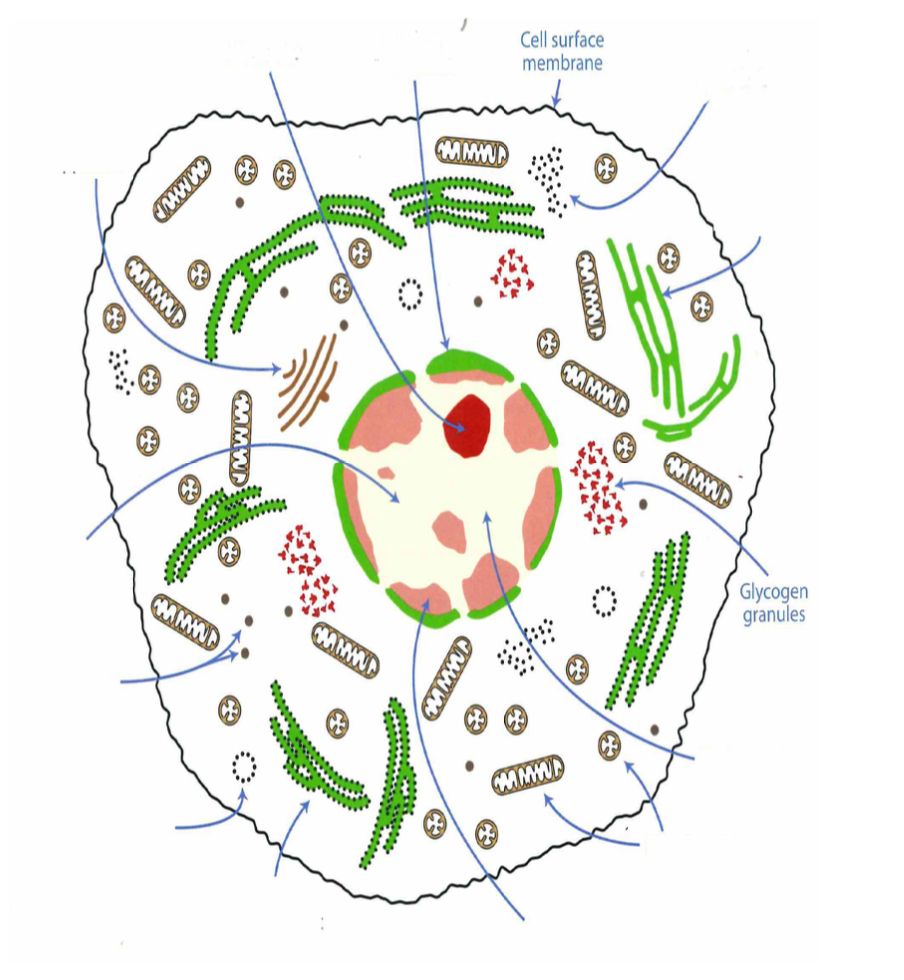
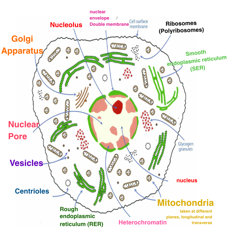
The nucleus
This is the largest and most obvious organelle Usually between 10-25 μm in diameter. Contains DNA in chromosomes.
What are histones and what are their role?
Found in close association with DNA. DNA wound around histones for support and protection.
What is chromatin? Describe its appearance.
Chromosomes condensed. In Heterochromatin the chromatin is densely packed appearing dark. Euchromatin less dense – appearing light.
Nucleus contains one or more nucleoli. A nucleolus appears even darker than densely packed chromatin when viewed under a microscope. What does the nucleolus contain and why is it important?
The nucleolus contains DNA that codes for ribosomes – rRNA. Ribosomes are organellesthat carry out protein synthesis. When produced the ribosomes move out of the nucleolus and out through the nuclear pores to the cytoplasm.
Why are nuclear pores found in the nuclear envelope necessary?
To allow ribosomes and mRNA to leave the nucleus and enter the cytoplasm.
Describe the structure of the nuclear envelope.
Double membrane. The outer membrane is encrusted in ribosomes and is the site of origin for the rough endoplasmic reticulum.
Describe the structure of the ER.
Folded sacs of membrane called cisternae
Distinguish between the rough and smooth ER.
RER is encrusted in ribosomes, SER is not.
What Is the site of origin of the rough ER?
It is an extension of the outer nuclear membrane.
Function of the rough ER (RER)
Provides a scaffold for ribosomes to carry out protein synthesis
Function of the SER
Synthesis of lipids and detoxification of drugs/poisons/carbohydrate metabolism
Ribosomes - SITE of Protein Synthesis
(polyribosomes lots)
Very small organelles visible as black dots on EM micrographs.
Found free in cytoplasm or attached to the outer surface of the rough ER.
Formed of a large and a small subunit – made of protein and ribosomal RNA (rRNA). Can be found in groups called polyribosomes = hotspots for protein synthesis
Describe the structure of the Golgi apparatus.
Folded sacs of membrane called cisternae
The Golgi modifies proteins, for example:
They may have carbohydrate added to form glycoprotein
Lipid may be added to form lipoprotein
They may have prosthetic groups or cofactors added
Different polypeptides can be joined together to form proteins with a quaternary structure
They can be labelled, packaged or sorted
What r lysosomes made from
Golgi apparatus
What do lysosomes contain
Hydrolytic enzymes
Role of lysosomes
Fuse with other vesicles that contain something that needs to be destroyed or digested.
Describe the structure of the mitochondria.
Bean shaped.
Have a double membrane separated by an inter-membrane space.
The inner membrane is highly folded to form cristae that extend into the matrix of the mitochondria.
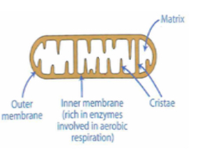
What is the function of the mitochondria?
Site of ATP synthesis during aerobic respiration
How does the structure facilitate the role?
Folding of inner membrane to form cristae increases the surface area which increases the number of enzymes that can be embedded within the membrane which increases the rate of respiration.
What are microtubules and what is their role?
Hollow cylinders form of the protein tubulin. They form the cytoskeleton that maintains the cell shape and keeps organelles anchored in place. They provide a network aiding movement within the cell.
Why are spindle fibres important?
Important in the movement of chromosomes during mitosis and meiosis. (formed of microtubules)
What is the role of centrioles?
Involved in the assembly of the spindle fibres during cell division and are important in cilia and flagella. (formed of microtubules)
Centrioles are not found in plant cells.
Cytoplasm, Location
part of the cell between the cell surface membrane and the nucleus
Protoplasm Location
all the cell within the cell surface membrane including the nucleus.
Cytosol - Location
part of the cell within the cell surface membrane but outside the nucleus and other membrane-bound organelles such as mitochondria
Actual size
Image size over magnification
must convert image size mm to um (x1000)
Actual size unit
um
Image size unit
Ruler/mm
Magnification
Image over actual .
mm(x 1000) over um
How do plant cells differ to animal cells?
Contain chloroplasts, cellulose cell wall, vacuole. Do not contain centrioles.
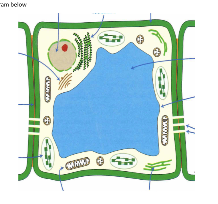
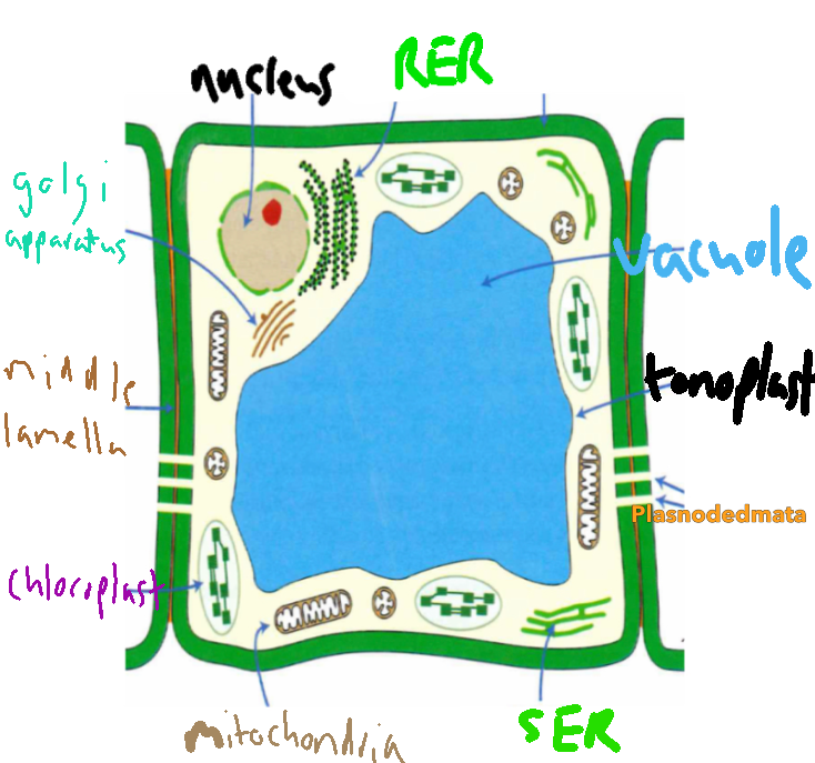
Describe the structure of the plant cell wall.
All plant cells are surrounded by a cell wall composed of cellulose.
Primary cell wall – made up of many microfibrils orientated in different and random directions. The loose arrangement allows the cell wall to expand as the cell grows.
Secondary cell wall -additional layers of cellulose can be deposited to form the secondary cell wall. Each layer of cellulose has the microfibrils orientated in the same direction. Other layers are orientated in different directions to give a lattice arrangement = strength.
Middle lamella – cell walls of adjacent cells are linked by the middle lamella.
Is largely made up of pectin.
Calcium pectate forms a gel or cement that acts as an adhesive and holds neighbouring cells together.
What is the function of the cell wall?
Provide support and tensile strength
Plasmodesmata
Describe the structure and function.
Strands of cytoplasm which extend between neighbouring plant cells. Allow molecules to pass through.
Describe the structure of the chloroplast.
Double membrane which encloses stroma. System of membranes called thylakoids which are stacked at intervals to form grana. Between grana are inter-grana. Starch grain is characteristic of chloroplasts.
What is the role of the chloroplast and how is it adapted for this role?
Site of photosynthesis. Thylakoids contain chlorophyll – stacking to form grana increase surface area to increase light absorption for photosynthesis
Large vacuole
Stores ions and water and has an important role in development of turgor for support. Surrounded by a tonoplast membrane.
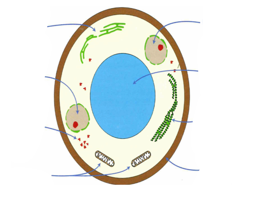
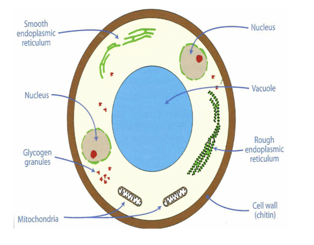
Are fungal cells prokaryotic or eukaryotic
Eukaryotic
How are fungi cells different from plant cells?
Fungi Cell wall composed of chitin not cellulose.
Dont carry out photosynthesis or have chloroplasts.
How are fungi cells similar to animal cells
Glycogen granules and lysosomes present.
Have more than one nucleus (multinucleate)
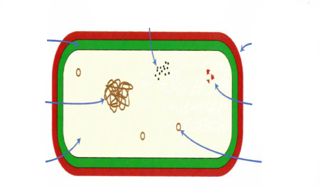
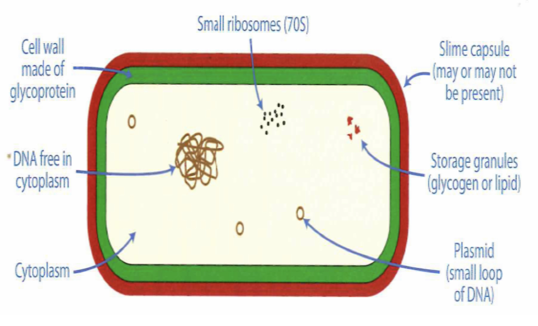
prokaryote cell diagram
prokaryotic cell | eukaryotic cell | |
|---|---|---|
size | ||
site of DNA | ||
DNA organisation | ||
Ribosomes | ||
Internal structure | ||
Cell walls | ||
Plasmids | ||
Microtubules |
prokaryotic cell | eukaryotic cell | |
|---|---|---|
size | Usually < 5 µm | 10-200µm |
site of DNA | DNA free in the cytoplasm | Found in membrane-bound nucleus |
DNA organisation | Circular (loops) without histones | Linear and in chromosomes. Contain histones |
Ribosomes | Small – 20nm | Large 25nm |
Internal structure | No complex organelles | Complex membrane bound organelles |
Cell walls | Peptidoglycan (glycoprotein) | Plants- cellulose, Fungi – chitin, Animals - absent |
Plasmids | Present | Not present |
Microtubules | Not present | Spindle fibres and other microtubules present |
What basic components do cell surface membrane consits of
phospholipid bilayer and protein.
Explain the arrangement of the phospholipid bilayer
Phospholipid phosphate heads are hydrophilic and fatty acid tails are hydrophobic. Tails orientate away from water and heads towards water.
Proteins can be attached to the membrane in two ways
1, Extrinsic – attached peripherally
2. Intrinsic – embedded within one of the two layers
Why is the cell surface membrane described as the fluid mosaic model?
Phospholipid molecules can flow sideways but always keep the bilayer arrangement with proteins floating between the phospholipid molecules.
Fluid mosaic model
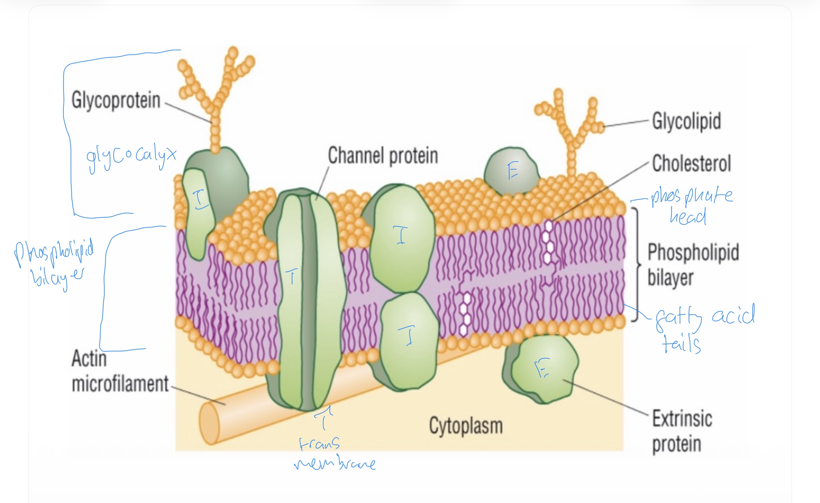
What is in the glycocalyx
Glycoproteins + glycolipids outside cell surface membrane, peripheral to protein or phospholipid
Glycocalyx
Invovled in cell to cell recognition allow cells to form tissues
Glycoprotein act as antigens/ receptor sites for hormones
Glycoprotein + glycolipids important as they form hydrogen bonds with water to stabilise membrane
Cholesterol function
Increases membrane stability - restricts sideways movement at high temp
Low temp- maintains fluidity acting as wedge between phospholipid molecules stops phospholipids from sticking together
Phospholipid bilayer function
Hydrophobic/ hydrophilic properties cause arrangement of phospholipids into bilayer
Provides membrane with selective / permeable properties
Extrinsic proteins
Attached peripherally to the membrane
Intrinsic proteins
Integrally embedded into one of 2 layers of phospholipids
Transmembrane proteins
Intrinsic proteins that extend across membrane
What do all viruses contain?
Protein coat and genetic material.
Examples of viruses
Bacteriophage, and HIV (human immunodeficiency
virus
Describe the structure of a bacteriophage
Contains DNA
Contains a protein coat
Has a base plate and tail fibres which allow it to attach to bacterial cells.
How do bacteriophages replicate in cells?
Infects bacterial cells
Their viral DNA codes for the production of proteins (coat)
Replicate in the bacterial cells and rupture it releases new bacteriophages.
Describe the structure of the HIV virus
Has a phospholipid bilayer with glycoproteins (obtained from human cell it invades)
Has a protein coat
Contains RNA
Contains reverse transcriptase enzyme
What is the role of reverse transcriptase?
To convert the RNA to DNA. The DNA then makes new viruses by synthesising protein coats and viral RNA.
HIV is an example of a retrovirus. What are retroviruses?
Viruses that contain reverse transcriptase and can covert viral RNA to DNA. Reverses normal transcription.
How does HIV invade the human body?
Invade a type of lymphocyte called helper T-cells. As more T-cells are infected and destroyed the immune system becomes compromised and the condition AIDS develops.
Why are viruses not ‘true’ cells?
Can not live without a host. No cytoplasm or organelles.
What similarities are found between bacteriophages and HIV?
Both contain genetic material. Both have a protein coat
What are the differences between bacteriophages and HIV?
HIV has a phospholipid bilayer, bacteriophage does not
Glycoproteins present in HIV but not phages
Bacteriophage normally contain DNA and HIV contains RNA
HIV contains reverse transcriptase, phages do not
Bacteriophage have tail fibres attached to a base plate – HIV do not.
Why is reverse transcriptase not required in most phages?
Most phages already contain DNA
How do viruses replicate in cells?
Bacteriophages invade bacteria/inject DNA into bacteria;
bacteria allow the production of new bacteriophage;
bacterial lysis occurs/new bacteriophage are released;