8A Class Reading Assessment: Embryology, Fetal Circulation
1/16
There's no tags or description
Looks like no tags are added yet.
Name | Mastery | Learn | Test | Matching | Spaced | Call with Kai |
|---|
No analytics yet
Send a link to your students to track their progress
17 Terms
The two cardiac tubes are developed at how many weeks?
3 weeks
7 weeks
4 weeks
10 weeks
3 weeks
The two cardiac tubes are completely fused as one tube at of the beginning of what week?
Week 6
Week 4
Week 5
Week 2
Week 7
Week 4
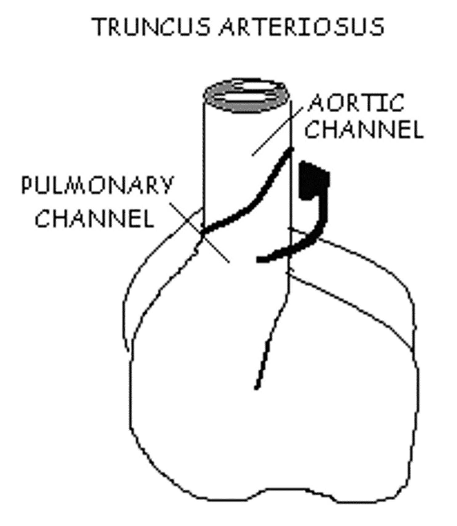
The fetal heart normally folds to the __ , known as d- looping, and the bulboventricular loop is formed.
No answer text provided.
right
medial and posterior
left
right
During cardiac embryonic development, the heart begins to beat at what week?
6 weeks
7 weeks
3 weeks
5 weeks
4 weeks
4 weeks
The fetal heart is fully developed at the end of what week?
7 weeks
4 weeks
3 weeks
10 weeks
7 weeks
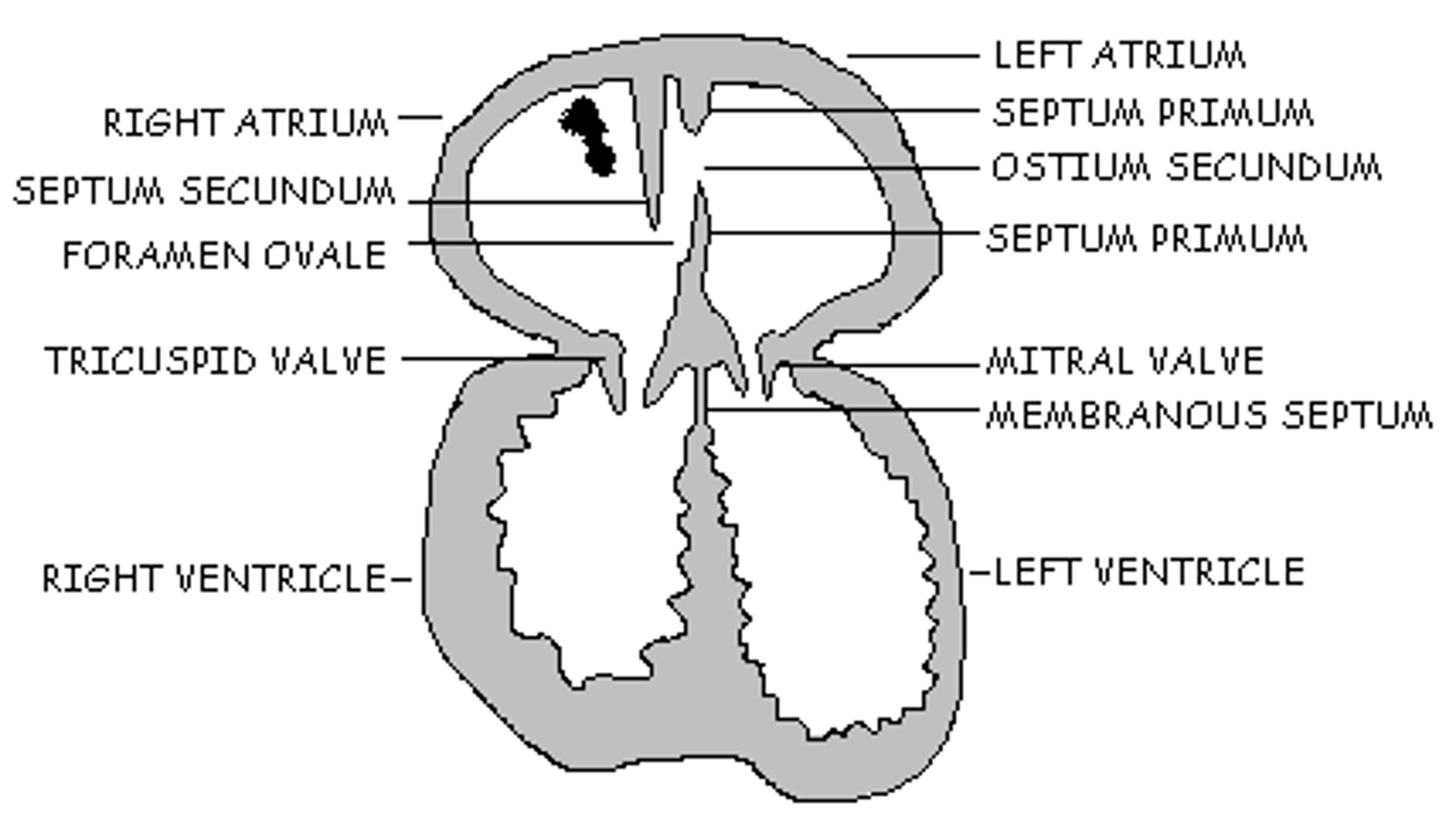
What fetal cardiac structure allows blood flow between the right and left atriums?
Ostium primum
Foramen ovale
Endocardial cushion
Ostium secundum
Foramen ovale
The fetus receives O2 and CO2 exchange through the:
mother's lungs
fetus' lungs
Aorta
placenta
placenta
The right-sided pressures are higher than the left-sided pressures in the fetal heart.
True
False
True
The fetal circulation pattern is designed to mix oxygen rich blood with oxygen depleted blood.
True
False
True
The fetal IVC is part of the circulation pathway.
True
False
True
All the following are adaptations or shunts in the fetal circulation, EXCEPT:
umbilical vein
ductus arteriosus
ductus venosus
foramen ovale
portal vein
portal vein
In the image to the right, at what week is this fetal development occurring?
Week 7
In the image to the right, at what week is this fetal development occurring?
Week 7
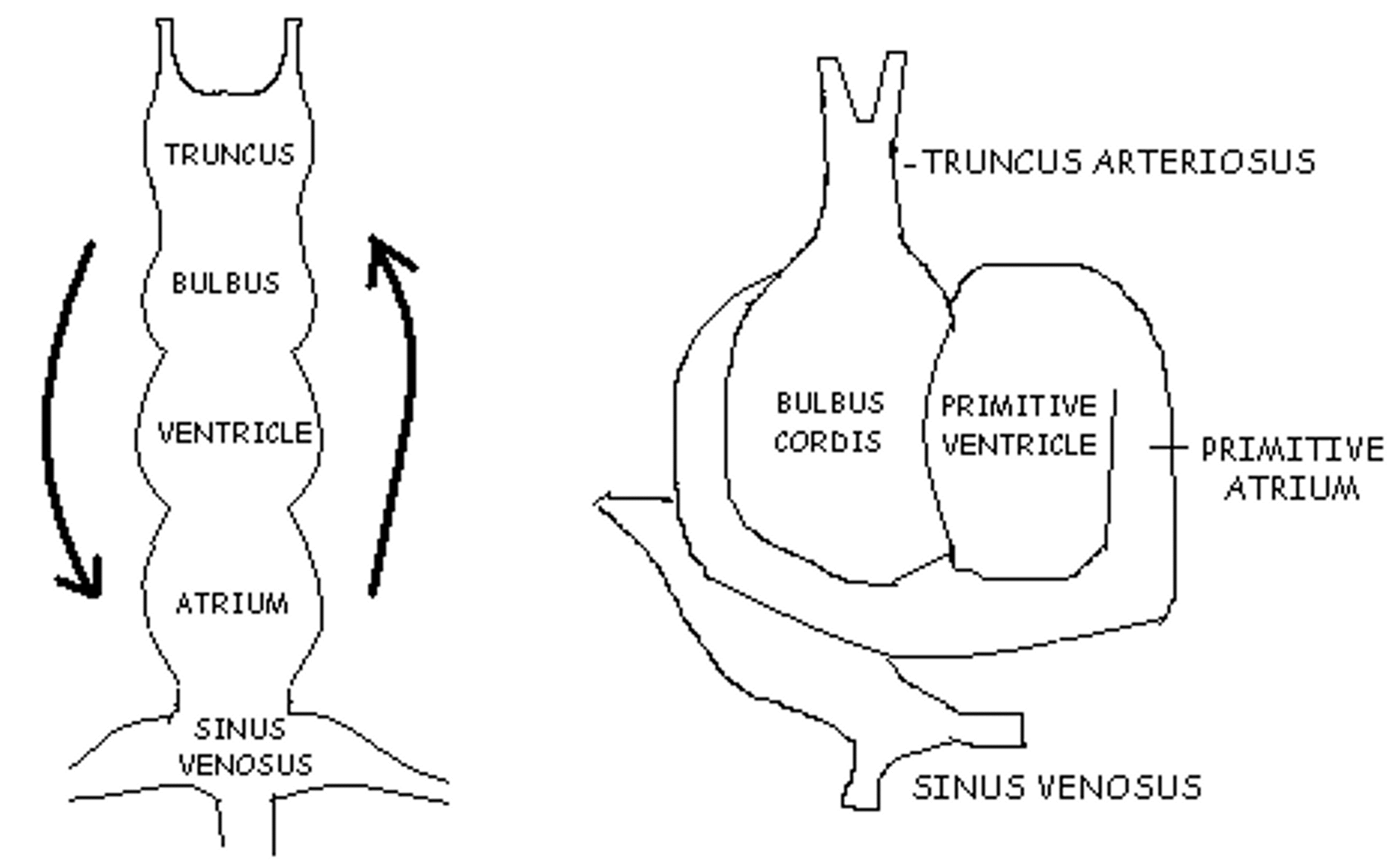
In the image to the right, at what week is this fetal development occurring?
Week 4
In the premature infant, it is common to have persistent fetal circulation, which is the result of pulmonary hypertension with persistent right-to-left shunting across the and .
ASD and PFO
PFO and PDA
ASD and VSD
PDA and VSD
PFO and PDA
The remnant of the ductus arteriosus is called the?
ligamentum arteriosum
fossa ovalis
patent ductus arteriosus
ligamentum venosum
ligamentum arteriosum
What causes the foramen ovale to close?
increased right atrial pressure
increased left atrial pressure
decreased left atrial pressure
decreased right atrial pressure
increased left atrial pressure