wk 3 (L3 cornea)
1/28
There's no tags or description
Looks like no tags are added yet.
Name | Mastery | Learn | Test | Matching | Spaced | Call with Kai |
|---|
No analytics yet
Send a link to your students to track their progress
29 Terms
what is the cornea
clear dome shaped covering that covers the iris + pupil
occupies 1/6 of the globe area
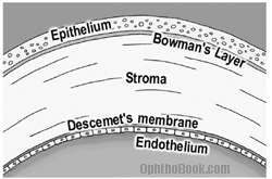
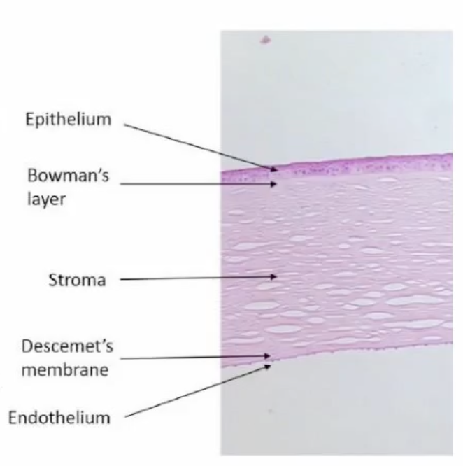
5 layers of the cornea
“Every Bad Sassy Dad Eats”
From front to back:
Epithelium – outer protective layer.
Bowman’s layer – tough layer between epithelium + stroma
Stroma – thickest layer, mostly collagen for strength and transparency.
Descemet’s membrane – thin basement protective layer.
Endothelium – innermost layer, pumps out excess fluid to keep cornea clear.
what is the transitional region where the cornea merges with the sclera/ conjunctiva
limbus
what is the central thickness of the cornea
550 um (microns)
thickness of cornea peripherally ?
700 um (microns)
size of central optical zone of cornea + function
3-4 mm
critical for image formation
what type of epithelium is the cornea
Stratified squamous non-keratinized epithelium
how thick is the human corneal epithelium
50-60 um (microns)
what are the 3 layers of Corneal epithelium (top layer of the cornea)
“Spicy Wings Bravo!”
Superficial (squamous) cells – top
Wing cells – middle
Basal (columnar) cells – deepes

top layer of the corneal epithelium (top layer of cornea) function + name
superficial squamous cells
2-3 layers thick
stick together using desmosomes
outer surface of microvilli + microplicae (ridges) that help the tear film stick
As the cells age, they loosen and fall off into the tear film, making way for new cells.'
✅ Basically: superficial cells are super flat they protect, hold tears, and naturally shed when old.
middle layer of the corneal epithelium (top layer of cornea) function + name
wing cells (wing shaped)
2-3 layers below superficial squamous cells
cells are not as flat as the surface cells above them
✅ In simple terms:
Wing cells are the middle cells in the corneal epithelium that are shaped like wings and work together closely to help move new cells up toward the surface securely and efficiently.
bottom layer of the corneal epithelium (top layer of cornea) function + name
basal columnar cells
single layer of tall columnar cells
They are held firmly to the basement membrane by special anchoring structures (like hemidesmosomes).
key idea:
Basal cells are the bottom “engine room” cells of the corneal epithelium — they divide to make new cells, stick together, and anchor the epithelium to underlying tissue so the surface stays intact and can renew itself.
corneal epithelium turn over time?
every 10 days
(entire surface layer of the corneal epithelium is completely renewed roughly every 7-10 days in a healthy eye.)
what’s the XYZ hypothesis in corneal epithelium turn over
New corneal stem cells are produced limbus (X), move across the cornea (Y), and replace cells that are lost at the surface (Z)
what is the Bowmans layer
werf
below last layer of the corneal epithelium (basal cells)
acellular (without cells) layer made of collagen
strong, protective “lasagne sheet” under the epithelium that helps keep the cornea smooth and intact
what is the corneal stroma
Lies beneath Bowman’s layer and above Descemet’s membrane.
mostly water and collagen arranged into lamellae
makes up 90% of corneal thickness
dense regular connective tissue
Contains keratocytes (special cells that maintain the collagen and extracellular matrix).
4 core proteoglycan proteins
decorin
lumincan
keratocan
mimican
2 types of glycosaminoglycans in the cornea + explanations
keratan sulfate (higher water affinity// water-loving)
dermatan sulfate (holds water loving)
ratio of both differs anterior compared to posterior
keratan (top of stroma under Bowmans membrane) = pressed sponge (loves water but can’t hold much)
dermatan (bottom of stroma above Descemet’s membrane) = loose sponge (hold more water)
purpose of more dermatan sulfate in anterior of corneal stroma vs less in posterior
prevent water evaporation from cornea
as it can damage the
2 layers of Descemet’s membrane
anterior lamina
posterior lamina
where does the descement’s membrane end
it ends abruptly at the limbus
in a thickened area of collagen called
schwalbe’s line
what’s the corneal endothelium
layer of the cornea
next to anterior chamber + interacting with aqueous humor
hexagonal
cells do not replenish
why is the cornea clear
because light is not scattered
even keratocyte spacing
no blood vessels
endothelial fluid pump (water is controlled)
where does cornea get nutrients
diffusion from aqueous humour + limbal vessels
when eyes are closed what supplies oxygen to cornea
palpebral conjunctiva
what is corneal neovascularisation
the cornea is normally avascular (no blood vessels)
corneal neovascularisation: when blood vessels grow on cornea it scatters light = blurry vision
causes: hypoxia (overhearing contacts), infection, trauma, chronic dryness
key points of corneal nerves
super sensitive
lots of nerves form a network
can lose sensitivity over time eg. contacts
what structures make up the uvea?
iris
ciliary body
choroid
what’s anterior uveitis
inflammation of the front part of the uvea