Week 5 - Viruses
1/50
There's no tags or description
Looks like no tags are added yet.
Name | Mastery | Learn | Test | Matching | Spaced | Call with Kai |
|---|
No analytics yet
Send a link to your students to track their progress
51 Terms
Ivanovski and Beijerinck
Isolated tobacco mosaic virus in the later 1800s, beginning viruses as a science
Walter Reed
Showed that yellow fever was caused by a virus transmitted by mosquitoes
Virus + size
Small intracellular obligate parasites between 10-100 nm
Size variation of viruses
Some viruses like CroV, Megavirus chilensis, and mimivirus are huge
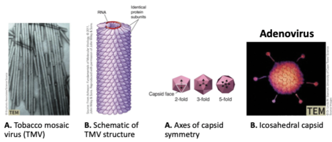
Structure of viruses
Use ssRNA or dsDNA
A capsid shell protects the virus
Some have an envelope of plasma membrane around the capsid shell
Nucleocapsid
Includes both the shell of capsomere proteins and the virus genome it surrounds
Shapes of capsids
Can be helical, icosahedral (20 faced polygon), irregular, or complex
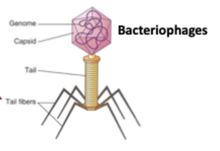
Bacteriophages + replication cycle
Tail fibers stick to cell surfaces, inducing a conformational change. Inner core tube proteins are injected into the cell wall.
Hemagglutinin and neuraminidase
Viral proteins on the surface of many nucleocapsids
Enveloped viruses vs naked viruses
Presence of a plasma membrane surrounding the nucleocapsid
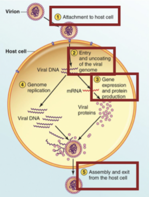
Replication cycle of every virus (4 steps)
Attachment to host cell
Entry and uncoating of the viral genome
Gene expression and protein production
Assembly and exit from the host cell
Entry into animal cells of non-enveloped and enveloped viruses
Non-enveloped: via endocytosis
Enveloped: via membrane fusion or endocytosis to fusion
Entry into plant cells
Often requires some type of damage to plant tissues since there is a protective cell wall
Entry into bacteria
Uses bacteriophages, injects the inner core tube proteins into the cell
Why are viruses considered acellular?
They have no ability to replicate independently
Coevolution hypothesis
Viruses evolved along with their host cells
Could explain RNA viruses, but there is overall little evidence
Regressive hypothesis
Viruses are cells that lost their replication and metabolism
Although examples of this exist, it doesn’t explain RNA viruses
Progressive hypothesis
Genetic material gained the ability to replicate semi-autonomously and move from cell to cell
Transposons and retrotransposons are evidence of this
Transposons vs retrotransposons
DNA transposons use a "cut-and-paste" method and can insert copies of themselves into the host cell
Retrotransposons use a "copy-and-paste" method via copying their genome into RNA, then converting it to DNA, and then integrating it into the host genome
Using the progressive hypothesis, how might bacteriophages and animal viruses evolved?
May have been genetic components that escaped bacteria/animal cells and moved between cells
Lytic cycle
Replicate in and lyse the host cell, killing it
Lysogenic cycles
Bacteriophages integrate their genome into the host cell genome, which is replicated each time the host cell replicates
Key properties of viruses
Cannot replicate independently
Use host cell processes (like transcription and translation) to make new virus particles
Can have DNA or RNA
Helical vs icosahedral viruses
Helical capsids tightly associate with the viral genome and form a cylinder-like structure. Icosahedral capsids are like balls that contain the viral genome
Contrast bacteriophages with animal and plant viruses
Bacteriophages typically do not enter the host cells and inject their genetic material in instead
What evidence does not support the regressive evolution of viruses?
Sequences of viral genomes do not seem like they were once cells that lost most of their genetic material. They resemble the genomes of their hosts, and share no features indicating their ancestors were bacteria or archaea
Plaques in a bacterial lawn
Zones of clearing where a phage has killed an infected cell and its adjacent cells. Can be counted to determine the concentration of phages in the solution applied to the agar plate.
Cultivating animal viruses
Must use a tissue culture of host cells to grow targets for the virus
HeLa cells
The first human cell line, isolated from Henrietta Lacks. Immortal due to them being cancerous
Cytopathic effects (CPE)
Changes in cells caused by viral infection, ranging from morphology to cell death
Syncytia
A cytopathic effect where cells fuse into a large, multi-nucleated cell structure
Viral purification
Filtering out large cells and cellular debris via differential centrifugation or gradient centrifugation
Differential centrifugation
A method of viral purification.
Low speed centrifugation, pellet of whole and broken cells forms at the bottom
Medium speed centrifugation, pellet of nuclei and other large organelles
High speed centrifugation, pellet of virus
Gradient centrifugation
Viral component and particles of different density settle into different bands, forming a density gradient
Methods of viral quanitification (4)
Direct count, hemagglutination assay, plaque assay, endpoint assays
Direct count + cons
Electron microscope is used to see a known volume of material, count the viruses, and scale it up to determine titer
Not easy and doesn’t differentiate between viable and nonviable particles
Titer
Concentration of a virus preparation
Hemagglutination assay + pros and cons
Engineer viruses to stick to RBCs. Lower number of viral particles forms a button, while high numbers form a shield.
Pros: cheap, fast, easy
Cons: not all viruses can do it, doesn’t differentiate live/dead viruses, doesn’t give a virus number
Plaque assay
Virus is diluted and placed on target cells, then plaques are counted to determine the number of viral particles.
Endpoint assays
Determine either TCID50 (amount of virus to induce CPE in 50% of the cells) or LD50 (amount of virus to kill 50% of the test animal subjects)
Explain how filtration and centrifugation can be used to purify viruses
Viral particles are very small and can be filtered out of larger cell counterparts. Centrifugation causes the largest particles to form a pellet, leaving viral particles only
Strategies used to name viruses
Location, disease they cause, physical characteristics, organism they infect, simple letter/number combinations
ICTV classification scheme (5)
Classify viruses into order, family, subfamily, genus, and species, based on morphology, genome structure, replication strategy, host range, and pathogenicity
Baltimore classification scheme
Separates viruses into seven classes based on how they generate mRNA
Electron microscopy in virus identification
First step in virus identification
Allows for visual observations of viral morphology
Some viruses are indistinguishable under a microscope
Nucleic acid analysis in virus identification
PCR and RT-PCR are used to identify viruses by genome sequence
Can study viral evolution patterns
RT-PCR + uses for viruses
Converts RNA to DNA, which can then be amplified via PCR. Can study RNA produced by infected cells
RT-PCR process (7)
Isolate RNA
Add primers, reverse transcriptase, and nucleotides
RT synthesizes DNA copy of RNA (now you have a DNA copy, proceed with normal PCR)
Heat sample to denature strands and deactivate RT
Add primers, Taq polymerase, and nucleotides
Taq makes a second DNA strand
Repeat PCR cycles to amplify DNA
How can viruses be used as molecular biology tools?
Can be engineered to deliver working copies of genes to replace damaged versions (gene therapy)
Oncolytic viruses
Replicate in and destroy cancerous cells while having limited effects on normal cells
Oncoviruses
Viruses that cause cancer