A/P II Lecture Exam 1
1/158
There's no tags or description
Looks like no tags are added yet.
Name | Mastery | Learn | Test | Matching | Spaced |
|---|
No study sessions yet.
159 Terms
How do all of the body’s tissues and varying body systems communicate and interact?
Central Nervous System (CNS)
Peripheral Nervous System (PNS)
List the parts of the Central Nervous System.
Brain
Spinal Cord
List the function of the central nervous system (CNS).
Interneurons integrate and process signals and then select and appropriate response.
List the parts of the peripheral nervous system.
Cranial Nerves
Spinal Nerves
Ganglia
List the function of the peripheral nervous system (PNS).
Carries information from sensory neurons TOWARDS the CNS for processing and relays processed signals from CNS to motor neurons so that the appropriate response can be executed,.
What cells make up the nervous system? List them.
Neurons
Glial Cells
What are neurons?
Specialized cells that send electrical signals to targets throughout the body.
Define: Soma/Cell Body
What does this structure do?
*Terms are used interchangeably.
The central part of the neuron that contains the nucleus and is responsible for maintaining the cell's functions (e.g protein synthesis and energy production).
Define: Dendrites
What does this structure do?
Branched extensions that RECIEVE signals (post-synaptic) from different types of stimuli, forming GRADED POTENTIALS.
Define: Axon
What does this structure do?
The larger/longer extension that TRANSMITS signals to other cells, in the form of ACTION POTENTIALS.
Define: Axon Hillock
What does this structure do?
Located at the junction where the axon connects to the soma, the structure acts as the trigger zone for nerve impulses (ACTION POTENTIALS).
What is a Graded Potential? Which part of the Neuron detects it?
Graded Potential: a change in a neuron’s membrane potential (carrying in magnitude based on the strength of the stimulus).
*Unlike action potentials, which are all-or-nothing responses, graded potentials can be of different sizes depending on how strong the stimulus is.
Dendrites
What is an action potential? Which part of the neuron detects it?
Action Potential: a rapid, large, and all-or-nothing electrical signal that travels along the length of a neuron, allowing it to communicate with other neurons, muscles, or glands.
Axon Hillock
Why do neurons make (neural) ciruits?
To relay sensory input and motor output.
What are the three general steps of neuronal processing?
Sensory Input
Integration
Motor Response
Neural Processing - Sensory Input
What happens? What type of neurons are present at this step? Is this step involved in the CNS or PNS?
Sensory neurons will detect stimuli (external or internal) and transmit a signal towards the CNS.
Types of Neurons Involved: Sensory Neurons
PNS
Neural Processing - Integration
What happens? What type of neurons are present at this step? Is this step involved in the CNS or PNS?
Signals are relayed to interneurons in the BRAIN and SPINAL CORD for analysis and/or processing, so an appropriate response can be selected.
Types of Neurons Involved: Interneurons
CNS
Neural Processing - Motor Response
What happens? What type of neurons are present at this step? Is this step involved in the CNS or PNS?
A processed signal is then relayed to motor neurons to trigger the needed response muscles, glands, organs, etc.
Types of Neurons Involved: Motor Neurons
PNS
Define: Sensory Neurons
Afferent or Efferent?
Def: Sensory neurons are responsible for carrying signals from sensory receptors (like those in the skin, eyes, ears, nose, etc.) to the central nervous system (CNS).
Afferent
Define: Motor Neurons
Afferent or Efferent?
Def: Carry signals from the CNS to muscles or glands to initiate movement or secretions.
Efferent
Define: Interneurons
Location (CNS or PNS)?
Def: Responsible for connecting sensory neurons to motor neurons.
CNS
Define: Ganglia/Ganglion
CNS or PNS?
A collection of neuron cell bodies in the PNS.
Define: Nerves
CNS or PNS?
A bundle of axons in the PNS.
Alternate Def: Groups of neurons with similar functions with axons bundled together.
True/False - Nerves cannot have the axons of both sensory and motor neurons moving in opposite directions in the fiber.
False
Explanation: The statement is false because axons are capable of sending signals in opposite directions; in fact, many nerves have BOTH afferent and efferent axons running through them.
Define: Nucleus/Nuclei
CNS or PNS?
A collection of neuron cell bodies in the CNS; where processing occurs.
Define: Tract
CNS or PNS?
A collection of axons in the CNS.
Alternate Def: Bundles of neuron fibers (dendrites or axons).
Are interneurons present in the peripheral nervous system?
No
Are interneurons present in the central nervous system?
Yes
Does the spinal cord have interneurons?
Yes
Do somatic afferents usually deal with voluntary responses or involuntary responses?
Voluntary Responses
Do visceral afferents usually deal with voluntary responses or involuntary responses ?
Involuntary Responses
The PNS further separates motor responses into two efferent divisions, list them.
Somatic System
Autonomic System
What is the somatic nervous system?
The system that relays voluntary control—carries signals from CNS to skeletal muscles.
What is the autonomic nervous system?
The system made up of efferent neurons that control involuntary actions—regulate activity of body processes: digestion, cardiac output, etc.
The autonomic system is further divided into two divisions, list them:
Parasynthetic Division
Sympathetic Division
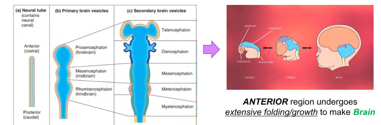
How do central nerves form (CNS)? Review the process.
*Everything starts out as a neural tube.
Neural tube forms along the LENGTH of the entire EMBRYO.
The ANTERIOR region undergoes extensive folding/growth to make the BRAIN.
How do peripheral nerves form? Review the process.
*Precursor cells (cell bodies) originate during neural tube formation.
During nerve development, the neurons must start as cell bodies and grow their axon projections to targets.
Motor neurons can have their cell bodies in the CNS (think: cortex and gray horns) and grow axons OUT towards the PNS [easier task].
Sensory Neurons: neurons grow by starting as cell bodies and grow axons [trickier task].
What if the cell body has to start in the PNS?
A border between the forming neural tube and outer layer forms a neutral crest, which has cells that delaminate/separate and migrate to different parts of the body.
How do neurons “wire” (i.e. connect their axons to their targets)?
Axons of ALL Neurons need to grow from neuron cell bodies to their targets.
During development, where do PNS cell bodies arise from, before growing axons?
Some from neural tube (motor neurons); some from neural crest (sensory neurons).
Define: Neural Crest Cells
What do they do during development?
Def: A border that forms between a neural tube and an outer layer.
Dlaminate/separate and migrate to different parts of the body.
What are some cell types Neural Crest Cells develop into? Is it only Neurons?
They can develop into neurons, glial cells, melanocytes, cartilage and bone cells, smooth muscle, adrenal medulla cells, and so forth.
Typically, can CNS neurons regenerate? Why?
NO—as a result of growth inhibitor proteins in oligodendrocytes membranes and the formation of scar tissue via astrocytes which blocks axon regeneration.
Typically, can PNS neurons regenerate? Why?
YES—as a result of Shwann Cell’s actively aiding in the regeneration process and satellite cells lack of impeding the regrowth process.
What part of sensory neurons detect stimuli?
Sensory neurons have specialized structures at their dendrites, called receptors, that are sensitive to specific types of stimuli.
Remember: “Stimulus-gated ion channels are on dendrites of sensory neurons.”
Remember: Sensory neurons detect specific changes in their environment (aka STIMULI).
Do sensory receptor neurons detect external stimuli? Do they detect internal stimuli?
They detect BOTH!
If the neurophysiology is generally the same, then what makes sensory receptor neurons different?
1) Different “stimulus-” gated ion channels that induce the graded potential on the dendrites.
2) Different dendrite and neuron morphologies.
What are the three ways we can classify a sensory receptor neuron?
Location
Structural Complexity
Stimulus Type
Touch on the location classification of sensory receptor neurons.
Where in the body they function.
General Senses v. Special Senses
Where are sensory receptors for general senses found? List the subtypes of stimuli detected by general senses.
Skin or Internal Tissues (usually not in specialized sensory organs).
Subtypes:
Touch
Pressure
Temperature
Pain
Where are sensory receptors for special senses found? List the subtypes of stimuli detected by special senses.
Head/Specialized Sensory Organs
*Specialized sensory organs include eyes, nose, ear, tongue, etc.
Subtypes:
Vision
Hearing
Eqillibrium
Smell
Taste
Touch on the structural complexity classification of sensory receptor neurons.
Their morphological features.
Can range from simple to complex.
True/False - Simple nonencapsulated nerve endings and complex/specialized nerve endings are usually a part of general senses; simple encapsulated nerve endings are usually a part of special senses.
False
Explanation: Simple nonencapsulated nerve endings and simple encapsulated nerve endings are usually a part of general senses; complex, specialized nerve endings are usually a part of special senses.
Define: Interoceptors
What types of stimuli do they usually detect?
Sense INTERNAL Stimuli
Define: Exteroceptors
What types of stimuli do they usually detect?
Sense OUTSIDE Stimuli
Define: Proprioceptors
What types of stimuli do they usually detect?
Sensitve to stimuli inside the body.
Touch on the stimulus type classification of sensory receptor neurons.
The type of stimulus they detect.
List subtypes/subclasses of stimuli detected by interoreceptors?
Chemoreceptors
Mechanoreceptors
Thermoreceptors
List subtypes/subclasses of stimuli detected by extoreceptors.
Chemoreceptors
Mechanoreceptors
Thermoreceptors
Photoreceptors
What type of stimuli do chemoreceptors usually detect? Where are these receptors found?
Respond to stimuli when a specific chemical binds to gated channels, opening them up and depolarizing the membrane.
Found:
General Senses: Blood Glucose, Toxins in Interstitial Fluid, Ions in CSF, etc
Special Senses: Tongue (Taste) & Nose (Smell)
What type of stimuli do mechanoreceptors usually detect? Where are these receptors found?
Mechanical Force Stimuli
Found:
Special Senses: Ear (Hearing & Equilibrium)
What type of stimuli do thermoreceptors usually detect? Where are these receptors found?
Temperature Change Stimuli
What type of stimuli do nociceptors usually detect?
Damaging Stimulus w/Pain
Ex:
Too Much Heat = Pain
Too Much Physical Strain = Pain
Too Much Chemical = Pain
Is vision a general sense or a special sense”?
Special Sense
How many cranial nerves serve structures of the eye? List them.
Four
CN 2 - Optic
CN 3 - Oculomoter
CN 4 - Trochlear
CN 6 - Abducens
Where are photoreceptors located in the eye?
Retina
Are photoreceptors located at the top or bottom of the neural layer in retina?
Bottom
What is the general order of signal relay, after light photons stimulate photoreceptors?
Photoreceptor —> Bipolar Cell —> Ganglion Cells (CNII-Optic Nerve)
What are rods? What do they detect? What are their general characteristics/functions?
Def: A type of photoreceptor that is numerous, very sensitive, only has one type of visual pigment, and detects dim light.
Functions:
Perceive Gray-Scale Images (no color-vision or sharp/high-resolution images).
What are cones? What do they detect? What are their general characteristics/functions?
Def: A type of photoreceptor that contains detects bright light and contains three pigment types: red, blue, and green.
Functions:
Perceive Sharp/High Resolution Color Images
What is the general cellular anatomy of photoreceptors?
Inner Segment & Outer Segment
Touch on important characteristics of the outer segment anatomy of photoreceptors.
Light-Recieving Region
Contains visual pigments that change shape when they absorb light.
How often do photoreceptors (rods and cones) regenerate their outer segment? Why?
Every 24 hours due to intense light exposure.
What is retinal?
A form of vitamin A and is a component of the photopigments found in the photoreceptor cells (rods and cones) of the retina. It plays a critical role in the phototransduction process, which is the conversion of light into electrical signals in the retina.
What is opsin?
A type of protein that is a key component of photopigments, the light-sensitive molecules found in the rods and cones of the retina.
What do retinal and opsin come together to make?
Visual Pigment
How do visual pigments absorb light, to open ion channels?
Change Shape
What is 11-cis-retinal?
“Bent” Retinal in the Dark
What is all-trans-retinal?
“Straight” Retinal, After Light Exposure
List the three steps of phototransduction.
1) Pigment Synthesis
2) Pigment Bleaching
3) Pigment Regeneration
Phototransduction Steps - What happens in pigment synthesis?
11-cis-retinal is made from Vitamin A and it binds to rhodopsin in a series of synthetic steps.
*Rhodopsin and “bent” 11-cis-retinal accumulate to high levels in the dark.
Phototransduction Steps - What happens in pigment bleaching?
Light absorption by the rhodopsin causes 11-cis-retinal to convert into ”straight all-trans-retinal.
*This causes a transducin protein to start a neural circuit that makes electrical signals.
Phototransduction Steps - What happens in pigment regeneration?
Enzymes will slowly convert “straight” all-trans-retinal back into the “bent” 11-cis-retinal. If levels of light exposure are NOT too high, regeneration can keep up with bleaching.
*Under these conditions, photoreceptors will remain functional and keep working.
Define: Transducin
Is a G-protein that plays a crucial role in the process of phototransduction; it is involved specifically in the rods of the retina, which are responsible for vision in low-light conditions.
What does transducin eventually lead to?
Depolarization Photoreceptor
Briefly describe how depolarization of photoreceptor affects bipolar cells.
Depolarization of photoreceptors STOPS the release of inhibitory neurotransmitters.
Briefly describe how depolarization of bipolar cells affects ganglion cells.
The depolarization of bipolar cells releases stimulatory neurotransmitters.
Briefly describe what depolarization of ganglion cells leads to.
Light-detection signals being sent down axons of CNII (Optic Nerve).
Define: Optic Disc
What does it cause?
Def: Where CN II (Optic Nerve) leaves the retina and heads to the brain.
Causes: Blind Spot—the bran “makes up” an image to fill in details.
Why?: Because there are no photoreceptors (rods and cones) present here!
What is color blindness? What cells have defects with this condition?
A condition in which one or more of the cones opsins is ABSENT.
What colors are most commonly affected with color blindness?
Red Cones
Green Cones
Define: Refraction
When light changes speed/direction as it passes through transparent media.
Define and list the function: Lens
Def: A transparent interface that has different media.
Function: Alter the path of light, allow for focusing.
Is the lens of the eye concave or convex?
Convex—-allows for light to be focused on a point (focal point).
What are the three locations where light path is altered moving through the eye.
Entering Cornea
Entering Lens
Leaving Lens
Where is the lens located in the eyeball? What is its function?
Location: Located behind the iris and pupil in the eyeball
Function: Changes shape to focus light on retina.
Why must the lens be precisely shaped?
Focus light on the retina.
What muscles contract to change shape of the lens?
Ciliary Muscles
What are ciliary zonules?
Ligaments that attach ciliary muscles to lens.