Digestion
1/7
Earn XP
Description and Tags
Name | Mastery | Learn | Test | Matching | Spaced |
|---|
No study sessions yet.
8 Terms
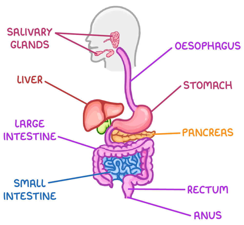
Structure of the Human Digestive System
Salivary glands - these secrete saliva containing amylase.
Oesophagus - this transports food to the stomach.
Stomach - this digests food and produces acid to destroy pathogens.
Liver - this produces bile salts to aid lipid digestion.
Pancreas - this secretes pancreatic juice containing enzymes.
Small intestine - this contains the ileum, a site of further digestion and absorption.
Large intestine - this absorbs water and stores waste.
Rectum - this stores faeces before removal via the anus.
Food travels in this order:
Mouth → Oesophagus → Stomach → Small intestine → Large intestine → Rectum → Anus
Physical and chemical digestion
Digestion takes place in 2 stages:
Physical breakdown - the breakdown of large food pieces into smaller ones to increase surface area for digestion.
Chemical digestion - enzymes catalyses hydrolysis reactions that break bonds in large insoluble molecules to form smaller soluble molecules.
Carbohydrate digestion
Carbohydrases are produced by the salivary glands, the pancreas, and the epithelial cells lining the ileum.
Starch digestion:
Salivary amylase breaks down the starch into the disaccharide maltose in the mouth.
Acid in the stomach denatures salivary amylase.
Pancreatic amylase continues starch digestion in small intestine.
The epithelial cells in the ileum lining produce maltase to break down maltose into α-glucose monomers. Maltase is a membrane-bound disaccharide.
Hydrolysis of other disaccharides:
Sucrose is hydrolysed to glucose and fructose by sucrase.
Lactose is hydrolysed to glucose and galactose by lactase.
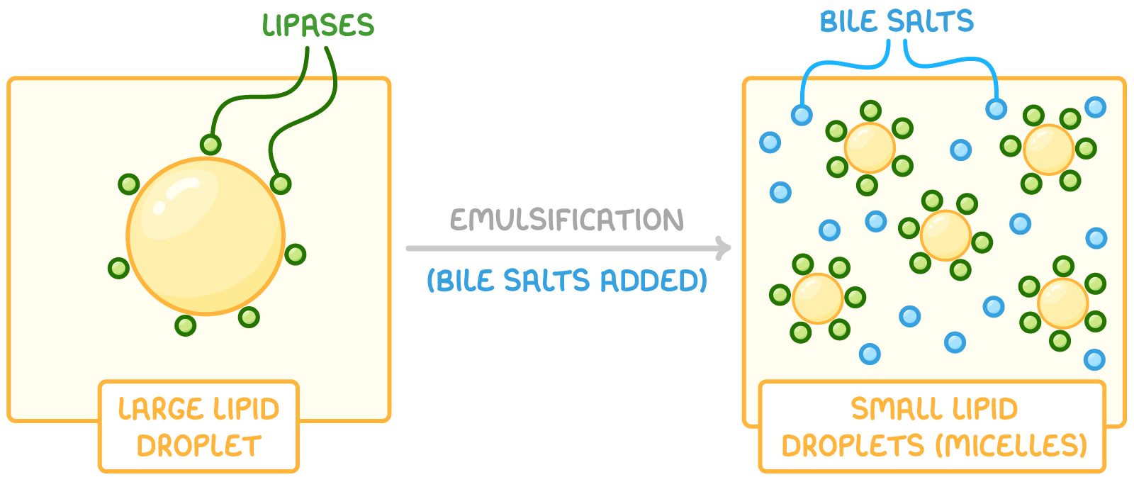
Lipid digestion
Lipases are produced by the pancreas and small intestine.
The digestion of lipids:
Bile salts emulsify lipids into tiny droplets called micelles, increasing the surface area of the lipids.
Pancreatic lipase breaks down micelles into fatty acids and monoglycerides.
Protein digestion
Some proteases (peptidases) are produced by and act in the stomach. Other proteases are produced by the pancreas and the epithelial cells lining the ileum and act in the small intestine.
There are 3 types of protease that achieve this:
Endopeptidases - these hydrolyse internal peptide bonds in the middle of protein to form shorter polypeptides, increasing the number of ends for other protease to work on.
Exopeptidases - these hydrolyse peptide bonds at the ends of polypeptides to remove terminal amino acids or dipeptides.
Dipeptidases - these break down any remaining dipeptides into amino acids.
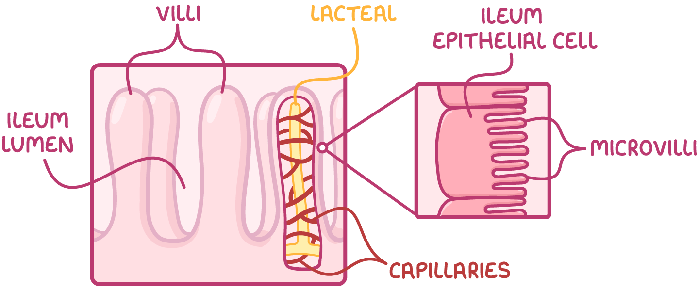
Structure of the ileum
The ileum is the final section of the small intestine. It is adapted to absorb digested food molecules across the epithelial cells that form its lining so nutrients can enter the bloodstream.
Adaptations of the ileum for absorption:
The walls are folded into finger like villi to increase surface area.
Epithelial cells have microvilli to further increase surface area.
It has thin walls to reduce diffusion distance.
It has an extensive capillary network and a good blood supply to maintain steep diffusion gradients.
Muscles in the ileum wall contract to mix content and bring new material into contact with villi.
Absorption of amino acids and monosaccharides
All monosaccharides and amino acids need to be absorbed, which could not be achieved by diffusion alone.
Co-transport allows monosaccharides and amino acids to be transported into ileum epithelial cells against their concentration gradient.
How AA’s and monosaccharides are absorbed into the blood:
Sodium ions are actively transported out of ileum epithelial cells.
This sets up a sodium ion concentration gradient between the ileum lumen and the epithelial cells.
Sodium ions are co-transported with amino acids or monosaccharides from the lumen into the epithelial cells.
Amino acids and monosaccharides move be facilitated diffusion from epithelial cells into the blood.
Absorption of triglycerides
Micelles are broken down to release fatty acids and monoglycerides.
As they are both non-polar, fatty acids and monoglycerides can diffuse into the epithelial cells that line the ileum.
Triglycerides reform inside the cells’ ER.
Triglycerides are packaged into chlyomicrons for transport.
Chylomicrons are released from the epithelial cells by exocytosis into lacteals, which are lymphatic vessels in the villi.
Chylomicrons are transported via lymph vessels in the lymphatic system to the blood.