Chapter 13: Cerebral Cortex
1/119
There's no tags or description
Looks like no tags are added yet.
Name | Mastery | Learn | Test | Matching | Spaced |
|---|
No study sessions yet.
120 Terms
What is the cortex considered as?
The brain’s most complex area (billions of neurons and trillions of synapses
What is the cortex responsible for?
Responsible for several cognitive/
mental activities:Consciousness
Executive functions
Free will
Personality
Skilled movements
Language
Emotions

What does the longitudinal fissure separate?
separates the two hemispheres

What does the lateral or sylvian fissure separate?
separates temporal from frontal and parietal

What does the parietooccipital sulcus separate?
separates occipital from parietal
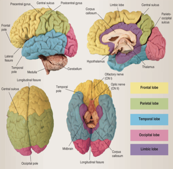
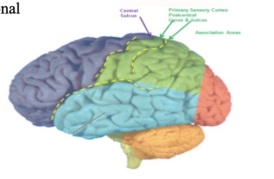
What does the central sulcus separate?
separates frontal and parietal

What does the precentral gyrus contain?
contains the primary motor areas

What does the postcentral gyrus contain?
primary somatosensory area

Where is the cingulate gyrus located?
medial surface frontal lobe

Where is the cingulate and parahippocampal gyri located?
In the limbic lobe

Define cytoarchitecture
The study of how cells, especially neurons, are arranged and organized within the central nervous system

Why was Korbinian Brodmann important?
Korbinian Brodmann conducted detailed comparative studies of numerous species, each of which displays common as well as varying patterns of cytoarchitexture and gyral folding
In simple, he was important because he mapped the brain based on how cells are organized and function (aka cytoarchitecture)
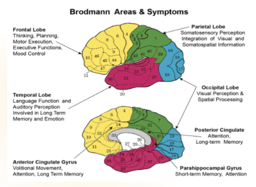
What are different areas of the brain called?
Brodmann areas
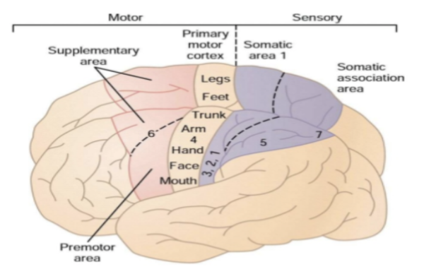
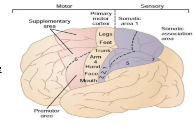
Different areas of the brain and what they represent (don’t have to memorize)
Areas 1, 2, 3 Primary somatosensory cortex (postcentral gyrus)
Area 4 Primary motor cortex (precentral gyrus)
Area 5 Somatosensory association cortex
Area 6 Premotor and supplementary motor cortex
Area 9 Dorsolateral/anterior prefrontal cortex (motor planning, and organization)
Area 10 Anterior prefrontal cortex (memory retrieval)
Area 17 Primary visual cortex
Area 22 Primary auditory cortex
Area 37 Occipitotemporal (fusiform) gyrus
Areas 22, 39, 40- Wernicke's area (language comprehension)
Areas 44, 45- Broca's area (motor speech programming)

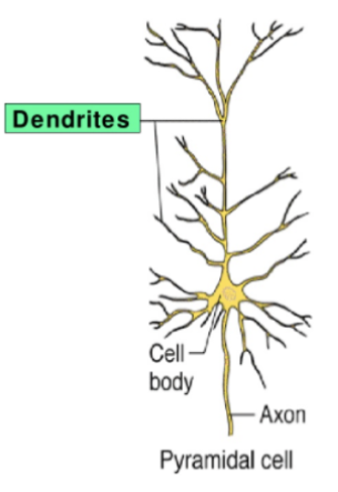
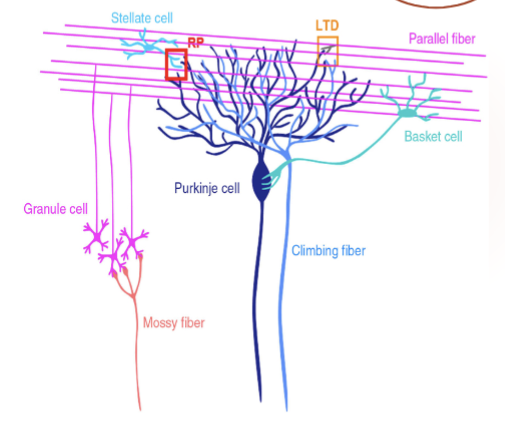
What are pyramidal cells (III and V)?
The main type of neuron that sends information out from the cortex.
How did pyramidal cells get their name?
Name comes from the triangular shaped soma, or cell body

Who discovered pyramidal cells?
First discovered and studied by Santiago Ramon’ y Cajal
What do pyramidal apical dendrites and axons look like/do?
The apical dendrites is the long upper branch. It has small spines where connections from other neurons attach (synapses)
Their long axons leave the cortex to reach other cortical areas or various subcortical sites
What are pyramidal cells important?
These cells control output and are key for learning and neuroplasticity (the brain’s ability to change and grow).
Their dendritic spines strengthen or weaken connections based on experience — that’s how learning happens.
Where are granular (stellate cells, II and IV) located?
These are small interneurons that stay within the cortex (they don’t send signals out).

Describe the granular cell’s shape, axon and dendritic trees
Have shorter axons and smaller dendritic trees
typically small (< 10micrometres) with multipolar neurons

What is the granular cells responsible for?
Their job is to process and pass information locally — basically they help communication between nearby neurons in the cortex.
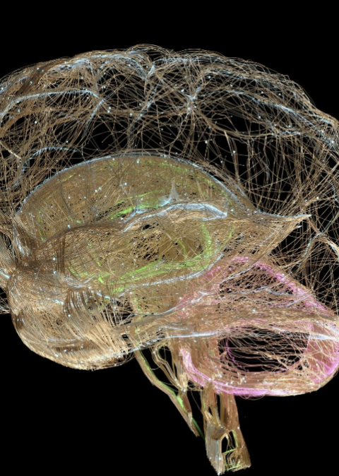
How do the brain communicate?
The brain communicates using fiber bundles, which are large groups of axons that connect areas together

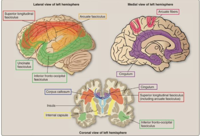
Define association fibers
connect different parts within the same hemisphere

Define commissural fibers
connect the left and right hemispheres.
Define projection fibers
connect the cortex to other brain or spinal cord areas

What type of fibers are the inferior fronto-occipital fasiculus and what do they connect?
Association fiber
connects the frontal, temporal, and occipital lobes

What type of fibers are the uncinate fasciculus and what do they connect?
Association fibers
a hook-shaped connection between the frontal lobe (orbitofrontal cortex) and temporal lobe (hippocampus & amygdala)
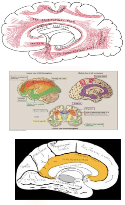
What type of fibers are the superior longitudinal fasciculus and what do they connect?
Association fibers
Connects the frontal lobe to the parietal and occipital lobes.

What type of fibers are the arcuate fasciculus and what do they connect?
Association fibers
links Broca’s and Wernicke’s areas (key for language).

What type of fibers are the cingulum and what do they connect?
Association fibers
links emotional centers — connects the cingulate gyrus to the parahippocampal gyrus (limbic system).

What are examples of commissural fibers
Corpus callosum
Anterior commissure
Posterior commissure
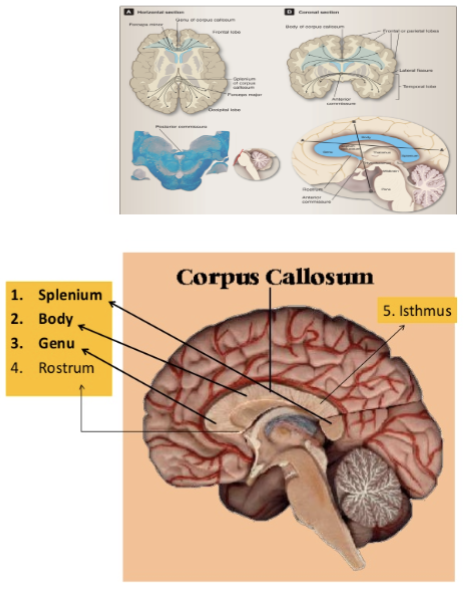
What do the corpus callosum do?
Connects the two hemispheres at the parietal lobes and the posterior parts of the frontal lobes

Why is the corpus callosum important and where is it located?
Largest connective white matter pathway in the brain
Made up of more than 200 million nerve fibers.
Sits in the center of the brain, measures around 10 centimeters (cm) in length, and is shaped like the letter “C.”

What are the parts of the corpus callosum and what do they connect?
Rostrum & genu- connect frontal lobes
Trunk/ body connects posterior frontal, parietal , and superior
temporal lobe
Splenium connects the occipital lobes

Describe the anterior commisure
Also known as precommissure
Small oval (about 5 mm wide).
Connects the temporal lobes on each side.
Also carries decussating fibers from the olfactory tracts (smell).
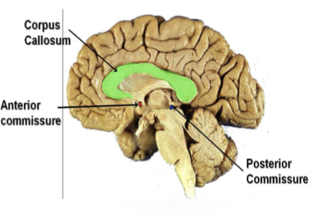
Describe the posterior commisure
Also known as epithalamic commissure
Located near the cerebral aqueduct (where CSF flows between the 3rd and 4th ventricles).
Important for the bilateral pupillary light reflex (how your pupils react to light).

What do projection fibers do?
These fibers send information in and out of the cortex.
Afferent fibers: bring sensory info to the cortex.
Efferent fibers: send motor commands from the cortex
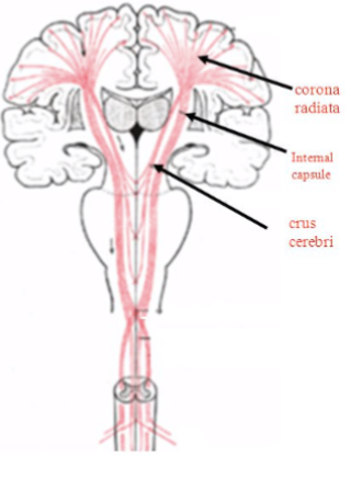
How are projection fibers arranged?
Deeper to the cortex, these fibers are arranged radially as the corona radiata.
Then gather together into the internal capsule (between the thalamus and basal ganglia).
From there, they go down through:
The crus cerebri (in the midbrain)
The pons
The pyramids of the medulla
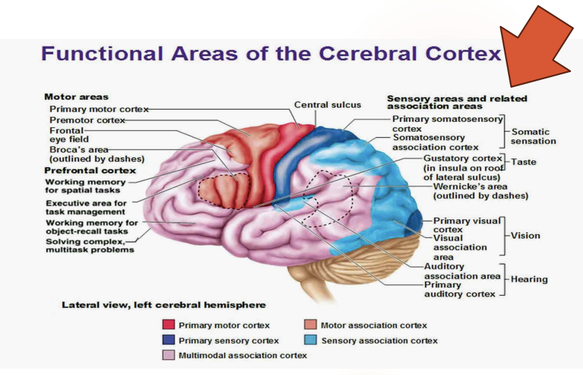
The cortex is divided into?
Primary areas and association areas
What is the primary area responsible for? Are they symmetrical? How are their lesions?
Handles motor, sensory, special senses (vision, hearing, taste, smell)
Largely symmetrical
Lesions here are more straightforward
What is the association area responsible for? How are their lesions?
Handle higher-level functions like emotion, personality, and reasoning.
Damage here causes complex issues (changes in behavior, mood, or judgment).

What are the three parts that control movement?
Primary motor cortex, premotor area, supplementary motor area
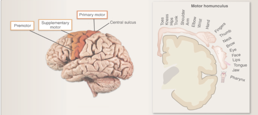
Where is the primary motor cortex located and what does it control?
Located on the precentral gyrus (front of central sulcus).
Controls voluntary movement on the opposite side of the body
More than one half of the entire primary motor cortex is concerned with motor control of the muscles of the hands and the muscles associated with speech
Damage to the primary motor cortex can result in what?
Upper motor neuron signs like paralysis, hypertonicity (muscle stiffness), and hyperreflexia (overactive reflexes)

What is the premotor area responsible for?
Plans and coordinates complex movement patterns
EX: We get an idea on how we will use scissors. It will send signals to the primary motor cortex to carry out the movement and stimulate specific muscles

How does the premotor area send signals to the motor cortex?
The anterior part of the premotor area forms the “idea” of a movement (if damaged → ideational apraxia)
The image will excite the posterior part, which will activates muscle sequences (if damaged → ideomotor apraxia)
It can either send information directly to the primary motor cortex or through the basal ganglia to the thalamus and to the primary motor cortex

Define praxis
Motor planning
Define ideational apraxia
Difficulty in planning and organizing complex sequences of movements, even when the patient understands the overall goal of the task.
EX: Recognizing a toothbrush but not knowing what to do with it (difficulty with planning and organizing movements)
Define ideomotor apraxia
Difficulty in carrying out simple, previously learned movements, even when the patient understands the task and has the physical ability to perform it.
EX: Recognizing a toothbrush and knowing what to do with it but can’t perform the movement (difficulty with executing movements)
Where is the supplementary motor area located?
Found inside the longitudinal fissure and upper frontal lobe.

What is the supplementary motor area responsible for?
Controls posture, bilateral coordinated limb movements, and movement sequences
stimulating the supplementary motor area (SMA) of the brain can cause muscles on both sides of the body to contract, and these contractions are often related to stabilizing body position
EX: reaching out to grab something, the SMA will ensure that the body is properly balanced and braced to prevent you from losing your balance
Lesions to the supplementary motor area can cause what?
Apraxia, inability to perform purposeful actions

Something to Remember
A supplementary motor complex is an association area = Premotor and Supplementary Motor Area
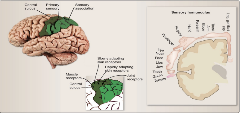
Where is the primary somatosensory cortex located?
In the postcentral gyrus, just behind the central sulcus.

What does the primary somatosensory cortex received?
This area receives touch and body sensation signals (pressure, temperature, pain, vibration, body position)

How does the sensory afferents travel to the primary somatosensory cortex?
Pathway:
Sensory info travels up the spinal cord (via posterior columns, spinothalamic & trigeminothalamic tracts).
It passes through the thalamus (a relay station).
Then travels through the internal capsule.
Finally reaches the postcentral gyrus (where the sensation is felt).
Each side of the brain receives info from the opposite side of the body
Define cortical plasticity
Area of brain that represents the body part can change over time
Amputees
Visually impaired
Taxi drivers
Define lateral inhibition
Area that receive ascending input sends inhibitory projections to adjacent areas which increases contrast between area receiving input and area that does not
Lesions to the primary somatosensory cortex can lead to what?
Deficit in awareness of sensory input and poor localization of sensory stimuli
Where is the somatosensory association cortex (Areas 5 and 7) located?
Located behind the primary sensory cortex in the superior parietal lobe.
What is the somatosensory association cortex responsible for?
Combines/recieves information from touch, pressure, and movement to understand what we’re feeling

Define tactile agnosia
Can feel the object but can’t recognize them
Define asterognosis
Can’t identify the objects by touch alone
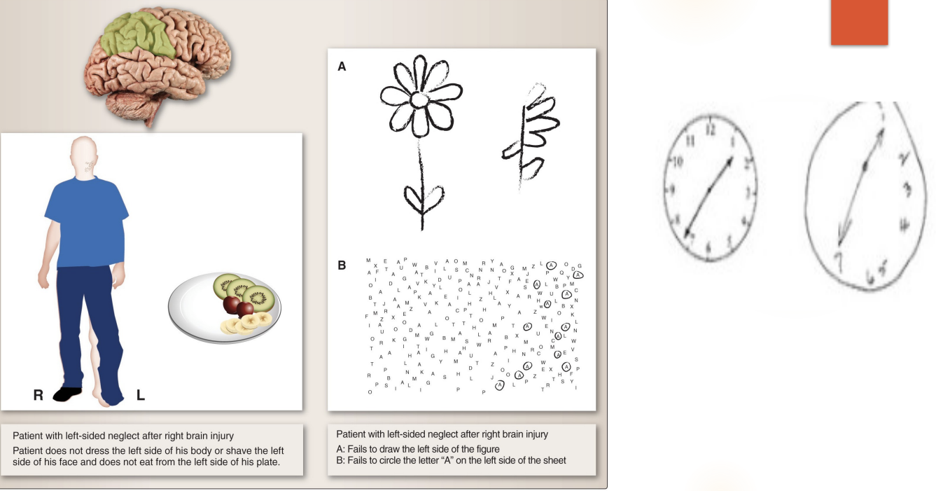
What can happen when the somatosensory association cortex is damaged?
When the non-dominant hemisphere (right) is damaged, there will be neglect to the contralateral side (lefT)
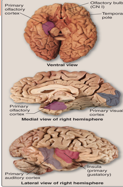
Where is the visual cortex located?
The primary visual cortex is in the occipital lobe.

What is the primary cortical region of the brain?
The Visual Cortex
What is the visual cortex responsible for?
Receives, integrates, and processes visual information from the retinas
Signals are interpreted and form is recognized (light, shapes, color)

Lesions in the visual cortex results in what?
Deficit in opposite (contralateral) visual field

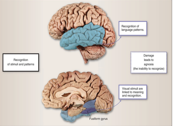
What is the visual association area responsible for?
adds meaning — recognizing what you see (faces, objects, motion).

Lesions to the primary visual area results in what?
Blindness or vision loss on the opposite side
Lesions to the visual association results in what?
Visual Agnosia
Inability to recognize objects in the opposite visual fields despite intact vision
Can also result in trouble tracking objects ipsilaterally
Where is the primary auditory cortex (A-I) located?
Located deep within the lateral sulcus, on the Heschl’s gyri in the temporal lobe, which is the primary representation of auditory information from the cochlea
Will receives sound info from both ears.

Where is the secondary auditory cortex (A-II) located?
Located more rostrally in the temporal lobe and contains Brodmann area 42)
Will process complex sounds (speech, rhythm, melody).
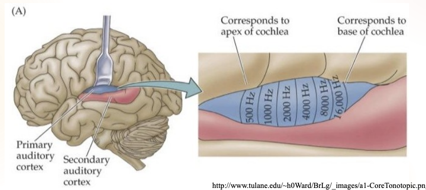
What is the auditory cortex responsible for?
INTERPRET AND GIVE MEANING TO THE SOUNDS WE HEAR
Leison’s to the auditory cortex will result in what?
WORD DEAFNESS OR ACOUSTIC VERBAL AGNOSIA (INTACT HEARING, UNABLE TO INTERPRET WHAT IS HEARD)
Damage to Wernicke’s area result in what?
Aphasia
a condition where a person has difficulty understanding spoken and written language
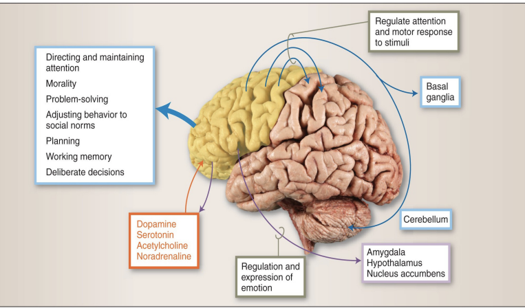
Where is the prefrontal cortex (PFC) located? What is it responsible for?
Behind the forehead and above the eyes
Dictates our personality, our goals, and our values

The superior and lateral parts of the prefrontal cortex are connected to and responsible for?
Connected with sensory and motor cortex, BG, and cerebellum
Regulate attention and motor responses to stimuli.

The inferior and medial portions of the prefrontal cortex are connected to and responsible for?
Interconnected with amygdala,hypothalamus, nuc. acumbens
Brainstem nuclei involved in arousal
Responsible for the regulation of emotions.
What is the dorsalateral PFC responsible for?
top part
Handles executive functions like planning, cognitive flexibility, problem solving, maintain attention to a task, and working memory

What is the orbitofrontal cortex responsible for?
Decision making particularly associated emotional and social
Close to the limbic system
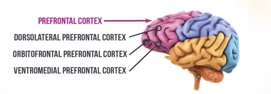
What is the ventromedial prefrontal cortex responsible for?
Connects to emotion areas (amygdala, thalamus, hippocampus).
Helps us make choices based on values, morals, and past experiences (“learning from mistakes”)

Damage to the prefrontal cortex can result in?
personality changes, poor judgment, impulsivity, emotional flatness, or inappropriate behavior.
Where is the parietal association area located?
Found in the posterior parietal cortex, where touch (somatic) and visional sensations combine
What is the parietal association area responsible for?
Helps you understand where your body and objects are in space (spatial awareness).
Helps with attention and awareness of self and environment.
Right hemisphere: awareness of space (attention in space).
Left hemisphere: awareness of time and sequencing (attention in time).

Lesions to the parietal association area results in?
Left side neglect, more common after the right side is damaged
How much of our brain is made up of the temporal lobe? and where is it located?
The temporal lobe makes up about 22% of the entire cortex.
Located under the Sylvian fissure (lateral sulcus) and in front of the occipital lobe

What are the two sulci in the temporal lobe?
superior and inferior temporal sulci (the grooves btw)

What are the three gyri in the temporal lobe?
Superior temporal (Area 22)
Middle temporal (Area 21)
Inferior temporal (Area 20)
(the ridges btw)
What are the deeper structures in the temporal lobe?
Fusiform gyrus
Hippocampal gyrus
Amygdala
Dentate Gyrus
Inferior Temporal Gyrus

Damage to the temporal association area results in?
Agnosia: can’t recognize familiar things even though senses work.
Prosopagnosia: can’t recognize faces.
Aphasia: problems with language comprehension or expressio
Where is the superior temporal gyrus located and what is it responsible for?
The top ridge; contains the primary auditory area.
Left side: processes words and speech sounds (language).
Right side: processes music, pitch, and tone.
What is the middle temporal gyrus responsible for?
Handles auditory processing and language understanding.
What is the inferior temporal gyrus responsible for?
Handles visual processing and recognition memory (recognizing people, objects, memory of what things look like).
What is the amygdala responsible for?
Handles emotional learning and memory modulations, especially fear and anxiety; linked to PTSD.

What is the hippocampus responsible for?
Structure located in the medial temporal lobe
In cross section resembles a ‘sea horse’
Short term storage of new learning.
Critical for long-term memory storage.
Declarative(Explicit)memory
How many areas in the brain are for language? What are they called?
There are two main areas for language in the dominant hemisphere (usually the left)
Wernicke’s and Broca’s area
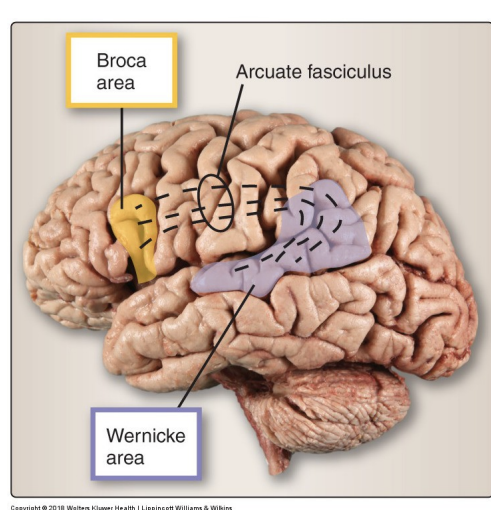
Where is wernick’s area located? What is it responsible for? Lesions here can result in what?
In the upper temporal lobe, extending to the angular and supramarginal gyri.
Handles understanding language (spoken and written).
Lesion → Wernicke’s (Receptive) Aphasia:
Speech sounds fluent but doesn’t make sense (“word salad”).
They can’t understand others or recognize their own speech errors.
Reading, writing, and repeating words are also impaired

Where is broca’s area located? what is it responsible for? lesions here can result in what?
Located in the inferior frontal gyrus.
Handles speech production (turning thoughts into words).
Lesion → Broca’s (Expressive) Aphasia:
Speech is slow, effortful, and broken.
Comprehension is mostly preserved.
They know what they want to say but can’t get the words out.