Animal Models
1/33
There's no tags or description
Looks like no tags are added yet.
Name | Mastery | Learn | Test | Matching | Spaced | Call with Kai |
|---|
No analytics yet
Send a link to your students to track their progress
34 Terms
what are the 3 aspects of study
levels
models
measures
Molecular level
structure and function of molecular aspects of biological pathways
genes and proteins
uses PCR, gel electrophoresis, blotting and microscopy
most common area of research
Cellular level
investigates structure, function and behaviour of cells
uses cell culture, microscopy and electrophysiology
whole organism level
investigates structure and function of systems using the entire organism
uses behavioural tests and imaging
4 models
In Vivo
Ex vivo
In vitro
In silico
In vitro
within the glass
uses cells from a repository (cell line) - Cell culture
use microscopy and electrophysiology
Ex vivo
Outside the living
cells and tissues from an organism (eg. brain slices)
uses PCR, microscopy and electrophysiology
In vivo
within the living
uses living organisms
uses animal models, microscopy, imaging and electrophysiology
In silico
computational models
simulations based on data collected from other models
two types of measurement
static and functional
static measurements
measurements at a point in time
examine the morphology, density and expression of genes
techniques: microscopy, PCR, gel electrophoresis, blotting, immunohistochemistry and imaging
functional measurements
measurements that are changing over time
activity
techniques: microscopy, electrophysiology, behavioural testing, imaging
define animal models
non-human species used in biomedical research to mimic aspects of a biological process or disease found in humans
anatomy, physiology or response to a pathogen are similar enough to that of a human that their results can be extrapolated to improve human understanding
allow the performing of experiments that would be impractical or ethically prohibited with humans
Naive animal
an animal with no changes made to it, used to understand normal function
animal model
has been idealised or modified to represent something (eg. disease)
reasons to use drosophila, c.elegans and zebra fish
less complex nervous system than mammals (easier to understand but harder to translate to mammals)
cheaper and faster to breed
good for fundamental questions about genetics
highly conserved genetics (homologues)
can do behavioural studies (drosophila in mazes)
reasons to use mice and rats in animal research
mammalian nervous system so more translatable to humans
mice are most common for genetic models
rats are best when size or easy training is important
cheaper and faster than large animals
reasons to use larger animals in animal research
brain structure needs to be more similar to humans
sheep have folded cortex and solid tentorium (dura mater) —> good for stroke research
UK legislation on selecting animals for research
must use lowest sentient being to address the question
what are the three criteria for animal models?
predictive validity
face validity
constructive validity
Predictive validity
behaviour
do outcome measures from the animal correlate with the condition
face validity
neurobiology
does the animal model correspond ecologically, biochemically and pathologically to the condition?
constructive validity
mechanisms
does the model hold up the theoretical rational regarding mechanisms?
types of animal models
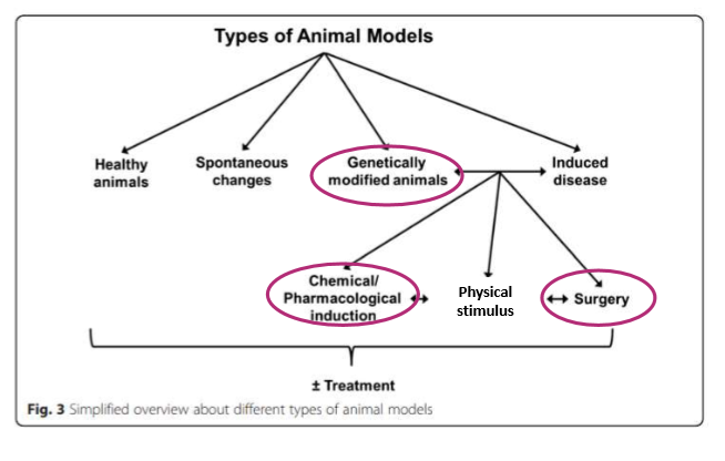
different ways to genetically modify animals
knock in
knock out
altering DNA to express characteristics
how might surgery be used on animal models?
causing lesions
causing pain
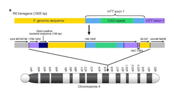
R6/2 model of Huntington’s disease genetic modification in animal models
first and most understood model of Huntingtons
take exon-1 CAG repeats from human and insert into chromosome 4 of mice to cause huntingtons
causes smaller straital volume and decline in motor function when compared to wildtype mice
MPTP model of Parkinson’s Disease in mice
MPTP drug can cross the BBB and is then taken up by glial cells
MPTP → MPP+ by MAO-B in the glia
MPP+ is then taken up by dopaminergic neurones via DAT (dopamine transporter)
MPP+ inhibits the electron transport chain in mitochondria
causes cell death
Is administered to the abdomen of mice via injection
administered gradually throughout the day to mimic human Parkinson’s
more frequent MPTP injection causes more striatal dopamine depletion → substantia nigra
stain for dopaminergic neurones with tyrosine hydroxylase
MPTP mice show anti-anxiety symptoms by spending more time in arm of maize with glass floor (open arm)
Ischemic stroke
most common (80-90% of cases)
obstruction of blood flow and so limited oxygen supply to brain
two types
global cerebral
focal cerebral
requiring craniectomy or not (remove part of skull to relieve pressure )
different models
haemorrhagic stroke
less common (10-20% of cases)
weakened blood vessel in the brain ruptures and bleeds into surrounding tissues
cam be modelled
MCAO model of stroke
surgical animal model
Middle cerebral artery occlusion model (MCAO)
filament inserted into carotid artery in the neck and up to the circle of Willis
this blocks the cerebral artery which feeds the medial section of the brain causing ischemia
cell death in the oxygen deprived areas
removal of filament causes reperfusion
done in controlled conditions, animals still undergo surgical conditions and anaesthetic which could have effects
stroke mice show difficulty seeing what is in front of them and MRI shows areas where cells have died
Lesion animal models
combine pharmacological and surgical models
canula inserted into brain so that drugs can be infused into particular brain areas causing cell death or damage
Parkinson’s model infuses 6-OHDA at the substantia nigra
Huntington’s model infuses ibotenic and quinolinc acids at the striatum
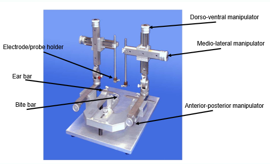
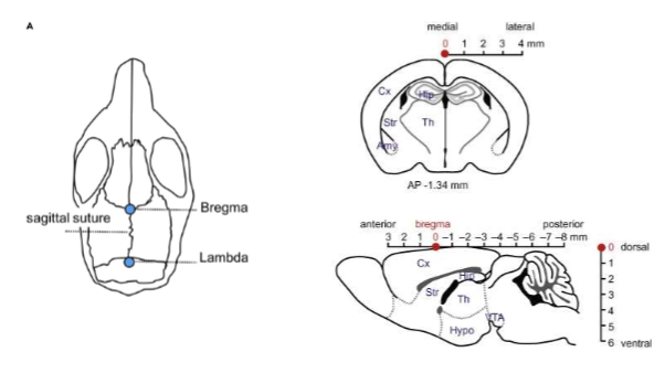
Brain surgery in mice
steriotaxic surgery uses 3D technique to locate neural locations with coordinates
uses the skull structure
Bregma is 0,0,0
done using a animal steriotaxic frame
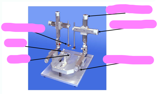
label the steriotaxic frame