Blood Supply and Innervation of the Lower Limbs
1/25
There's no tags or description
Looks like no tags are added yet.
Name | Mastery | Learn | Test | Matching | Spaced | Call with Kai |
|---|
No analytics yet
Send a link to your students to track their progress
26 Terms
Blood Supply: Thigh
1. Femoral artery
2. Profunda femoris
3. Medial circumflex femoral artery
4. Lateral circumflex femoral artery
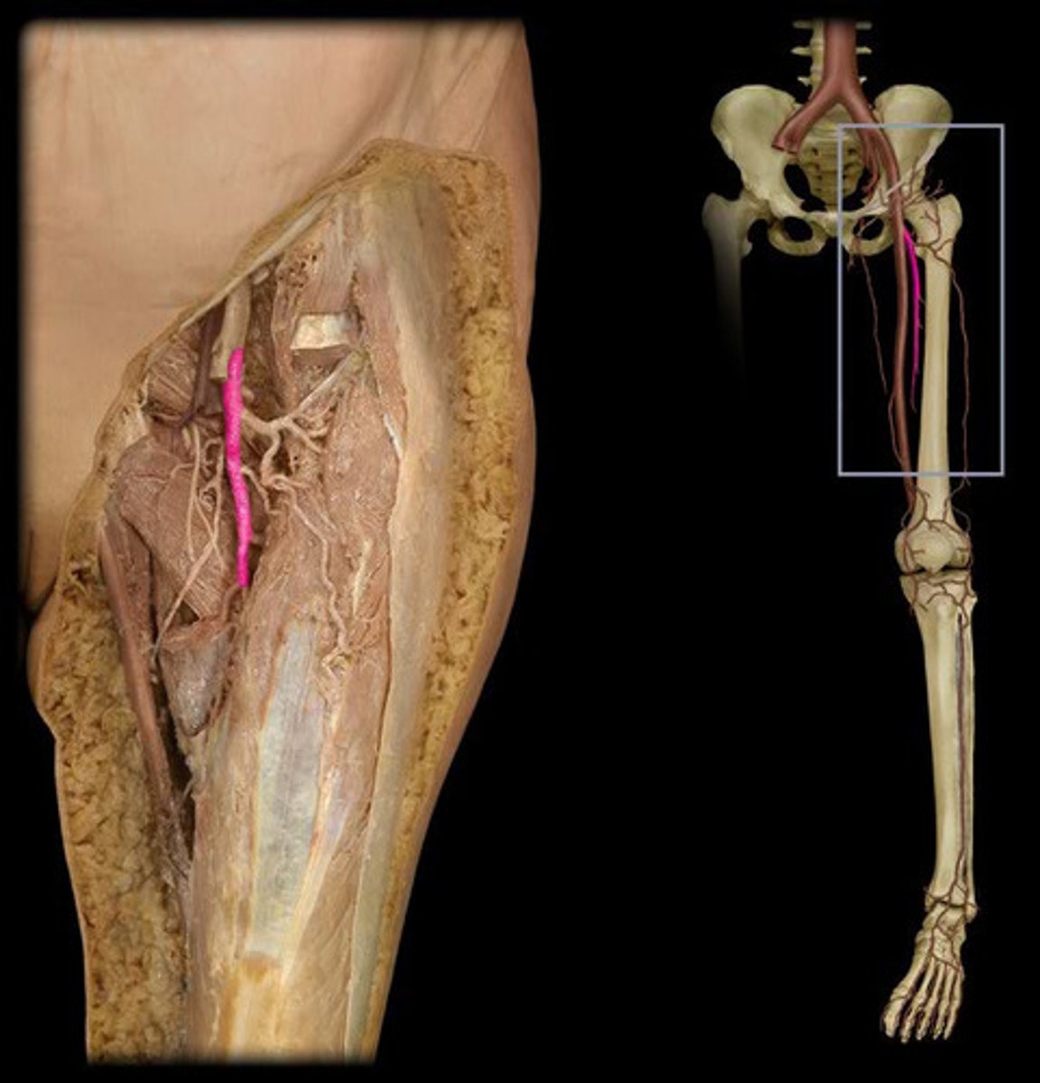
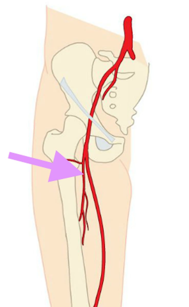
Femoral Artery
- Enters the lower limb at inguinal ligament
- Travels down the anteromedial aspect of the thigh
- Passes through the adductor canal
- Supplies the anterior thigh
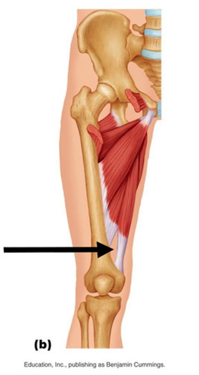
Adductor Hiatus
(gap) between the adductor magnus muscle and the femur that allows the passage of the femoral vessels from the anterior thigh to the posterior thigh and then the popliteal fossa.
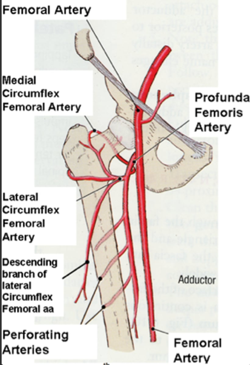
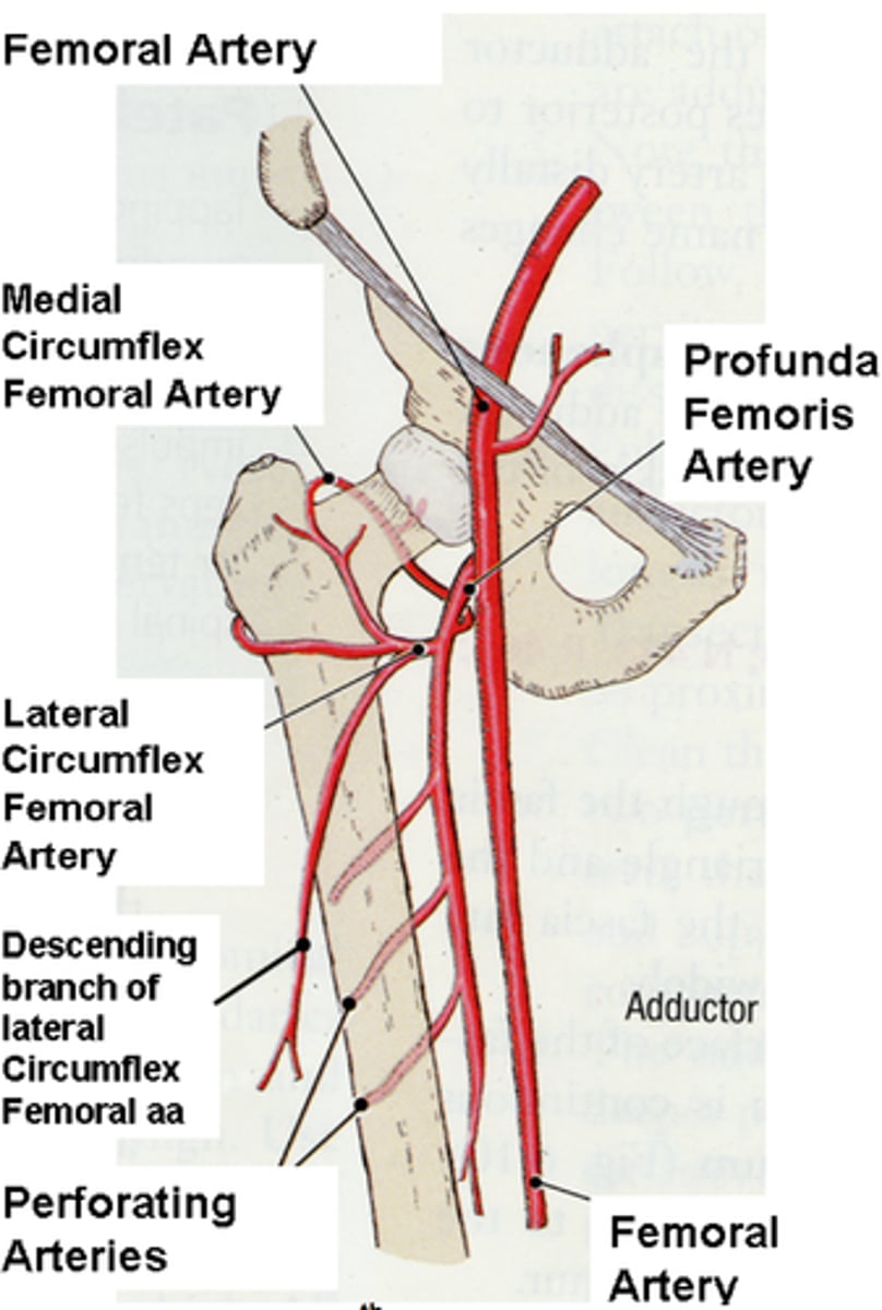
Femoral Artery Branches
- Profunda femoris branches off femoral artery
- Medial and lateral circumflex branch off profunda femoris

Profunda Femoris
- Deep artery
- Passes between muscles of the medial compartement
- Pierces the medial comaprtement muscle
- Supplies blood to the posterior thigh
Lateral Circumflex Femoral Artery
- Short branch of the deep femoral artery
- Curves around the neck of femur = "circumflex"
- Contributes blood to anterior thigh and hip joint

Medial Circumflex Femoral Artery
- Posteromedial branch of deep femoral artery
- Courses around neck of femur
- Emerges in gluteal region
- Supplies blood to posterior thigh and hip joint
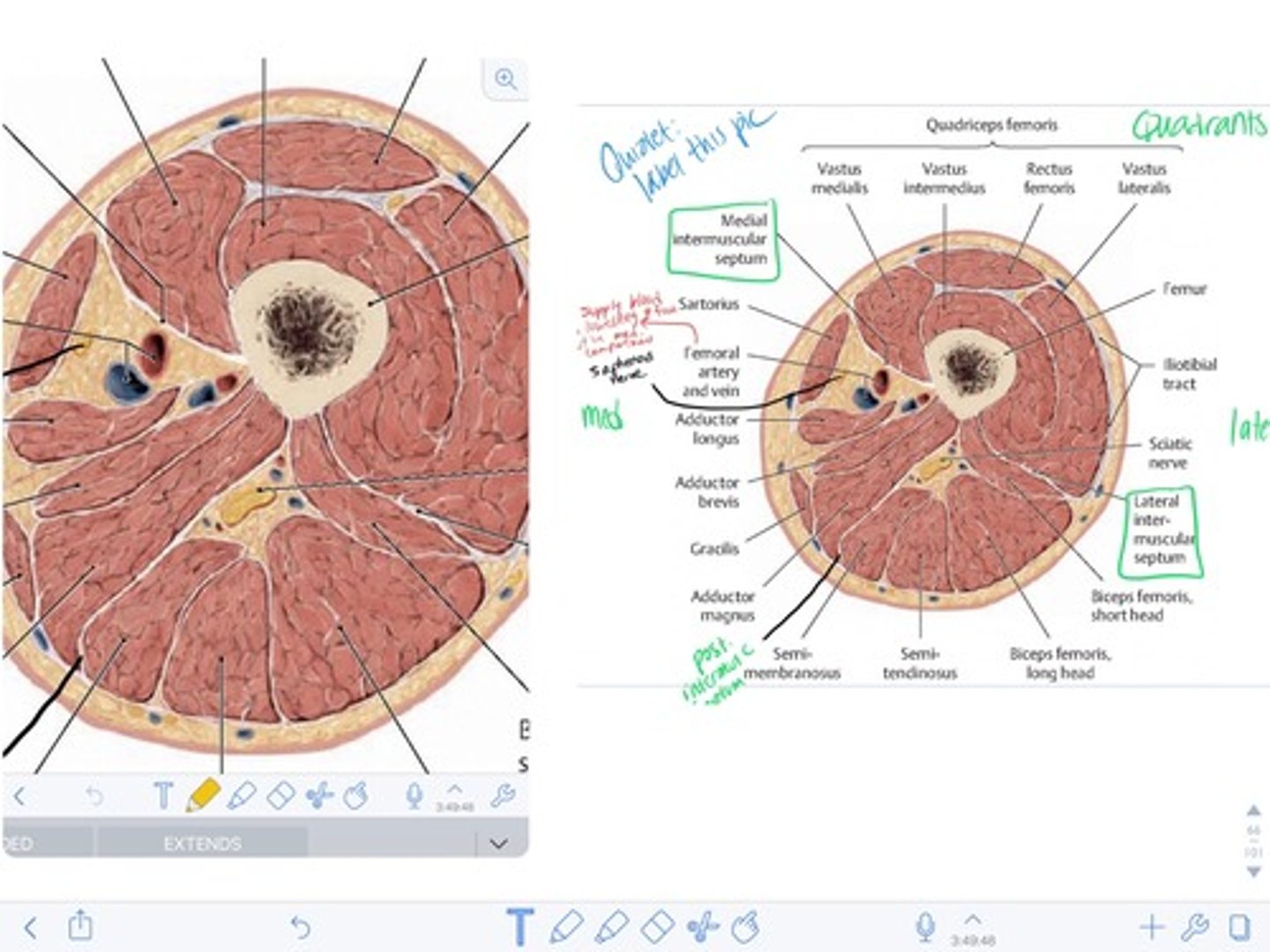
transverse section of thigh
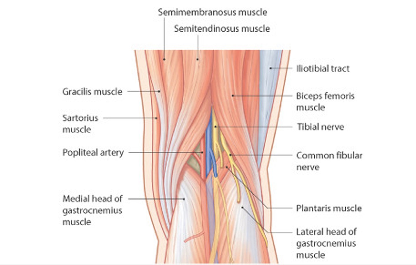
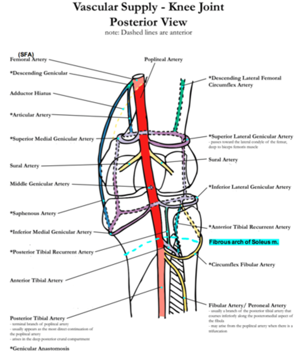
Blood Supply: Knee/Leg
Popliteal Fossa (posterior view):
1. Popliteal artery
2. Geniculate arteries
3. Posterior recurrent tibial artery
4. Anterior tibial artery
5. Posterior tibial artery
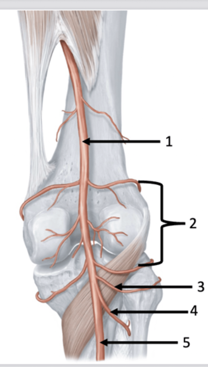
Popliteal Artery
- Enters leg at the base of the popliteal fossa
- Supples blood to the knee joint and superficial posterior leg
Posterior Tibial Recurrent Artery
- Branches laterally from popliteal artery
- Moves superior = "recurrent"
- Supplies blood to tibia and proximal end of fibula
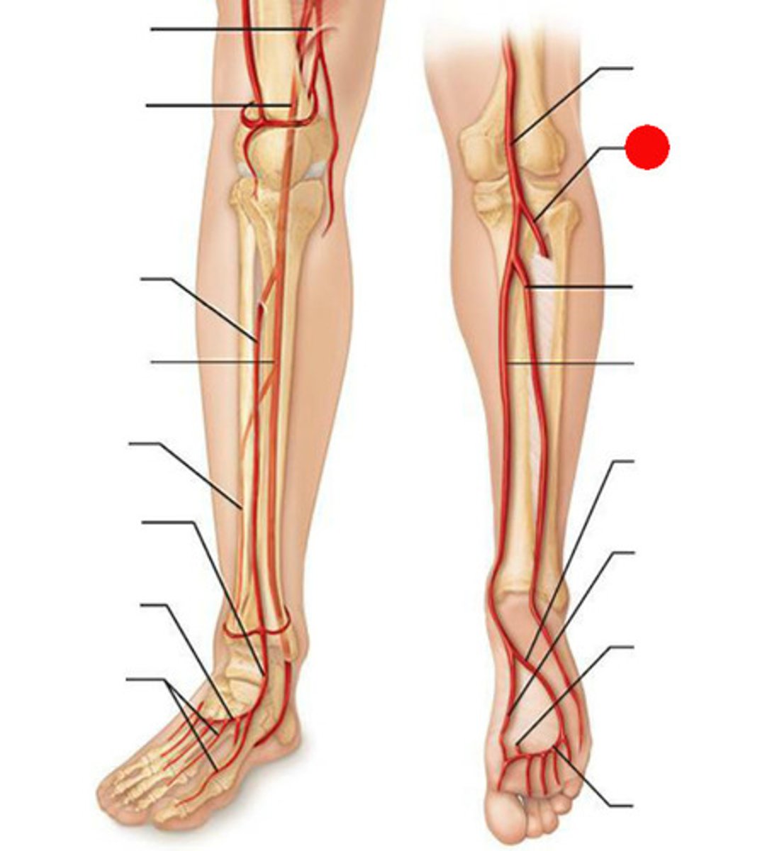
Anterior Tibial Artery
- Originates posteriorly
- Branches laterally
- Pierces interosseous membrane and runs on top anterior side
- Terminates in foot
- Supplies blood to anterior leg
Anterior Tibial Recurrent Artery
- Branches from anterior tibial artery
- Terminates superiorly at knee joint
- Supplies blood to knee joint
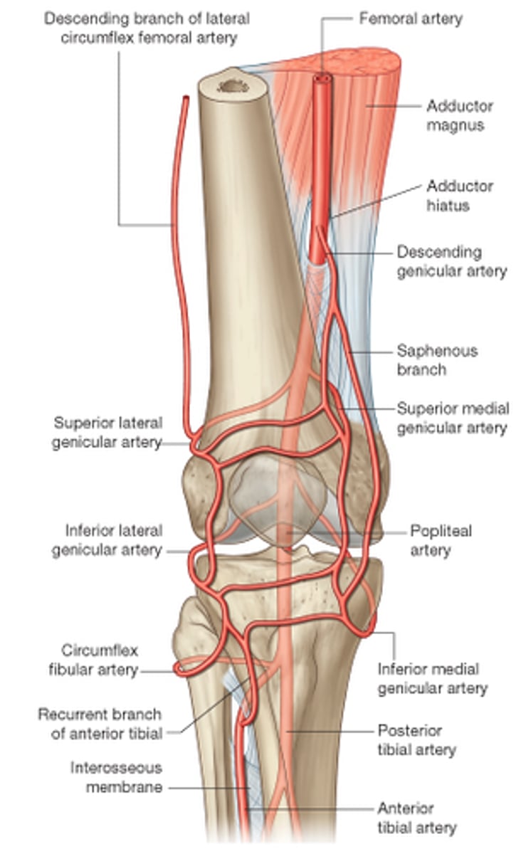
Posterior Tibial Arteries
- Runs between the superficial and deep posterior muscle groups
- Terminates in foot
- Supply blood to deep posterior leg
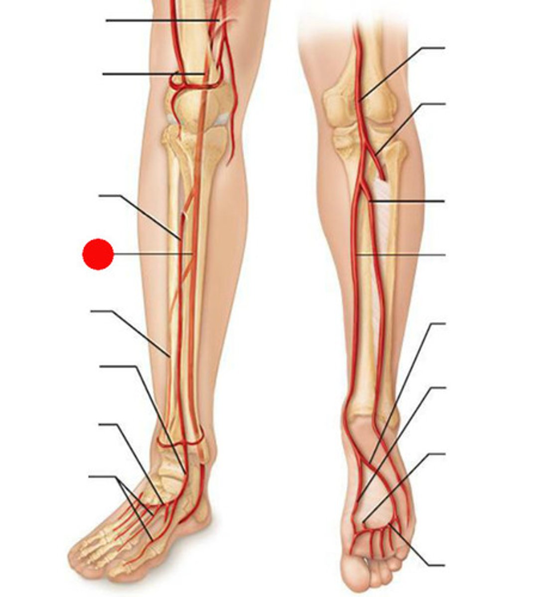
Fibular Artery
- Runs down parallel and lateral to tibial artery
- Supplies blood to lateral leg and superfical posterior
Blood Supply Dorsum of Foot
- Enters via anterior tibial artery
- Bifurcates at the dorsum of the foot into:
- Dorsalis pedis and lateral tarasal aretery
- Deep plantar artery branches posteriorly
- Arcurate artery (pinky)
- Supples dorsum/digits
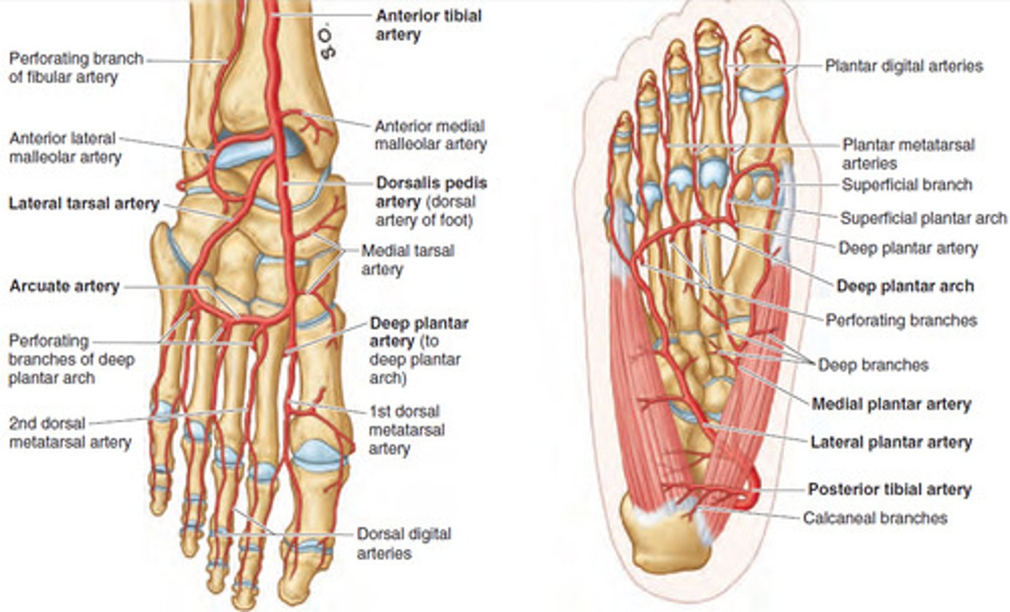
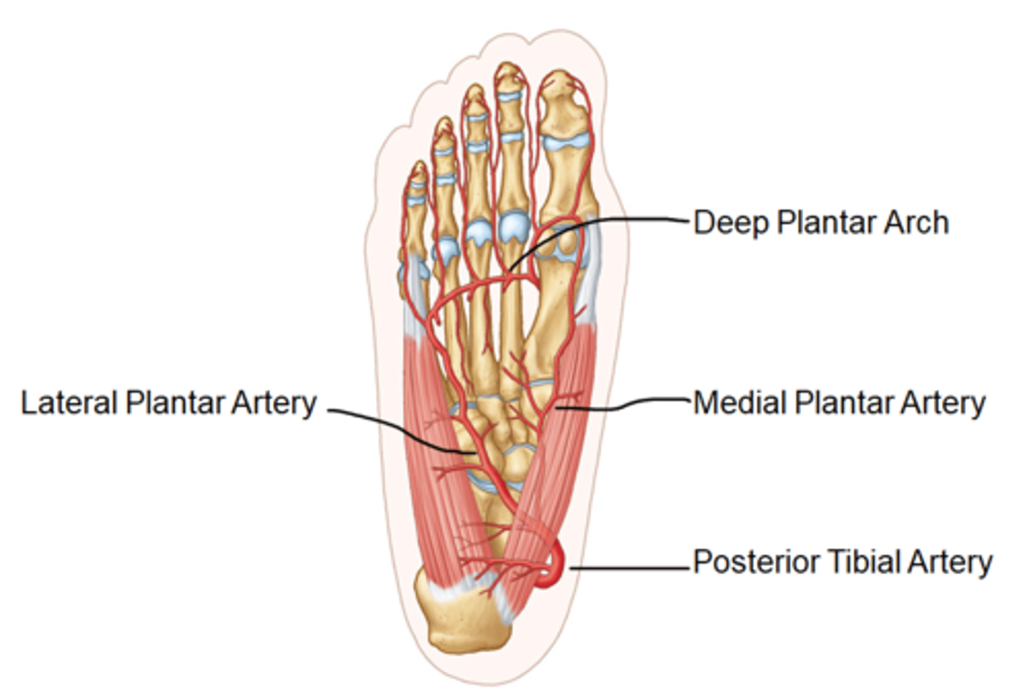
Blood Supply Plantar Surface of Foot
- Enters via the posterior tibial artery
- Curves under the medial calcaneus
- Bifuractes into medial and lateral plantar artery
- Deep planter arch, Deep planter artery
- Supplies: plantar surface of the foot and digits
Venous Drainage
Anterior foot:
- Dorsal venous network
- Great saphenous vein
- Anterior tibial veins
Posterior foot:
- Plantar venous network of the foot
- Posterior tibial veins
Leg veins:
- anterior + posterior tibial
- great saphenous
- popliteal
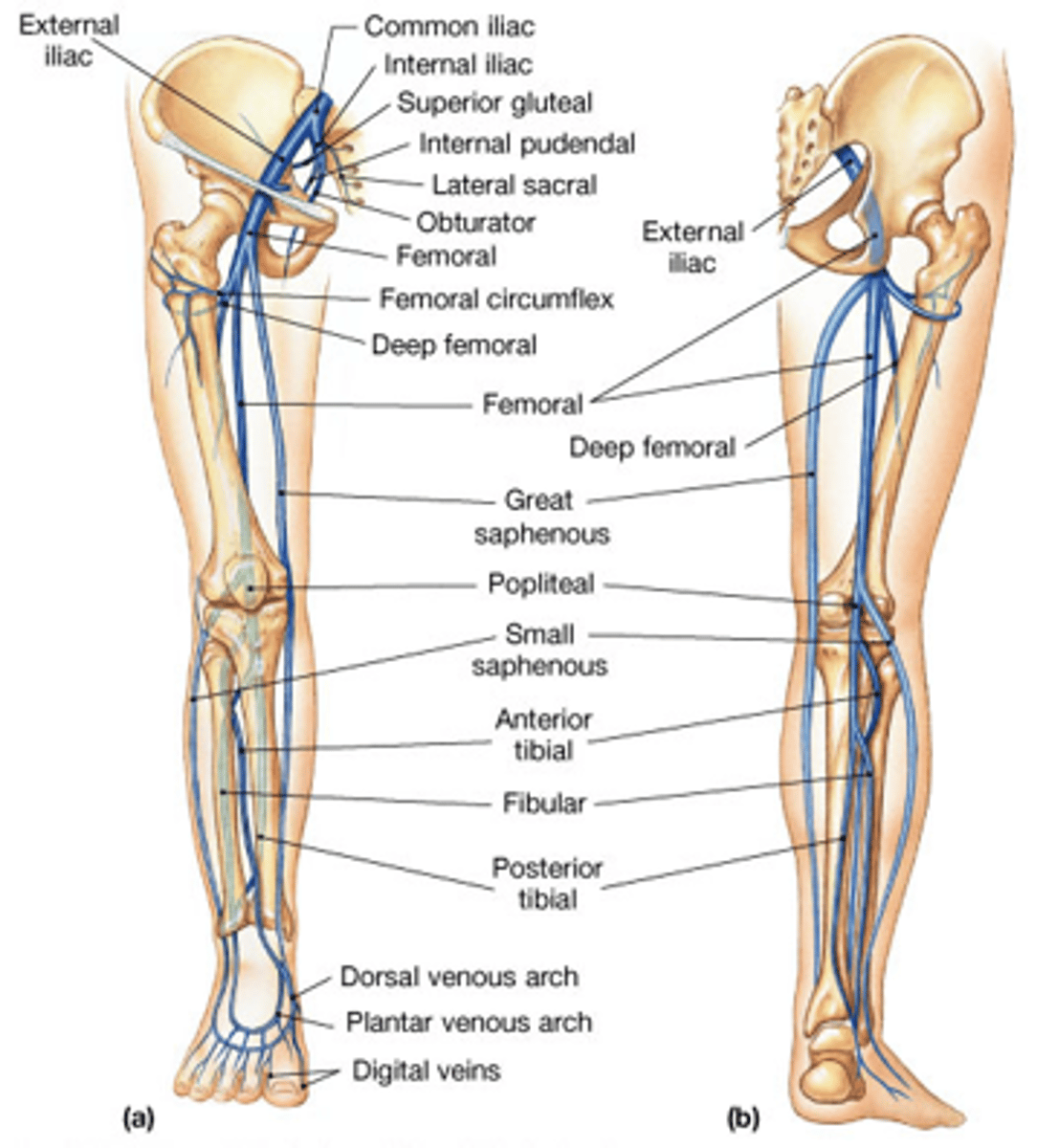
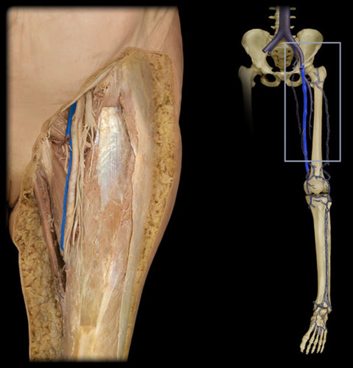
Great Saphenous Vein
- Empties into femoral vein
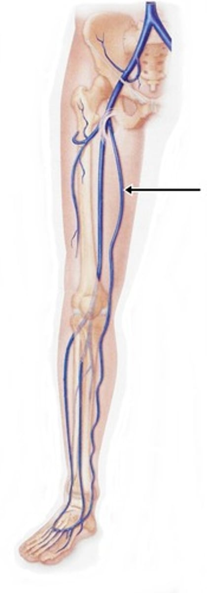
Femoral Vein
- Ascends from popliteal vein
- Ascends to inguinal (groin) region
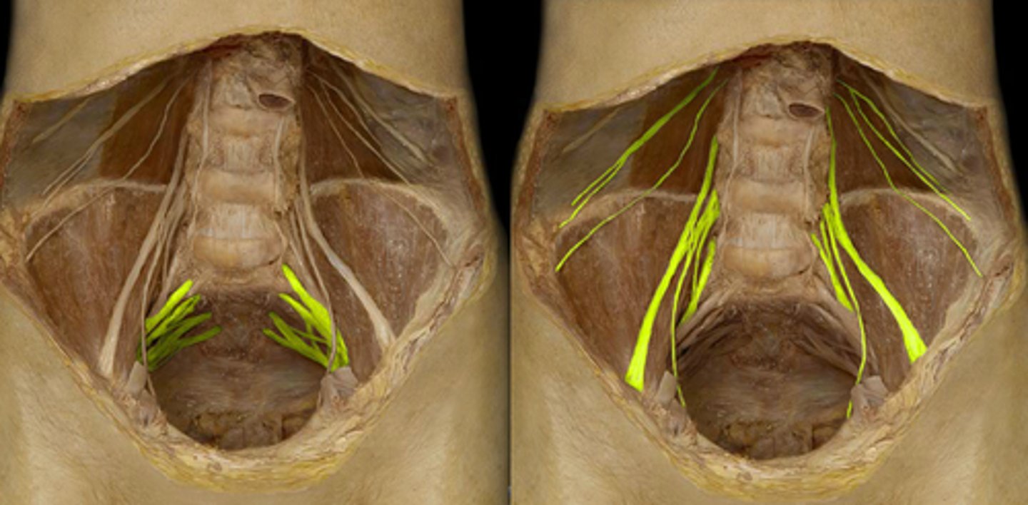
Lumbrosacral Plexus
- Formed from L2-S3
- Obturator nerve (L2-L4)
- Femoral nerve (L2-L4)
- Sciatic nerve (L4-S3)
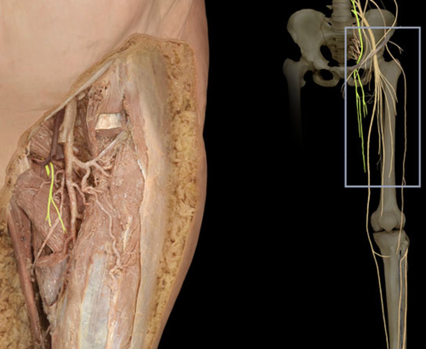
Obturator Nerve
- Runs down the anterior medial aspect of the thigh
- Terminates in thigh
- M: medial thigh muscles
- S: skin over the proximal part of the medial thigh
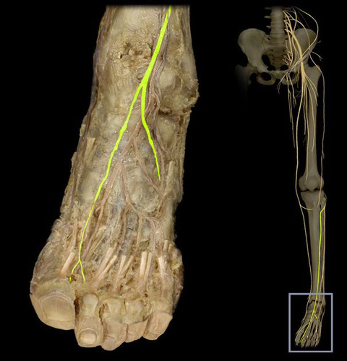
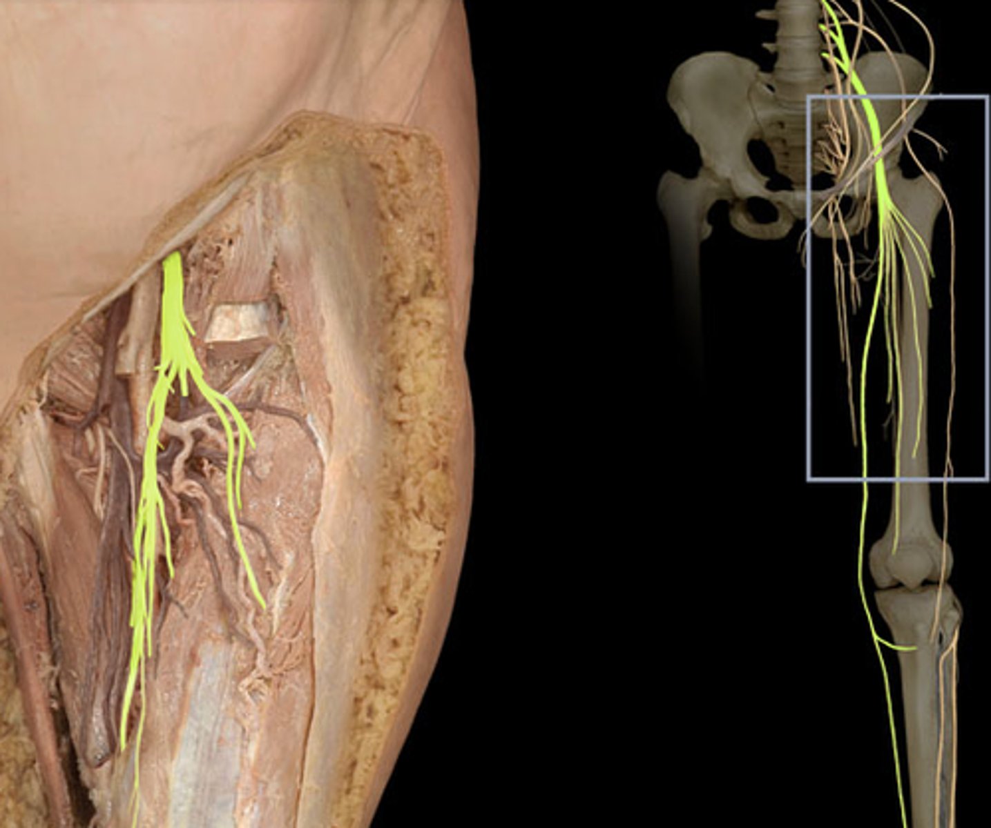
Femoral Nerve
- Descends the anterior thigh to leg
- Divides into branches
- M: anterior thigh Muscles
- S: skin of anteromedial thigh and medial aspects of knee, leg and foot
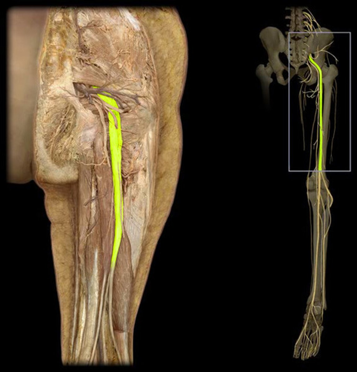
Sciatic Nerve
- Runs inferiorly between the medial and posterior thigh compartments
- M: posterior thigh muscles
- Terminates at the popliteal fossa
- Divides into 2 branches:
- Tibial/Fibular nerve
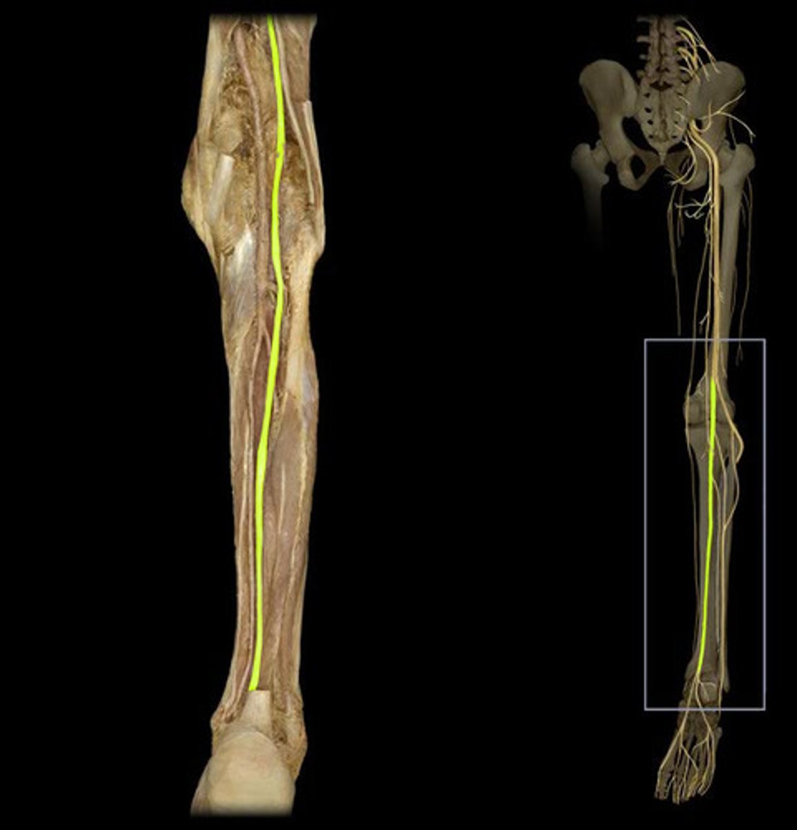
Tibial Nerve
- Continues down the posterior aspect of the leg
- Terminates at the heel of the foot
- M: posterior leg muscles and majority of foot muscles
- S: sole of the foot and posterior leg
Fibular (Peroneal) Nerve
- Curves around the head of the fibula
- Once in anterior compartment of the leg it divides into 3 branches
- Terminates in the foot
- M: lateral and anterior leg muscles
- M: medial aspect of foot muscles
- S: lateral leg and dorsum of foot