Muscle Physiology
1/54
There's no tags or description
Looks like no tags are added yet.
Name | Mastery | Learn | Test | Matching | Spaced |
|---|
No study sessions yet.
55 Terms
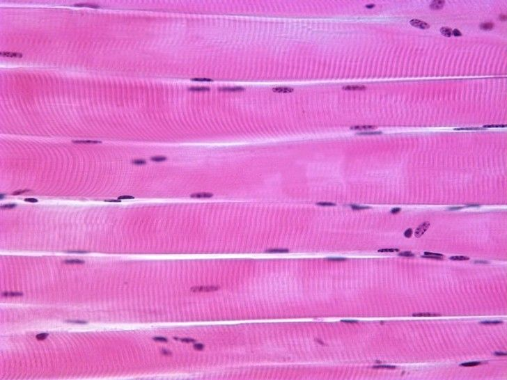
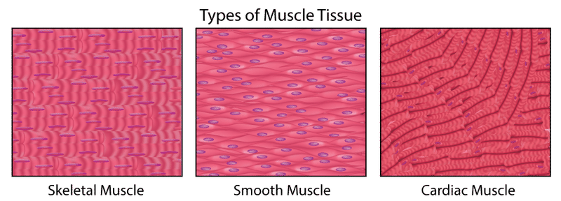
Skeletal Muscle
Striated
Attached to bones (voluntary movement, posture, and stabilization)
Innervated by somatic motor neurons that stimulates contractions
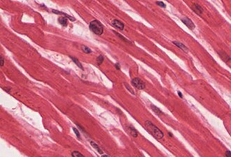
Cardiac Muscle
Striated
Heart (pumps blood)
Does not require nervous stimulation to contract (autorhythmic)
Smooth Muscle
Non-striated
Found in walls of visceral organs and blood vessels
Not subject to voluntary control
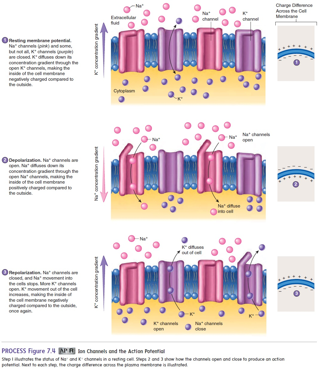
Electrical excitability
In response to AP, muscle contracts (excitation-contraction coupling)
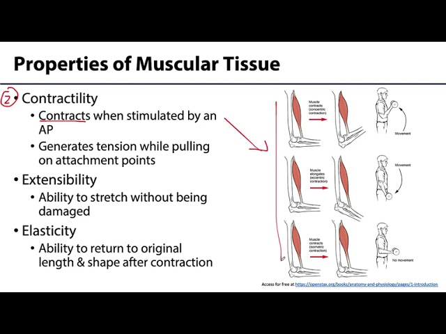
Contractility
Ability to generate tension/force in response to AP; if tension generated is greater than load placed on muscle, movement occurs

Extensibility
Ability of muscle to be stretched without damage

Elasticity
Ability to return to original shape and length after extension
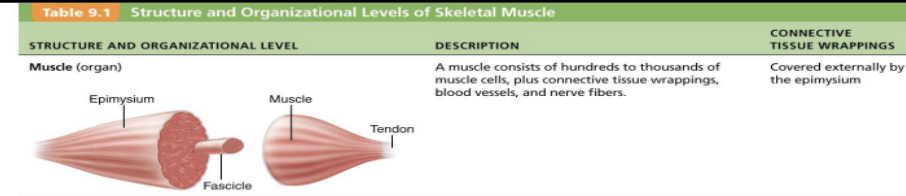
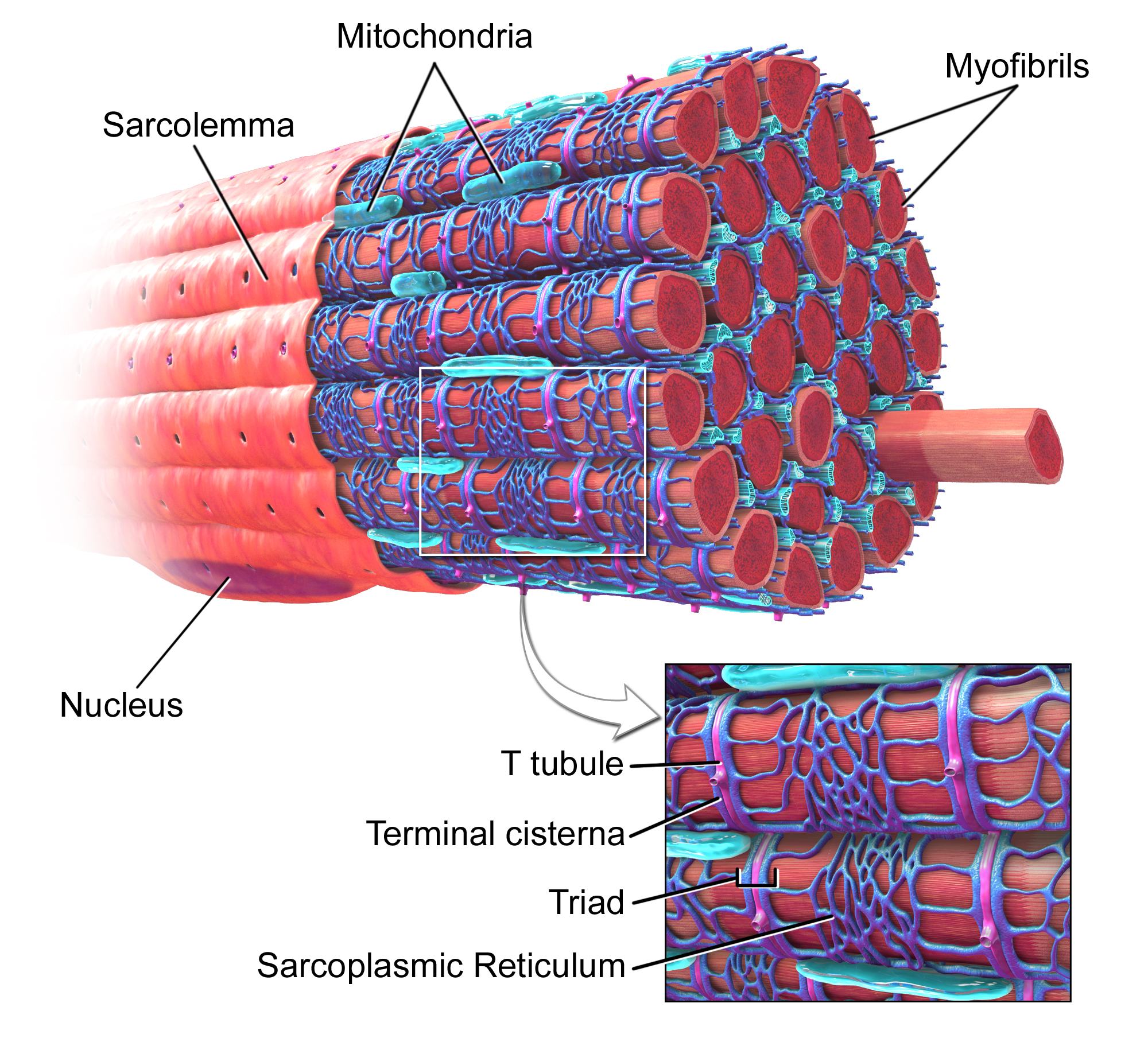
Structure of skeletal muscle
Encased by connective tissue (epimysium)
Comprised of bundles of fascicles
Each fascicle = bundles of muscle fibers
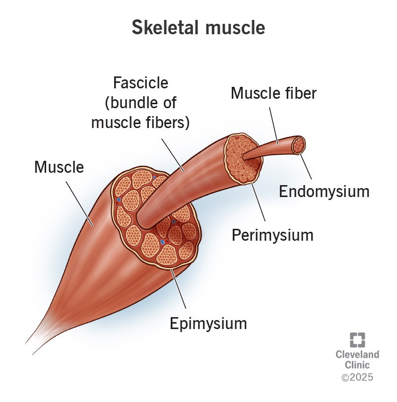
Muscle (organ)
consists of hundreds to thousands of muscle cells, plus connective tissue wrappings, blood vessels, and nerve fibers
What are its connective tissue wrappings/what surrounds it?
Covered externally by epimysium
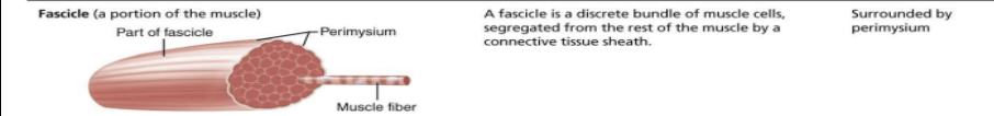
Fascicle (a portion of the muscle)
a discrete bundle of muscle cells segregated from the rest of the muscle by a connective tissue sheath
What are its connective tissue wrappings/what surrounds it?
surrounded by perimysium
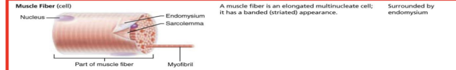
Muscle fiber (cell)
an elongated multinucleate cell (has banded/striated appearance)
What are its connective tissue wrappings/what surrounds it?
surrounded by endomysium
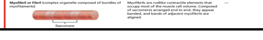
Myofibril/fibril (complex organelle composed of bundles of myofilaments)
rodlike contractile elements that occupy most of the muscle cell volume; made of chains of sarcomeres
composed of sarcomere arranged end to end (appears banded) and bands of adjacent myofibrils are aligned
singular muscle cell = muscle fiber
Sarcolemma = cell membrane
Sarcoplasm = cytoplasm
Multinucleate = many nuclei per fiber
Sarcoplasmic reticulum = modified smooth ER: stores Ca2+
Myoglobin = red pigments similar to hemoglobin
Contains Fe2+ that binds O2 = O2 reservoir
Myofibrils = bundles of contractile and elastic proteins that run length of muscle fibers (80% of volume of muscle)
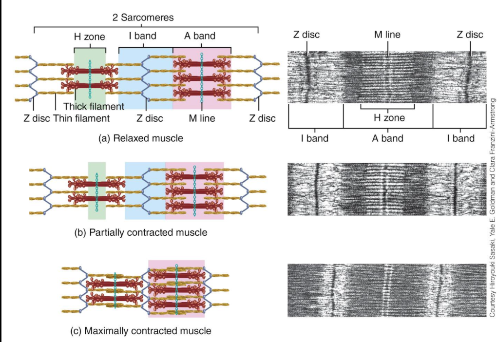
Sarcomeres
Units of contraction in skeletal and cardiac muscle
Made of proteins
“Thick” filaments = myosin (myosin proteins form thick filaments)
“Thin” filaments = actin (actin proteins form thin filaments)
Creates “striated” appearance
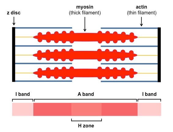
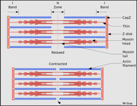
Regions of sarcomere
I-band
Z-disk
A-band
H-zone
M-line
Z-disk
Dark area in middle of I-band
appears light under microscope
anchors thin filaments; marks boundary between adjacent sarcomeres
when muscle contracts, Z-disks move closer together, shortening sarcomere

I-band (Isotropic = light)
region occupied by thin filaments only
appears light under microscope
I-band shortens as thin filaments slide in
A-band (Anisotropic = dark)
Dark-colored band under microscope
runs entire length of thick filaments
A-band stays the same length during contraction as length of thick filaments don’t change during contraction

H-zone
Located in middle of A-band, where thick filaments are present but no overlap with thin filaments
only thick filaments (no actin)
visible in relaxed fibers only; gets smaller/disappears in contraction

M-line
Dark line in middle of A-band (middle of H-zone/center of sarcomere)
seen as the “anchor point” for myosin proteins
holds thick filaments in place and aligns them

Myofilaments
“actual” contractile threads that slide past each other to allow contraction to occur
made up of two types:
thin filaments
actin
tropomyosin
troponin
thick filaments
myosin
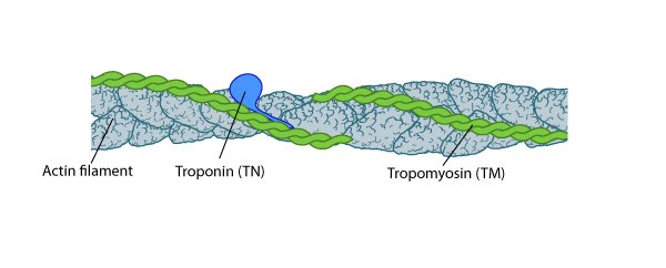
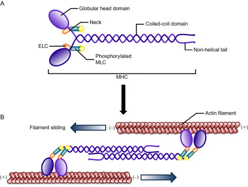
Actin (thin filament)
Globular protein molecules (G actin)
Each G actin molecule has binding site for myosin (thick filament)
along actin filament contains:
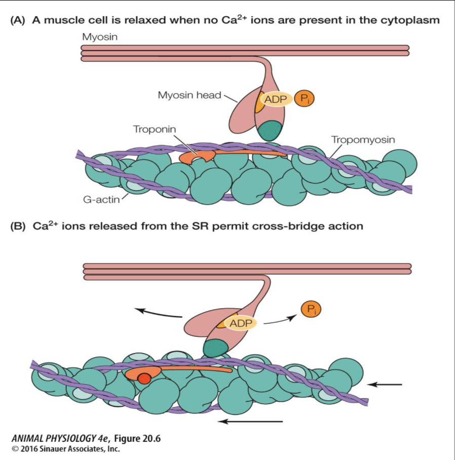
tropomyosin (covers binding sites when muscle is relaxed)
troponin (binds calcium & moves tropomyosin out of the way when contraction happens)

Tropomyosin (2 strands)
Elongated protein that spirals around actin strands
In relaxed muscle, blocks myosin binding site on actin

Troponin (thin filament)
Attached to tropomyosin
has binding sites for Ca2+
When calcium is bound, troponin and tropomyosin move out of the way to expose myosin binding site on actin
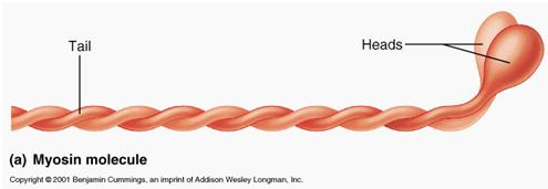
Myosin (thick filament)
Motor protein that pulls on actin to make muscle contract
Contains:
Myosin tails
Pointed toward each other
Myosin heads
Pointed away from one another
Can move at point of attachment to tail
Two binding sites
For ATP (part of myosin is ATPase)
For actin
Sliding filament theory
Muscle contraction occurs when thin filaments (actin) slides over thick filaments (myosin), shortening sarcomere (filaments do not change length however)
myosin heads pull on thin filaments, causing them to slide inward toward M-line
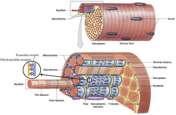
Sarcoplasmic Reticulum (SR)
modified version of smooth ER found in muscle cells
system of tubules that stores/releases Ca2+ ions
surrounds each myofibril
at rest, stores Ca2+ ions in terminal cisternae
during contraction, releases Ca2+ ions into sarcoplasm
during relaxation, reabsorbs Ca2+ to stop contraction

Terminal cisternae
Enlarged areas of SR adjacent to t-tubules that help with the storage of Ca2+ ions at rest and the release of Ca2+ ions during contraction
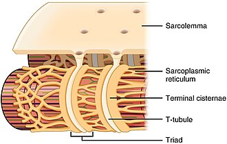
Triads
Specialized junction formed by T-tubules that aids in excitation-contraction coupling
helps transmit nerve signal deep into muscle fiber
when signal reaches t-tubule, triggers terminal cisternae to release Ca2+ ions
consists of:
a t-tubule
two terminal cisternae
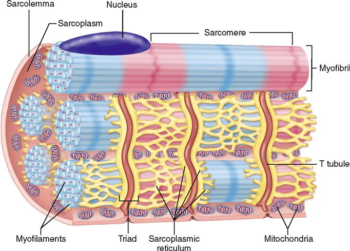
Transverse tubules (t-tubules)
Channels that are continuous with extracellular space; inversions of sarcolemma
Penetrates deep into cell; runs between terminal cisterns, encircles each myofibril
Function:
Conducts AP to every sarcomere, including those in center of muscle fiber
Two forms of myosin heads?
low energy form
high energy form
Cross-bridge cycling
repeated attachment, “pivot”, and detachment of myosin heads on actin filaments that shorten sarcomeres during muscle contractions
1 ATP consumed per cycle
Ca2+ released from SR and binds troponin
Several binding/unbinding cycles for single contractions
Cross-bridges work independently and asynchronous
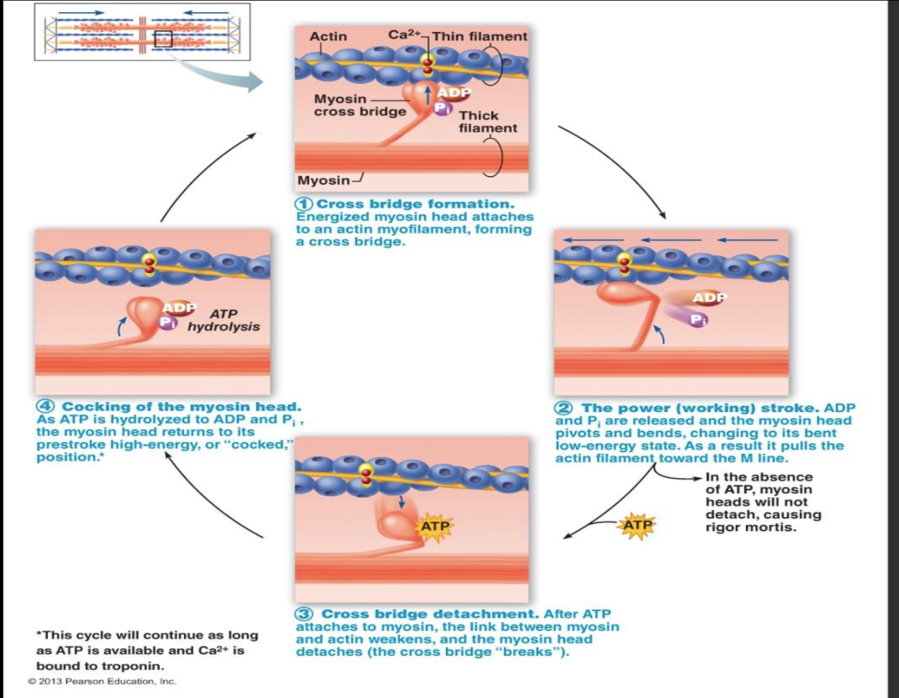
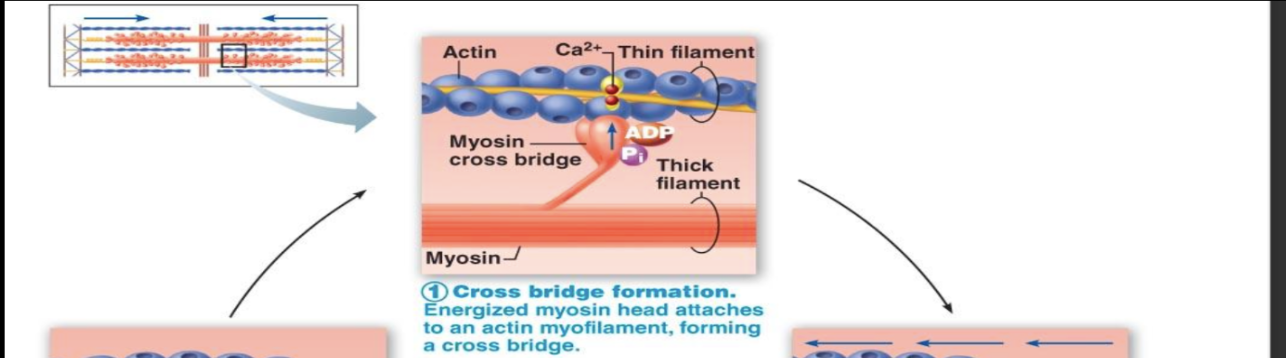
Cross-bridge formation
post-actin sites being exposed for myosin to attach
energized myosin head attaches to an actin myofilament, forming a cross bridge
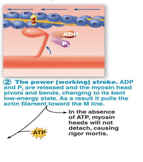
Power/working stroke
ADP and phosphate are released and the myosin pivots and bends, changing to it’s low-energy state, pulling the actin filament toward M-line
In absence of ATP, myosin head will not detach, causing rigor mortis
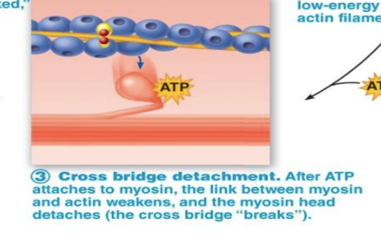
Cross-bridge detachment
After ATP attaches to myosin, link between myosin and actin weakens, making the myosin head detach/cross bridge “breaks”
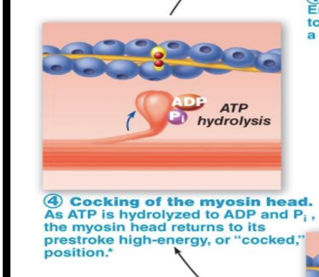
Cocking of the myosin head
As ATP is hydrolyzed to ADP and phosphate, myosin head returns to high-energy/”cocked” position and cycle can continue
Rigor mortis
stiffness/rigidity of muscle postmortem (after death)
Cells die, membranes lose integrity, Ca2+ leaks in, causing cross-bridge formation
Actin and myosin can’t detach from one another as body has run out of ATP, therefore actin cannot “unlock” myosin and, as a result, great muscle stiffness occurs
Role of calcium
Relaxed muscle cross-bridges are “primed” (ADP + P) but are unable to generate power stroke
Ca2+ from sarcoplasmic reticulum is required to expose myosin binding sites on actin via troponin/tropomyosin complex
An AP must be generated and travel alongside the sarcolemma and into t-tubules (excitation-contraction coupling)
Excitation-Contraction Coupling
“Series of events that link muscle excitation from a nerve impulse into contraction”
Nerve signal to Ca2+ release to muscle contraction (in short)
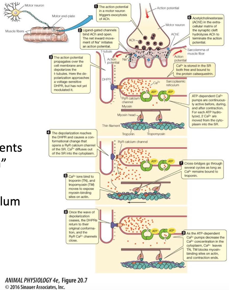
Excitation-Contraction Coupling Step 1
action potential from presynaptic motor neuron triggers exocytosis of ACh
at neuromuscular junction, acetylcholine is released
(
Excitation-Contraction Coupling Step 2
ACh binds to ligand-gated channels on sarcolemma and causes a net inward movement of Na+, initiating an muscular action potential
Excitation-Contraction Coupling Step 3
Action potential propagates over cell membrane and depolarizes t-tubules, triggering terminal cisternae of SR/sarcoplasmic reticulum to release Ca2+ into sarcoplasm
Excitation-Contraction Coupling Step 4
Ca2+ ions bind to troponin and tropomyosin, exposing myosin-binding sites on actin
Cross-bridge cycling occurs as long as Ca2+ remains bound to troponin
myosin heads attach to actin, sliding filament cycling
myosin pulls actin toward M-line, shortening sarcomere and contracting muscle
Excitation-Contraction Coupling (Relaxation)
Ca2+ ions are pumped back into SR
tropomyosin covers actin binding sites, detaching myosin (muscle relaxes)
AChE (acetylcholinesterase) ensures sarcolemma isn’t stimulated by breaking down remaining ACh in synaptic cleft
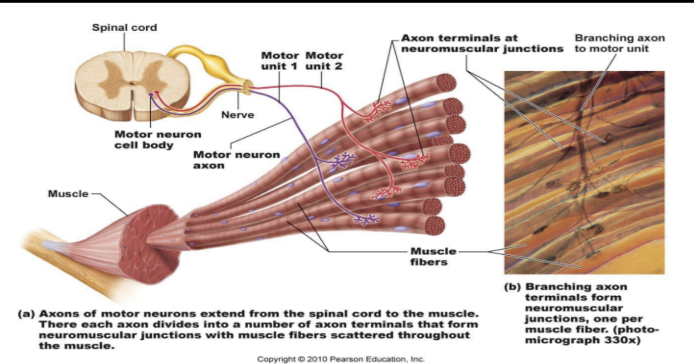
Neural Control
Each somatic motor neuron branches to innervate two or more skeletal muscle fibers
Each fiber receives input from only one motor neuron (even if motor neuron innervates multiple fibers)
Each motor unit can tell any number of muscle fibers to contract (as long as it innervates multiple)
Muscles which control precise movements tend to have?
Many small motor units
EX: larynx/hand muscles: 2-3 muscle fibers per motor unit
EX: muscles which move eyes: ~10-20 fibers per unit
Muscles which control larger powerful movements tend to have?
Larger motor units
EX: gastrocnemius of calf/biceps in arms: 2000 fibers per unit
Fibers in a motor unit are spread throughout muscle, so when one motor unit is stimulated, the result is?
A weak contraction of entire muscle (twitch)
structure is designed to allow whole muscle to contract as a “designated unit”
Motor unit
Somatic motor neuron with the fibers it innervates
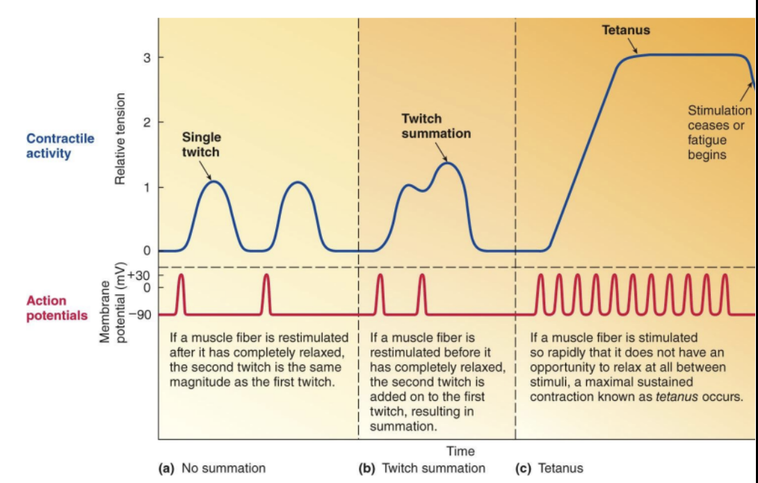
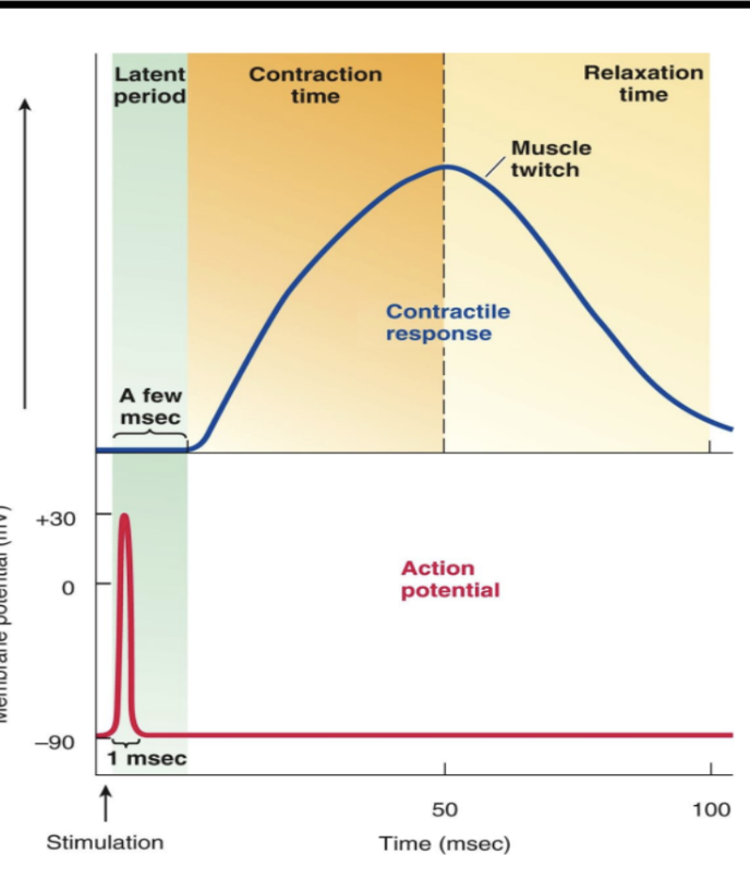
Muscle twitch
Response of a motor unit to a single AP in its motor neuron
usually only observed experimentally
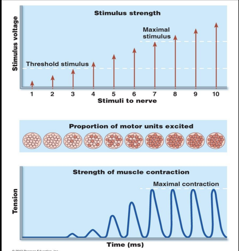
How can muscles generate different amounts of tension depending on the load?
Increasing the number of fibers/motor units participating in the contraction (motor unit recruitment) (response of a muscle to increased stimulus strength)
Increasing the tension developed by each contracting fiber (twitch/wave summation and tetanus) (response of muscle fiber to increased frequency of stimulation)
Motor Unit Recruitment
Process of increasing the # of motor units involved in the contraction (Controlled by CNS)
As the load placed on a muscle increases, muscles respond in the following way:
The number of somatic motor neurons firing increases which then…
increases number of motor units activated which then…
increases number of muscle fibers participating in contraction which then…
causes a generation of an increase in muscle tension
Asynchronous recruitment
Motor units alternate between contraction & relaxation
Why is asynchronous recruitment important?
Prevents fatigue (allows to rest)
Helps maintain posture
Helps maintain muscle tone
Muscle twitch
Each fiber participating in the contraction can increase the tension it generates via twitch/wave summation and tetanus
Summation & tetanus
Results from increased frequency of stimuli (increased firing rate of somatic motor neurons)
Keeps calcium in sarcoplasm for longer time period
Leads to smooth, continuous contraction and an increase in force generated by the fibers in a motor unit
Permitted by sustained, high Ca2+