BIOL 1410 (Anatomy)
1/149
There's no tags or description
Looks like no tags are added yet.
Name | Mastery | Learn | Test | Matching | Spaced | Call with Kai |
|---|
No analytics yet
Send a link to your students to track their progress
150 Terms
Anatomy
The study of internal and external structures -- the relationships among body parts.
Example. Bone and muscle
Gross Anatomy
A subdivision of anatomy that deals with structures visible to the unaided eye.
Example. Bones
Micro anatomy
A subdivision of anatomy which requires the use of a microscope.
Cytology
Subdivision of micro anatomy:
The study of internal structures of individual cells. Cytologists study internal structures of cells.
Histology
Subdivision of micro anatomy:
The study of tissues (groups of cells).
Chemical Level of Organization (1)
-Basic level
-Atoms (Ex. H and O atoms)
-Atoms join to form molecules (Ex. H2O, water)
Cellular Level of Organization (2)
-Molecules form organelles which form cells
-The cell is the basic unit of structure and function of life
•All cells contain chemicals but functions may differ (Ex. Red blood cells, neurons, muscle cells)
Tissue Level of Organization (3)
-A group of structurally similar cells with a common function
-4 major tissues:
a) Epithelium
b) Connective Tissue (CT)
c) Muscle
d) Nervous
Organ Level of Organization (4)
2 or more tissues working together for a common function
Ex. The stomach uses all 4 major tissues; it's function is digestion.
Organ System Level of Organization (5)
Several related organs that work together to accomplish a common purpose.
Ex. The respiratory system uses the trachea, bronchi, lungs and more to accomplish the function of respiration.
Organism Level of Organization (6)
All organ systems function together to maintain life (human being).
Note: Levels of organization is a hierarchy--each level contains those below.
Structure of an Atom
The nucleus contains protons (p^+): positive charge, and neurons (n^0): neutral charge. Electrons (e^-): negative charge, orbit the nucleus.
Atoms are electrically neutral --> the number of protons is equal to the number of electrons. Note that the number of neutrons may not be equal to these.
Ions
If an atom gains or loses an electron, it is no longer electrically neutral and becomes an ion. Atoms that gain(s) an electron(s) become negative ions and are called anions. Atoms that lose an electron(s) become positive ions and are called cations.
Important Ions (Electrolytes) (5)
1) Calcium (Ca^++)
2) Sodium (Na^+)
3) Potassium (K^+)
4) Hydrogen (H^+)
5) Chloride (Cl^-)
Chemical Bonds
Bond (holds) atoms together to form molecules. They allow the formation of chemical compounds that may be organic or inorganic.
Ionic Bond
Type of chemical bond where ions are formed--electrons transfer from one atom to another.
Ex. In NaCl, Na loses an electron while Cl gains an electron. The positive (Na+) and negative (Cl-) ions attract, forming the bond. In water, NaCl dissociates (dissolves/separates/ionizes) back into it's ions (Na+, Cl-).
Covalent Bond
Type of chemical bond where electrons are shared between atoms.
Ex. CH4
Organic Substances
Contain covalently bonded carbon (C) atoms.
Ex. Carbohydrates, lipids, proteins, and nucleic acids
Inorganic Substances
Usually lack carbon.
Ex. NaCl, H2O, O2
The exceptions to the carbon ruling include:
Carbonic acid (H2CO3)
Bicarbonate (HCO3^-)
Carbon dioxide (CO2)
Monoxide (CO)
Water (inorganic)
-Water is the most abundant substance in cells.
-It contains two hydrogen (H) atoms and one oxygen (O) atom.
-Many reactions of the body either take place in or involve water.
-It is a polar molecule which transports chemicals like O2 and nutrients.
-It maintains body temperature at 37°C.
Polar Molecules
Contain an unequal sharing of electrons which leads to a slight charge difference.
Acids
-May be organic or inorganic.
-Acids dissociate in water, releasing hydrogen atoms (H^+).
•As [H+] increases, pH decreases
•Ex. Hydrochloric acid (HCl) in water ==> H+ + Cl-
Bases
-May be organic or inorganic.
-In water, bases bind to free H+ atoms
•As [H+] decreases, pH increases
•Ex. NaOH in water ==> Na+ + OH- (hydroxyl ion); then OH- (base) + H+ ==> water
•Ex. HCO3- (bicarbonate; base) + H+ ==> H2CO3 (carbonic acid)
•Note: OH- and HCO3- act as bases by binding to the free H+ ions
pH Scale
-Reflects the concentration of free H+ in a solution.
-Basic --> acidic: increasing [H+]
0 ==========> 7.0 ==========> 14
acidic neutral basic(=alkaline)
-Ex. pH of blood ~7.35 - 7.45
Carbohydrate
-An important organic substance consisting of C, H, O
-Chemical formula: C(H2O)n (n=#)
-Functions:
•Source of energy for cells Ex. Glucose (C6H12O6)
•Used to build cellular structures Ex. DNA and RNA
Monosaccharides
Simple sugars which are the basic building blocks of other carbohydrates (CHO).
Ex. Glucose, fructose, ribose, deoxyribose
Disaccharides
The result of two monosaccharides covalently bonded together.
Ex. Glucose + fructose ==> sucrose
Polysaccharides
The result of many monosaccharides (basic building blocks) bonded together.
Ex. Glycogen (animals) and starch (plants)
Lipids
-Composed of C, H and O atoms (different ratio than carbohydrates), they are insoluble (non-polar) in water.
-Ex. Fats, oils, waxes, cholesterol, fatty acids (FA)
-Function:
a) Protect organs (padding)
b) Component of cell membranes
c) Source of stored energy
Glycerides
The most common type of lipid in the body, diet. They are composed of two building blocks:
i) Glycerol (backbone)
ii) Fatty acids (FAs)
Monoglyceride: glycerol + 1FA
Diglyceride: glycerol + 2FAs
Triglyceride: glycerol + 3FAs
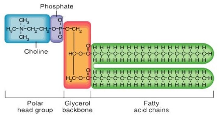
Phospholipids
A type of lipid composed of three building blocks:
i) Phosphate "head" group -- polar and hydrophilic (water soluble)
ii) Glycerol backbone
iii) 2 fatty acid tails -- non polar and hydrophobic (water insoluble)
Ex. Image of a diglyceride with a phosphate head group
Cholesterol
A type of lipid found in cell membranes and used to synthesize steroids.
Steroids
A type of lipid derived from cholesterol.
Ex. Bile salts, hormone (vitamin D, testosterone, estrogen, etc.)
Proteins
An organic substance composed of C, H, O, N (also sometimes S).
Ex. Albumin
Functions:
a) Structural material Ex. Collagen
b) Enzymes, hormones, transporters
c) Antibodies
Structures:
i) Amino acids (aa) -- basic building blocks of all proteins
ii) Dipeptides (2aa)
iii) Polypeptides (many aa)
iv) Protein - one or more polypeptides, folded in the final shape
Nucleic Acids
-An organic substance consisted of C, H, O, N, P
-Takes on two forms: DNA (deoxyribonucleic acid) or RNA (ribonucleic acid)
Nucleotides
-The building blocks of nucleic acids
-Contains:
•PO4- (phosphate)
•Monosaccharide (simple sugar)--ribose in RNA or deoxyribose in DNA
•An organic base
Organic Bases of Nucleotides (5)
1) Adenine (A)
2) Thymine (T) - DNA only
3) Uracil (U) - RNA only
4) Cytosine (C)
5) Guanine (G)
DNA (structure & function)
Structure:
Composed of phosphate, deoxyribose, A/T/C/G bases, the bases bind to form a double stranded helix (twisted ladder).
A-->T or C-->G
Alternating phosphates and sugars form "uprights" while bases form "rungs".
Functions:
Used in cellular reproduction or used as a template to make RNA.
RNA (structure & function)
Structure:
Composed of phosphate, ribose, A/U/C/G bases, RNA is single stranded.
A-->U or C-->G
Function:
Used in protein synthesis.
ATP
-Adenosine triphosphate
-An organic substance composed of 3 phosphates, ribose, and adenine, it has a modified RNA nucleotide
-A form of energy stored in covalent bonds
-Powers cellular activities; broken bonds release immediately usable energy
-Note: "~" = high energy bond
ATP --> ADP + Pi
(adenosine diphosphate) (inorganic phosphate)
The Cell
The basic structural and functional unit of the body which can perform all basic life functions.
Cell Membrane
Also referred to as Plasma membrane or Plasmalemma, it is composed of a phospholipid bilayer, cholesterol, membrane proteins, membrane carbohydrates, and microvilli.
Phospholipid Bilayer
-Contains a phosphate head group (hydrophilic)
-Contains FA tails (hydrophobic)
-Divides ICF (intracellular fluid/ inside the cell) from ECF (extracellular fluid/ outside the cell)
Cholesterol
Scattered throughout the membrane, it adds stability to the plasmalemma.
Membrane Proteins
2 Types:
i) Integral Proteins - Within the membrane, therefore have a hydrophobic region. Some extend across the plasma (called transmembrane proteins).
ii) Peripheral Proteins - Attached to either surface region of integral proteins.
Functions:
-Enzymes
-Transporters
-Channels
-Receptors
-Anchors
-Identity markers (for immune system)
Membrane Carbohydrates
Only on the outer surface, they bind to proteins (glycoproteins) or lipids (glycolipids).
Functions:
i) Cell recognition Ex. Egg and sperm
ii) Anchor cells together
Microvilli
Small projections of the cell membrane that functions to increase surface area. Best seen on cells of small intestine and kidney.
Fluid-Mosaic Model
Fluid--> Constituents can move around (phospholipids and some proteins).
Mosaic--> Proteins dot the surface like tiles in a mosaic.
Cytoplasm
The area inside the cell membrane and outside of the nucleus.
Cytosol
-A division of cytoplasm described as gel-like intracellular fluid
-Contains water and a suspension of carbohydrates, proteins, and lipids
-May contain inclusions
-Ex. Melanin (pigment), glycogen (stored glucose)
Organelles
-A division of cytoplasm, organelles are structures that perform a specific function and are essential for life.
-May be:
•Non-membranous--indirect contact with cytosol, or
•Membranous--surrounded by membrane that isolates them from the cytosol
Ribosomes
A non-membranous organelle that contain ribosomal RNA and proteins. Ribosomes are sites for protein synthesis and may be:
i) Free in cytosol--make proteins destined for cytosol, mitochondria, nucleus
ii) Attached to endoplasmic reticulum (ER)--makes proteins destined for all other sites
Centrosomes
-Dense area of cytoplasm which usually contains 2 centrioles (function uncertain; some cells lack them)
-Organizes microtubules to form spindle apparatus (for cell division)
-Organizes cytoskeleton
Cytoskeleton
-Determines cell shape (cell support)
-Formed from proteins
-Important in cell movement, cell division, movement/ anchoring of organelles and proteins (Ex. Receptors, enzymes)
-3 components: microfilaments, intermediate filaments and microtubules
Microfilaments
Made of actin, they function in:
-Muscle contraction (with myosin)
-Cell locomotion
-Maintenance of cell shape and projections (Ex. Microvilli)
-Cytokinesis
Intermediate Filaments
-Composition of tissues is specific (Ex. Keratin)
-Function: support the cytoplasm (scaffolding for the cell)
Microtubules
-Description: hollow tubes made of tubulin
-Function:
•Form centrioles, spindle apparatus, cilia (short), flagella (long)
•Structural
•Move or secure organelles in place
Mitochondria
A membranous organelle, it:
-Is the site of ATP synthesis
-Contain their own DNA, rRNA and proteins
-Has a double membrane
Endoplasmic Reticulum
-Membranous network throughout cytoplasm
-2 types:
i) Rough ER (RER)
•Ribosomes attached
•Synthesis of secretory, lysosomal, and membrane proteins
ii) Smooth ER (SER)
•Lacks attached ribosome
•Continuous with RER
•Synthesizes lipids and steroid hormones
Golgi Apparatus
A membranous organelle that looks like stacks of membrane discs. It modifies (Ex. Trims or adds CHO groups), sorts, packages and delivers proteins/ lipids to cell membrane, lysosomes or for secretion (cellular post office)
Lysosomes
Membranous organelle filled with digestive enzymes.
Nucleus
-Largest structure within the cell
-Cell control centre
-Cells may have 1 or more nuclei
-Parts: Nuclear envelope, nucleolus, chromosomes/ chromatids
Nuclear Envelope
Part of the nucleus, it contains a double membrane with nuclear pores, and is connected to the ER.
Nucleolus
Part of the nucleus, it is the dense (less light gets through) region of DNA, RNA and proteins, where ribosomes are made and assembled. It is non-membranous and more than one may be seen in some plants.
Chromosomes/ Chromatids
Part of the nucleus, it is made of DNA and histone proteins.
Can be:
i) Dispersed = chromatin
-DNA uncoiled chromosomes not individually visible
-In this form MOST of the time (when cell is not dividing)
ii) Condensed and chromosomes individually visible
-Found in dividing cells (during mitosis/ meiosis)
Gametes
A cell type based on chromosome content, it contains 23 different chromosomes = haploid (n), and includes ova (eggs) and sperm.
Somatic Cells
A cell type based on chromosome content, it includes all cells of the body except cells undergoing/ resulting from meiosis.
Ex. Muscle cell, nerve cell, etc.
These contain 46 chromosomes = diploid (2n)
-Each somatic cell contains 23 different Xms pairs: 23 Xms from Mom (ovum) paired with 23 Xms from Dad (sperm).
Homologous Chromosomes
These are chromosome pairs (one from each parent). Both chromosomes are identical in length, centromere position, and have genes for the same trait (Ex. Eye colour) in the same location (locus) BUT may have different alleles.
Gene
A unit of heredity or a region of DNA which contains information for synthesis of proteins.
Alleles
Different versions of a gene that code for different versions of a trait.
Ex. Blue eyes (Xms from Dad) or brown eyes (Xms from Mom)
OR
Genes occupying the same locus on homologous chromosomes. May be:
1) Homozygous - same allele of a particular gene on homologous chromosomes i.e. both would express the same version of a trait. Ex. Blond hair (bb) or black hair (BB)
2) Heterozygous - different alleles of the same gene i.e. alleles code for different expressions of the same trait Ex. Bb
Autosomes
Chromosomes and chromosome pairs are numbered 1-23. The first 22 are called autosomal chromosomes or autosomes. These contain genes for somatic characteristics.
Ex. Hair and eye colour, height, etc.
Each pair is not identical, but equivalent (=homologous Xms).
Sex Chromosomes
The 23rd pair of chromosomes which contains genes that determines sex (XX = F; XY = M).
-X or Y from Dad
-X from Mom
Cell Cycle
-Occurs for the growth and repair of tissues.
-Somatic "parent" cell (2n) become two genetically identical somatic "daughter" cells (2n).
-2 stages: Interphase and Mitotic (M)/cell division phase
Interphase
Chromosomes are present as chromatin and normal cellular metabolic activities are occurring.
Ex. Protein synthesis
G1 Phase
First stage of interphase, growth and metabolism are occurring. In the end, centrosomes are replicated.
Note: Most cells do not divide again once mature--> remain permanently in G1; in these cells it is termed G0.
S Phase
-Second phase of interphase
-Chromosomes replicate, but are still present as chromatin (not individually visible)
-ALWAYS occurs before division (mitosis and meiosis)
-Replicates are called sister chromatids
•Attach to each other at area of DNA called the centromere
Kinetochore
When kinetochore proteins (produced during S Phase) attach to each centromere of sister chromatids, kinetochore is formed (= protein + DNA complex, 1/chromatid). This is the site of attachment of single microtubules.
G2 Phase
Third phase of interphase, this is a period of growth, metabolism, and production of enzymes and other proteins needed for cell division.
Mitosis
First part of Mitotic (M) Phase, division of nuclear material (Xms) occurs. There are four phases: Prophase, Metaphase, Anaphase and Telophase, but it is a continuous process.
Cytokinesis
Second part of Mitotic (M) Phase, division of cytoplasm occurs.
End of M Phase
-Both mitosis and cytokinesis complete
-Daughter cells go into interphase (G1)
-Cycle starts over
Prophase
-Chromosomes condense (thicken, coil) from chromatin form and become individually visible
-Nucleoli/ nuclear envelope disappear
-Centrosomes move to either pole
-Microtubules form spindle apparatus (starting at centrosomes) and attach to kinetochore proteins (called kinetochore microtubules)
•Spindle moves chromosomes to cell equator
Metaphase
46 chromosomes line up at the equator of the cell.
Anaphase
-As the spindle microtubules shorten, the kinetochores are pulled away from each other, causing separation of centromeres, which results in the separation of the chromatids
-46 chromatids migrate to each pole
-Cytokinesis begins
Telophase
-Chromosomes de-condense to chromatin
-Nucleoli and nuclear envelope reappear
-Spindle disassembles
-Cytokinesis ends (or may end after telophase ends)
Meiosis
-"Reproductive nuclear division"
-Produces gametes (ovum and sperm)
-Involves interphase followed by two cell divisions: Meiosis I immediately followed by Meiosis II
-Interkinesis occurs between meiosis I and II, NOT interphase (no DNA replication)
Meiosis: Similarities to Mitosis
1. Prophase I, II
-Chromosomes condense
-Nuclear envelope, nucleoli disappear
-Spindle apparatus forms
-Kinetochore microtubules capture chromosomes
2. Anaphase I, II
-Cytokinesis begins
3. Telophase I, II
-Chromosomes de-condense to chromatin
-Nuclear envelope, nucleoli reappear
-Spindle disassembles
-Cytokinesis continues--may end after telophase ends
Meiosis I: Differences to Mitosis
Reduction division (2n to n)--
a) Prophase I: Homologous chromosomes attach together as tetrads (4 Xms in a row) and crossing over occurs.
b) Metaphase I: 23 tetrads line up along the cell equator.
c) Anaphase I: Tetrads separate and migrate to opposite poles, i.e. 1 homologous chromosome (with 2 sister chromatids) goes to each pole--chromatids DON'T separate.
d) Telophase I: After meiosis I and cytokinesis ends, each cell has 23 different chromosomes (n). These cells DO NOT have homologous chromosomes. The chromosomes (1-23) can be from either the Mom OR the Dad (randomly segregated).
Crossing Over
The piece of one chromatid exchanges with a matching piece from a chromatid of the homologous chromosome, i..e. non-sister chromatid. This acts to increase genetic variety.
Ex. You pass on your Dad's black hair with your Mom's blue eyes.
Meiosis II: Differences to Mitosis
Same as mitosis except with 23 chromosomes instead of 46 --> 23 chromosomes line up at the equator in metaphase II and 23 chromatids migrate to each pole in anaphase II. Cytokinesis is the same as well.
Purpose of Meiosis
Diploid germ cells need to turn into haploid gametes.
Germ cell: (primary (1^o) oocyte/ spermatocyte) 2n
----------->Meiosis I
Intermediate: (secondary (2^o) oocyte/ spermatocyte) n
----------->Meiosis II
Gamete: (ovum, spermatozoan) n
Note: 2^o oocyte is ovulated and spermatozoa is ejaculated
Fertilization
-Spermatozoon contacts and penetrates 2^o oocyte (n) which causes fertilization
-Triggers completion of meiosis II in oocyte-->becomes ovum (egg), then male and female pronuclei unite
-New cell is called a zygote (2n)
Pronucleus
The nucleus of sperm/ egg post-fertilization but prior to fusion.
Pre-embryonic Period
-First two weeks
-Broken down into 3 parts: Zygote, morula and blastocyst
Zygote
-A single, diploid cell
-Undergoes rapid mitotic divisions called cleavage divisions
Morula
A solid ball of 16-32 cells (cells called blastomeres), it is the same size as an ovum or zygote. This stage lasts for 72 hours.
Blastocyst
Stage duration is 5 days.
Features:
a) Fluid filled cavity develops (called blastocoel/ blastocyst cavity).
b) Inner cell mass - Cells will divide and differentiate (undergo more changes to become specialized) to form the embryo.
c) Trophoblast (made of trophoblast cells) - Cells will divide and differentiate to form the chorion. It also provides nutrients to the developing embryo.
Implantation
The blastocyst moves from the uterine tube and attaches to the endometrium of the uterine wall approximately 5 to 7 days after fertilization.
After Implantation
Inner cell mass form a two-layered (bilayered) embryonic disc composed of epiblast and hypoblast.
Epiblast
Will undergo mitosis to form:
i) Amnion --> with a fluid filled amniotic cavity
ii) 3 germ layers of the embryo