Role of the Cytoskeleton in Cell Migration
1/10
There's no tags or description
Looks like no tags are added yet.
Name | Mastery | Learn | Test | Matching | Spaced | Call with Kai |
|---|
No study sessions yet.
11 Terms
Why is cell movement important?
body plan/tissue/organ development
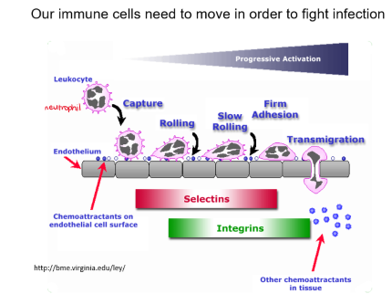
immune cells need to move to fight infection
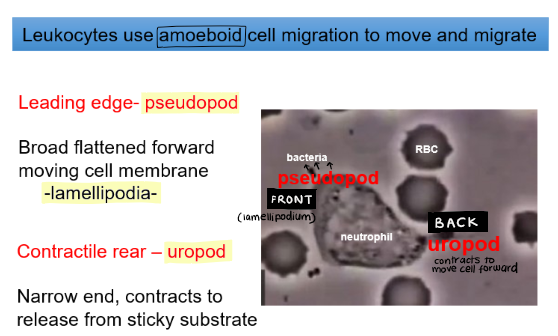
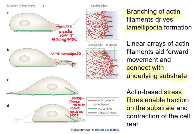
Cell movement requires force generation in different parts of the cell. What are the two ends of the migrating cells?
leading edge (pseudopod)
branched F-actin generates the pseudopod (lamellipodia) to move front of cell forwards
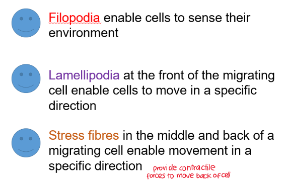
linear F-actin generates filopodia which links to lamellum
contractile rear (uropod)
actin-myosin stress fibres generate cone-shaped rear which contract to move back of cell forward
role of filopodia, lamellipodia and stress fibres in migrating cells
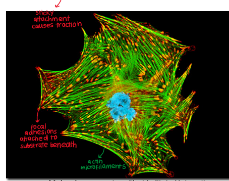
How do migrating cells exert traction on the substrate they are migrating on?
cell-substrate adhesions called focal adhesions
when migrating, new adhesions via vinculin and talin form at the leading edge form under the lamellipodium
stress fibres at the back are connected to rear focal adhesions and when they contract, focal adhesions are released
Attachment of the cytoskeleton to the cell membrane is needed for force generation to move the cell membrane. How is this facilitated?
WASp protein links cell membrane to actin
attaches to cell membrane and also Arp2/3 which generates lamellipodia
What happens in Wiskott-Aldrich Syndrome?
mutated WASp gene
abnormal cytoskeleton reorganisation leading to cell dysfunction and impaired cell migration
reduced force generation to move cell membrane
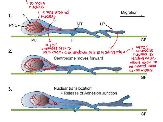
How does the nucleus move?
MT nuclear cage moves nucleus as cells migrate
MTOC connects nuclear cage to MTs in leading edge
as leading edge pushes forward, MTs at leading edge pull the MTOC forwards, which pulls the nucleus forwards as well
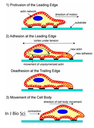
process of cell migration in a specific direction
filopodia sense chemoattractive signal (e.g. chemokines) and is stabilised by the skeleton
lamellipodium forms and is stabilised
new focal adhesions (via vinculin and talin) are made under the lamellipodium
focal adhesions are lost at the rear of the cell
MT nuclear cage pulls nucleus forward via MTOC anchor
contraction of actin-myosin stress fibres (myosin drives contraction of actin) at rear, stress fibres are connected to lamellum at the front, helps bring back of cell forwards
What are the three elements of the cytoskeleton that must work together to move eukaryotic cells?
actin
microtubules
actin-myosin stress fibres

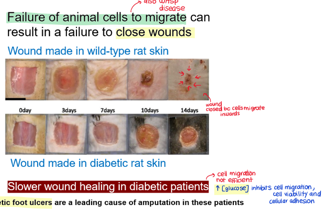
cell migration and wound healing
cells migrate inwards to close wound

Why is wound healing slower in diabetic individuals?
high glucose concentrations inhibits cell migration and cellular adhesions