Microscopy
1/19
There's no tags or description
Looks like no tags are added yet.
Name | Mastery | Learn | Test | Matching | Spaced | Call with Kai |
|---|
No analytics yet
Send a link to your students to track their progress
20 Terms
How do compound light microscopes work
they use visible light to illumnate specimens
Types of light microscopy
brightfeild (what we use)
dark feild
phase-contrast
fluorescene
Lenses
light is refracted when passing from one medium to aother
F = focal point
f = focal length
shorter focal length = greater magnifcation
Brightfeild microscope
specimens are vidualized because of diffrence in contrast (denstity) between the speciment and its surondings
total magnification = mag. of ocular lens * mag. objectve lens
upper limit of magnification = 1000x
upper limit of resolution = 0.2um (with blue filter)
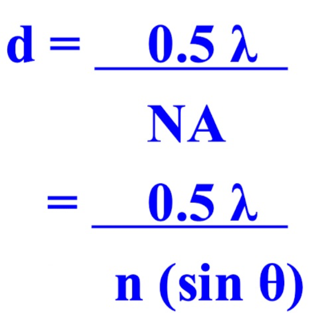
Microscope resolution
resolution (d) ability to distinguish two objects as distinct and seperate when viewed under a microscope clarity
wavelength of light (shorter wavelength) leads to better resolution (or smaller d)
Why do we use blue light
decreases light
leads to better reoslution
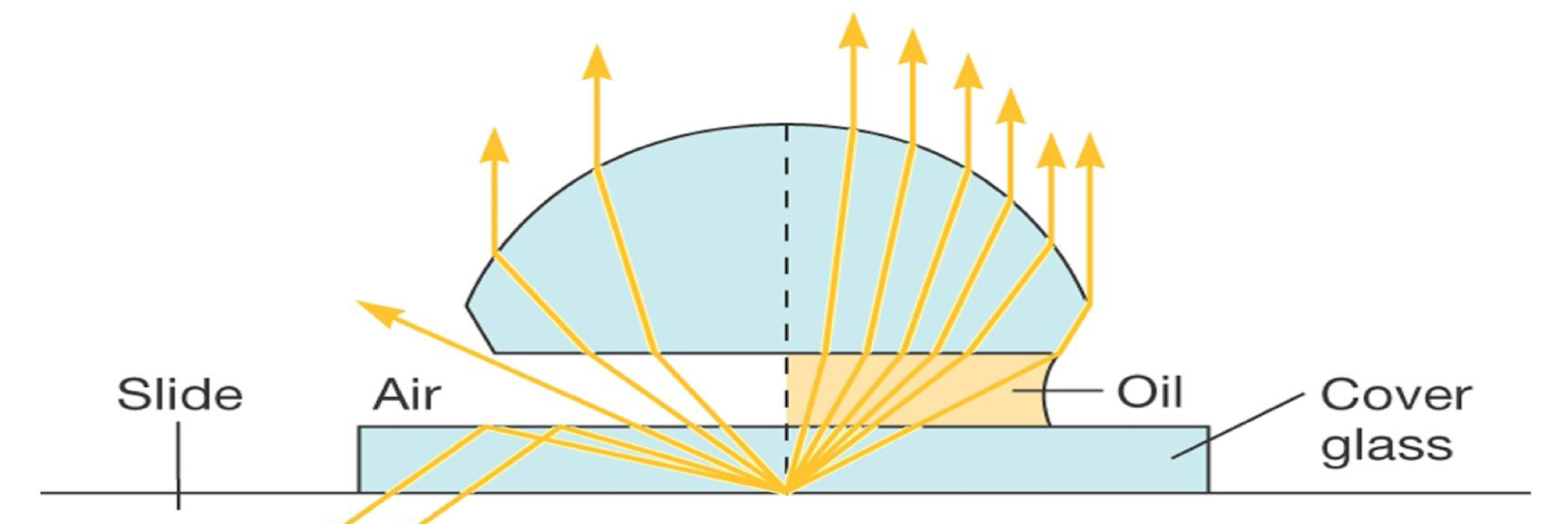
Numerical aperature (NA)
measure of light gathering ability
higher NA —> better resolution
Increase refraction index —> icnrease NA
Formula for resolution (D)
1st step for improving contrast in light microscopy
Fixation
preserves specimens and fixes them own position
organisms usually killed and firmly attached to microscope slide
heat fixation; used for bacteria and archaeons
chemical fixation; used with larger more delicate organisms
2nd step for improving contrast in light microscopy
staining
make internal and eternal structure of cell more visible by increasing (contrast) with backround
two common features of cells
chromophore groups
chemical groups with conjugated double bonds
give stain its color
ability to bind cells
2 types of stains
Basic stains
dyes with postive charges
bind to negativly charged structures
Acidic stains
dyes with negative charges
bind to postiviely charged structures
Define simple stain
a single staining agent is used
Differential staining
use more than one dye to preferentially stain features
used to detect presense or absence of structures
divides microorganisms into groups based on their staining properties
gram stain
acid-fast stain
endospore stain
Gram staining
most widely used differential staining procedure
divides bacteria into two groups based on diffrences in cell wall structure
gram postive, gram negative
Gram staining steps
flood the heat-fixed smear with crystal violet for 1 min (all cells purple)
add iodine solution for 1 min (all cells purple)
decolorize with alcohol briefy for 20 seconds (Gram postiive cells are purple, Gram negative cells are colorless
Counterstain with safranin for 1-2 mins (Gram positive are purple, gram negative are pink to red)
Acid fast staining
helpful for staining members of the genus mycobacterium
high lipid myotic acid contect in cell walls is responsible for thier staining characterists
Endospore staining
heated, double staining technique
bacterial endospore is one color and vegatative cell is a diffrent color
Capsule staining
negative stain - capsules are colorless against a stained background
Flagella staining
mordant applied to increase thickness of flagella