Ch. 10 Cell Reproduction
1/50
There's no tags or description
Looks like no tags are added yet.
Name | Mastery | Learn | Test | Matching | Spaced | Call with Kai |
|---|
No analytics yet
Send a link to your students to track their progress
51 Terms
what are the two functions of cell division?
• Multicellular organisms(humans) use cell division for growth, maintenance, and repair of cells and tissues.
• Single-celled organisms (bacteria) use cell division to reproduce.
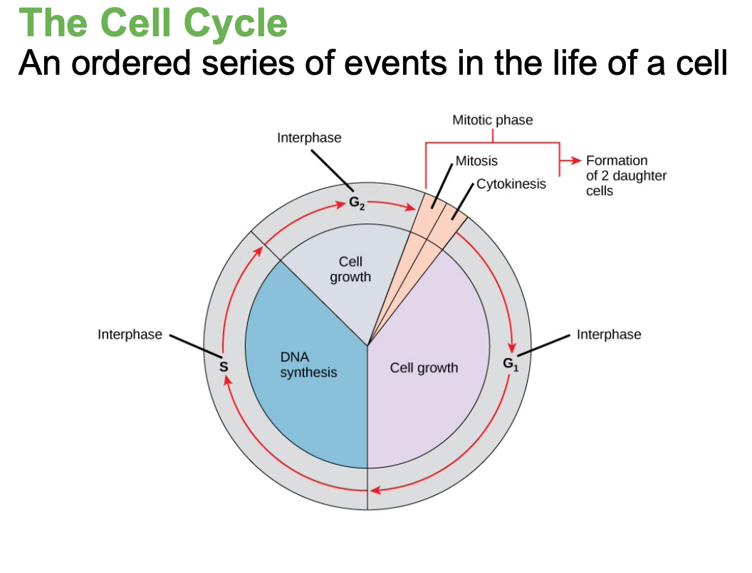
cell cycle
an orderly sequence of events that describes the stages of a cell’s life from the division of a single parent cell to the production of two new genetically identical daughter cells.
genome
A cell's genetic information is stored in the DNA, packaged as a double-stranded DNA molecule,
nucleiod
The region in the cell containing this genetic material is
Prokaryotic Genomes
(bacteria) consists of a single, double-stranded DNA molecule structured in the form of a loop or circle in an area called the nucleoid.
Plasmids: extra DNA, antibiotic resistance transfer
Plasmid Exchange
Bacteria can exchange plasmids with other bacteria, enabling the spread of traits like antibiotic resistance.
Eukaryotic Genomes
(like humans),Consists of several double- stranded DNA molecules in the form of chromosomes inside a nucleus.
somatic
Human body cells contain 46 chromosomes
Body cell
Not reproductive; skin, muscle, and nerve cells
diploid
Responsible for body’s growth, repair, and maintenance
gametes
(sperm or eggs) (sex cells) contain 23 chromosomes each.
haploid
have half the number of chromosomes
diploid
cell, nucleus, or organism containing two sets of chromosomes (2n)
46 chromosomes
haploid
(Gamete)cell, nucleus, or organism containing one set of chromosomes (n)
23 chromosomes
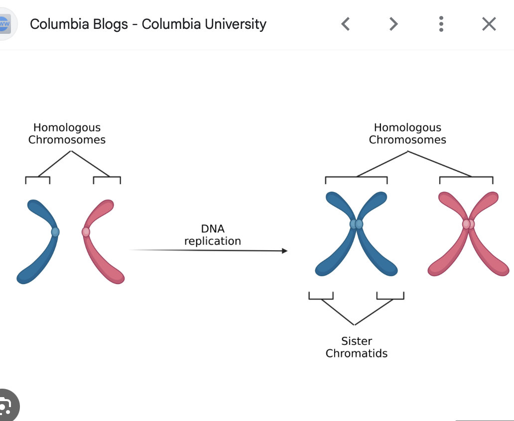
Homologous Chromosomes
Pairs of chromosomes (in diploid cells) that are similar in shape, size, and genetic content, one inherited from each parent.
Locus
position of a gene on a chromosome
heterologous pairs
chromosome pairs that do not match in size, shape, or genetic content.
In humans, the best example is the sex chromosomes:
Females have XX (a homologous pair).
Males have XY — this is a heterologous pair because the X and Y chromosomes are different.
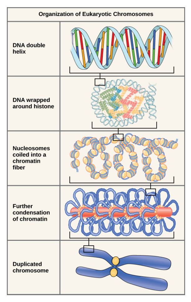
Organization of eukaryotic chromosomes
Alleles
different versions of the same gene that control a specific trait.
Each person inherits two alleles for each gene — one from each parent.
The alleles may be the same (called homozygous, like AA or aa) or different (called heterozygous, like Aa).
Alleles can produce different forms of a trait.
Example:
The gene for eye color has different alleles such as brown or blue.
chromatin fibers
long strands of DNA wrapped around proteins that help package it inside the nucleus.
-nucleousome that coils and folds
nucleosome
The histone-DNA complex (the bead)
It forms when a short stretch of DNA wraps around a core of eight histone proteins, creating a structure that looks like “beads on a string.”
help organize and compact DNA so it can fit inside the nucleus
histones
small proteins found inside the nucleus that DNA wraps around to stay organized and compact
linker DNA
connecting (string) DNA between nucleosomes
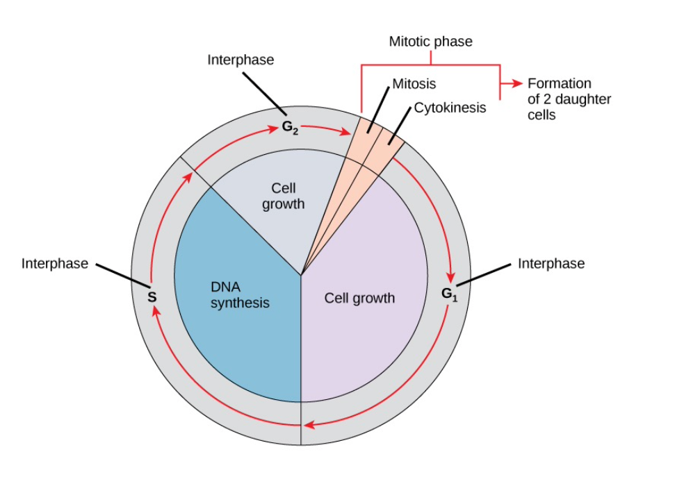
What are the two major phases of the Cell Cycle ?
Interphase (growth + DNA replication, preparation for cell division)
Mitotic Phase (nuclear & cytoplasmic division) the replicated DNA and cytoplasm are split and the cell divides
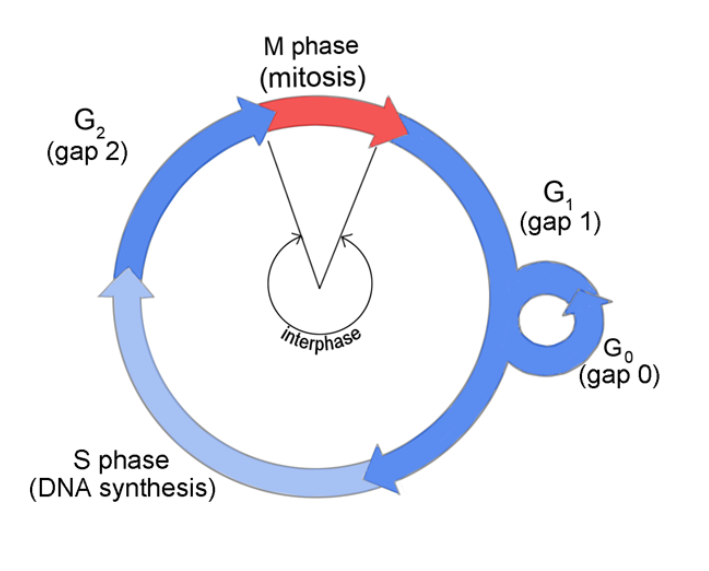
interphase
where the cell grows and DNA replication occurs/ prepares for division.
It consists of three distinct stages: G1, S, and G2 phases

G1 (first gap)
The cell grows, builds proteins, and stores energy.
It checks if it has everything needed to copy DNA.
S phase
DNA synthesis occurs
The cell copies its DNA, making two identical sister chromatids that are joined at the centromere
Centrosome duplication begins
Centrosomes produce the mitotic spindles to
move the chromosomes
• In animal cells, centrosomes are associated
with centrioles which help organize cell
division

G2 Phase (second gap)
Cell continues to grow
Cell prepares for mitosis, synthesizing(producing) proteins and duplicating organelles, while replenishing energy stores.
cytoskeleton breaks down

Mitotic Phase
where the cell divides into two identical daughter cells
Involves two processes:
Karyokinesis (mitosis) -Division of the nucleus, divided into five stages: Prophase, Prometaphase, Metaphase, Anaphase, Telophase
Cytokinesis -when the cytoplasmic components physically separate into 2 daughter cells
Karyokinesis (mitosis)
Division of the nucleus, divided into five stages: Prophase, Prometaphase, Metaphase, Anaphase, Telophase
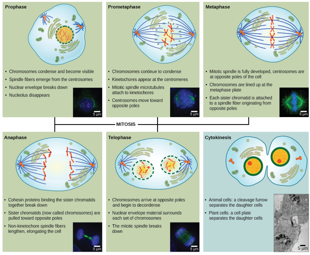
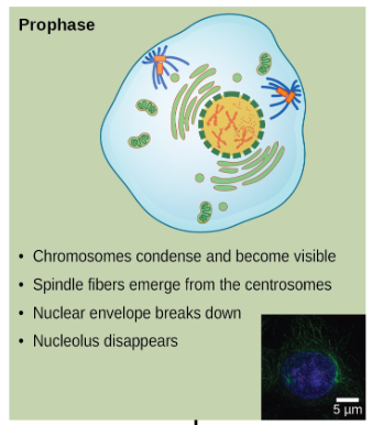
1.Prophase
Chromosomes condense and become visible;
nuclear envelope breaks down
spindle fibers form from centrosomes
centrosomes migrate
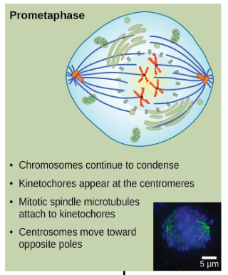
2.Prometaphase
Chromosomes continue to condense
Kinetochore forms at centromeres
Spindle fibers attach to kinetochores
Centrosomes move toward opposite poles
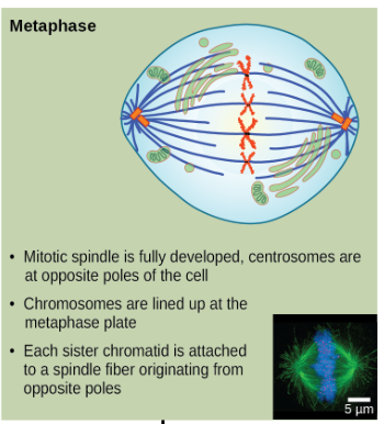
3.Metaphase
Mitotic spindle is fully developed,cetrosomes are at opposite poles of the cell
Chromosomes line up in the middle of the cell(metaplate).
Each sister chromatid is attached to a spindle fiber originating from opposite poles
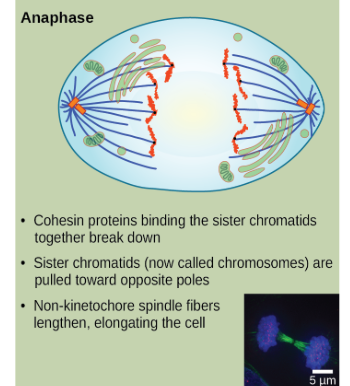
4.Anaphase
cohesion proteins binding the sister chromatids together break down
Sister chromatids (Chromosomes) separate and are pulled to opposite poles.
cell elongates
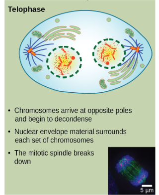
5.Telophase
Chromosomes arrive at opposite poles and begin to uncoil and relax again (decondense)
Two new nuclei form
nuclear envelope material surrounds each set of chromosomes
The mitiotic spindle breaks down
Cytokinesis
Animals cells: a cleavage furrow separates the daughter cells
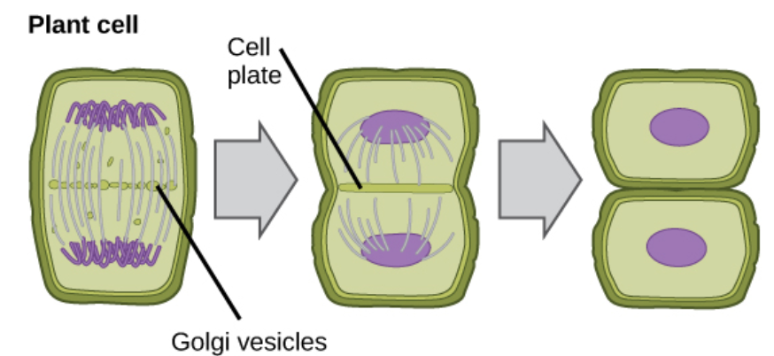
Plant cells: a cell plate separates the daughter cell
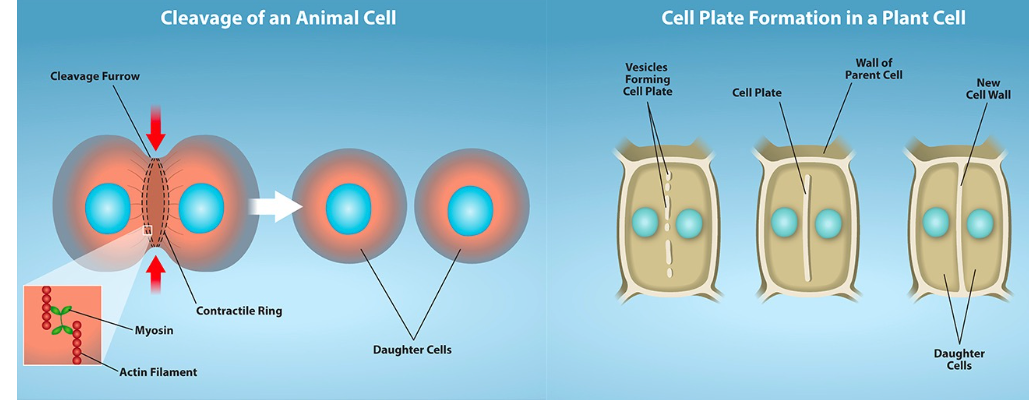
cytokinesis in Plant cells
a cell plate separates the daughter cell
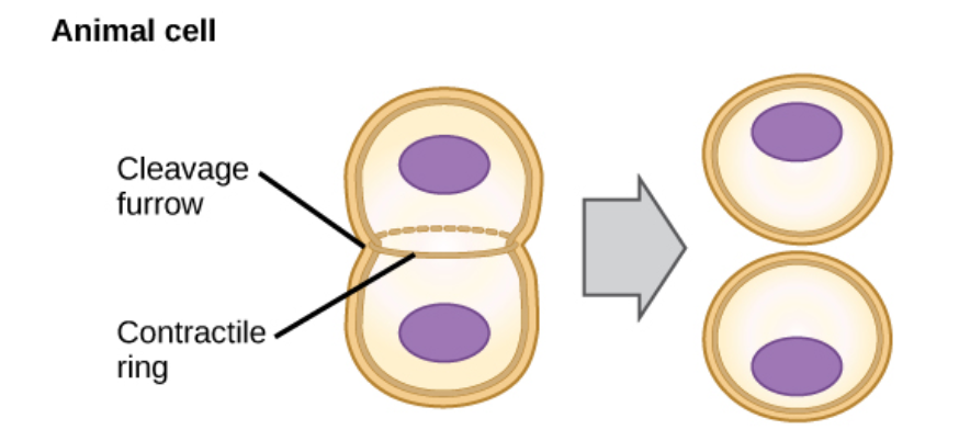
cytokinesis in Animal cells
typically starts during late anaphase
a cleavage furrow separates the daughter cells
Regulation at Internal Checkpoints
• New cell must duplicate the original
• Mistakes affecting function (such as mutated chromosomes or the wrong number of chromosomes) are regulated at 3 checkpoints in the cell cycle
(1) Near the end of G1
(2) At the G2 to Mitosis transition
(3) In metaphase of mitosis
1. G₁ Checkpoint (at the end of G₁ phase)
Purpose: Checks if the cell is ready to divide.
It looks at:
Cell size (is the cell big enough?)
Nutrients and energy available
DNA condition (is there any damage?)
If conditions aren’t right, the cell can:
Pause to repair damage
Or go into a resting stage (G₀) until conditions improve
A cell that does not meet all the requirements
will not be allowed to enter the S phase
Example:
If a cell’s DNA is damaged by radiation, the G₁ checkpoint stops division until the DNA is repaired.

G₂ Checkpoint (before mitosis starts)
This checkpoint prevents entry into the mitotic phase if certain conditions are not met
Purpose: Makes sure DNA replication was completed correctly and the cell is ready to divide.
It checks:
Cell size again
Protein supply
Whether all DNA was copied and not damaged
If there’s an issue, the cell waits to finish copying DNA or fix the damage
openstax_biology2e_ch10
Example:
If a chromosome didn’t copy fully, the G₂ checkpoint will stop mitosis until it’s fixed.

M Checkpoint (Spindle Checkpoint, during metaphase)
Occurs near the end of metaphase
Purpose: Ensures that each sister chromatid is properly attached to the spindle fibers before being pulled apart.
This prevents uneven chromosome numbers in the daughter cells (called nondisjunction)

Regulators of Cell Cycle
Positive Regulators promote movement to next step of the cell cycle
Negative Regulators stop advancement of the cell cycle
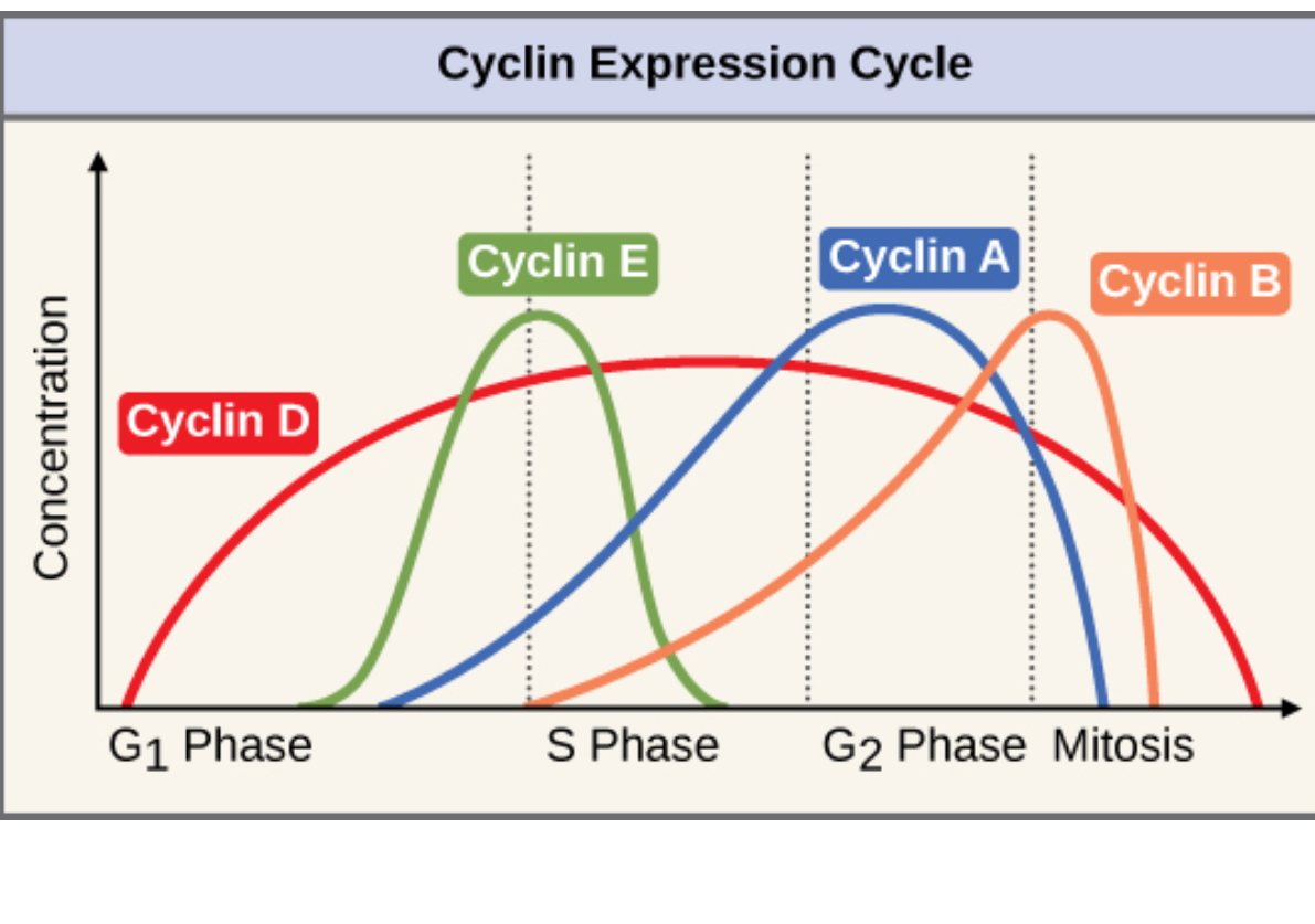
Positive Regulators
Cyclins and Cdks (cyclin-dependent kinases).
They work together to move the cell forward to the next phase.
Cyclin levels rise and fall with each stage.
Internal and external signals can trigger increases in cyclin protein levels
Cdks (Cyclin-dependent kinases)
• Only active when bound to cyclin & phosphorylated
• Phosphorylate target proteins → checkpoint progression
Negative Regulatory
• The best understood are retinoblastoma protein (Rb), p53, and p21
Rb, p53, and p21 proteins act as brakes.
They stop division if DNA is damaged or conditions are bad.
If these regulators fail, cells can divide uncontrollably (cancer).
• These act primarily at the G1 checkpoint
1. p53
• Detects DNA damage
• halts the cell cycle and then recruits specific enzymes to repair the DNA.if the DNA cannot be repaired, p53 can trigger apoptosis, or cell suicide, to prevent the duplication of damaged chromosomes.
- Stimulates p21
2. p21-enforces the halt in the cycle dictated
• Blocks Cdk/cyclin complexes
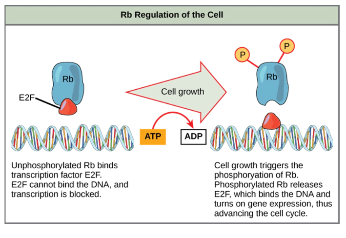
3. RbMonitors cell size and regulates cell cycle progression at G1.
• Binds E2F (gene expression blocker)
• Releases E2F when phosphorylated → S-phase transition
Oncogene
mutated version of a normal gene involved in the positive regulation of the cell cycle
a mutated/ damaged version of a proto-oncogenethat causes uncontrolled cell growth., which can cause cancer.
Proto-Oncogene
normal growth gene that helps control cell growth and division., but when changed, it can turn into an oncogene that causes uncontrolled cell division (cancer).
Tumor suppressor genes
segments of DNA that code for negative regulator proteins, the type of regulators that, when activated, can prevent the cell from undergoing uncontrolled division.
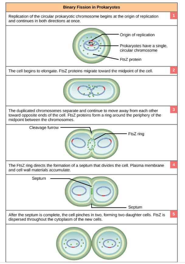
Binary Fission
prokaryotic cells (like bacteria) reproduce by splitting into two identical cells.(cell division)
a simpler and faster process than eukaryotic division.
A. Key differences
– No nucleus → no mitosis required
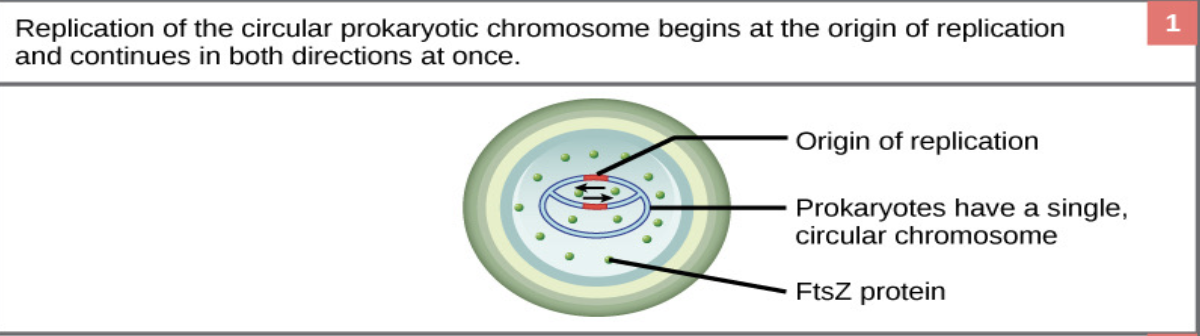
– Single circular chromosome1. DNA Replication:
The single circular DNA molecule in the bacterial cell is copied.
Each copy will go to one of the new cells.2. Attachment to Membrane:
The two DNA copies attach to different parts of the cell membrane
3. Cell Elongation:
The cell grows longer, pulling the two DNA molecules apart toward opposite ends of the cell4. Septum Formation:
A protein ring (FtsZ) forms in the middle of the cell and helps create a division wall, called a septum
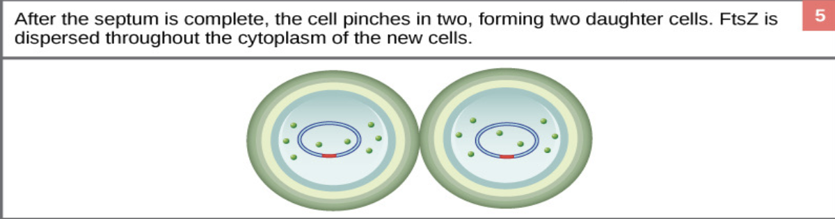
5. Cell Splits:
The septum finishes forming, dividing the cytoplasm and cell wall — now there are two identical daughter cells
1. DNA Replication
The single circular DNA molecule in the bacterial cell is copied.
Each copy will go to one of the new cells.
2. Attachment to Membrane:
The two DNA copies attach to different parts of the cell membrane
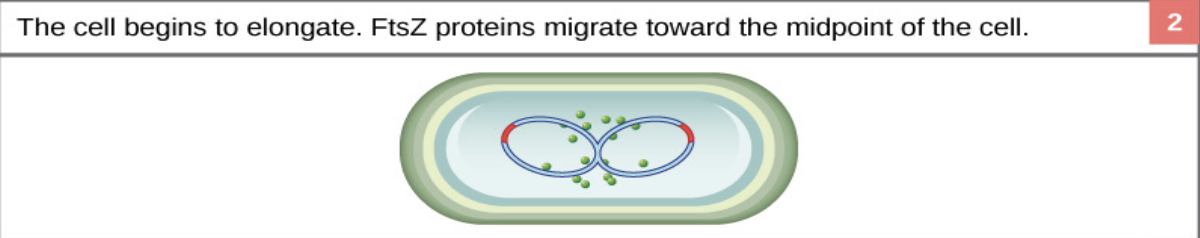
3. Cell Elongation:
The cell grows longer, pulling the two DNA molecules apart toward opposite ends of the cell
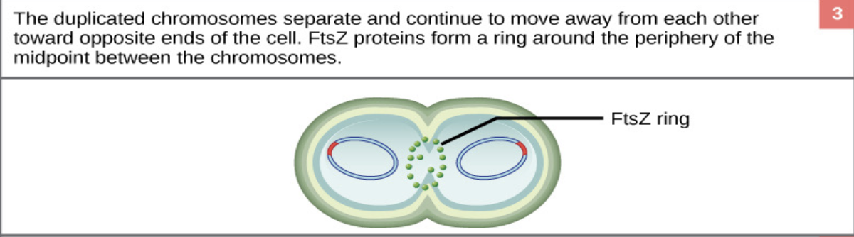
4. Septum Formation:
A protein ring (FtsZ) forms in the middle of the cell and helps create a division wall, called a septum
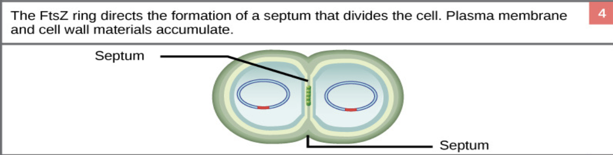
Cell Splits:
The septum finishes forming, dividing the cytoplasm and cell wall — now there are two identical daughter cells