ANHB2212 - Axial Skeleton
1/72
There's no tags or description
Looks like no tags are added yet.
Name | Mastery | Learn | Test | Matching | Spaced | Call with Kai |
|---|
No analytics yet
Send a link to your students to track their progress
73 Terms
How are somites formed?
Paraxial mesoderm of the trunk undergoes segmentation to form somites. (day 20 – day 30)
Are somites permanent or transient?
transient
How many somites form initially? What does this change to and why?
initially = 42-44 pairs
several cauda somites disappear = 37 pairs
What 3 progenitors do the somites contain?
axial skeleton (vertebral column + part of the occipital bone)
trunk musculature + associated tendons
spinal cord meninges
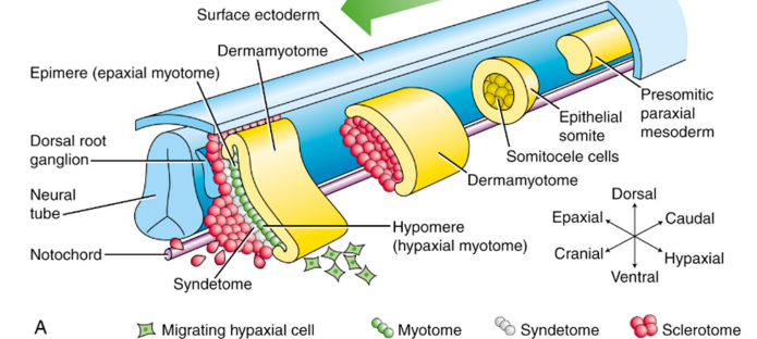
Describe the structure of a somite
Somites are balls of cells with a central cavity that contains somitocoele.
Describe the development of somites.
Somites reorganise into dermamyotome and sclerotome.
Dermamyotome forms the dermatome (> dermis) the myotome (> segmented muscles).
Myotome splits into epimere (> epaxial muscles) & hypomere (> hypaxial muscles)
Sclerotome forms the segmented bony parts (> vertebrae & ribs)
Syndetome forms between the myotome and the sclerotome (tendon progenitor cells > axial tendons)
Sclerotome cells that surround the notocord become what structure?
vertebral body
Sclerotome cells that surround the neural tube become what 4 structures?
vertebral arch
vertebral spine
transverse process
ribs
What 2 structures does ventral sclerotome form?
vertebral bodies and their intervertebral discs
What 2 structures does dorsal sclerotome form?
dorsal portion of neural arch and spinous process
What 3 structures does central sclerotome form?
pedicles and ventral parts of neural arch
proximal ribs
transverse process of vertebrae
What 2 structures does lateral sclerotome form?
distal ribs and some tendons
What 2 structures does medial sclerotome form?
spinal meninges and blood vessels of meninges
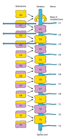
Describe resegmentation of sclerotome to form the vertebrae.
sclerotome splits into cranial and caudal segments along the intrasegmental boundary/von Ebner’s fissure.
caudal portions of one somite coalesce with the cranial portion of the next somite to form a vertebral rudiment (vertebral body)
segmental muscles extend across intervertebral joints and are supplied by spinal nerves that grow between cranial and caudal halves of somites.
Why are there 8 cervical nerves but 7 cervical vertebrae?
• Cranial half of the first cervical sclerotome fuses with the occipital bone of the skull.
• As a result, the nerve projecting to the first cervical somite is now located cranial to the first cervical vertebrae.
Klippel-Feil Syndrome
• Caused by failure of segmentation in the embryological period.
• Results in the fusion of two or more cervical vertebrae.
• Characteristic physical features include a short neck.
• Limited cervical mobility that can result in chronic headaches and pain.
What are the 2 methods of bone formation?
intramembranous ossification
endochondral ossification
Intramembranous Ossification
Bones form directly within mesenchymal condensations of osteoblasts. Osteoblasts form membranous sheets and secrete calcified matrix, then turn into osteocytes.
What are 4 examples of intramembranous ossification?
flat bones of the skull
facial bones
part of the mandible
clavicle
Endochondral Ossification
Mesenchyme first forms a hyaline cartilage model that is then replaced by bone. Here, chondrocytes die, leaving spaces than osteoblasts and blood vessels penetrate. Osteoblasts differentiate into osteocytes that form bone matrix that is remodeled into parallel arrays. Spaces coalesce to form bone marrow cavities.
What are 5 examples of endochondral ossification?
vertebral column
ribs
sternum
cranial base
limbs
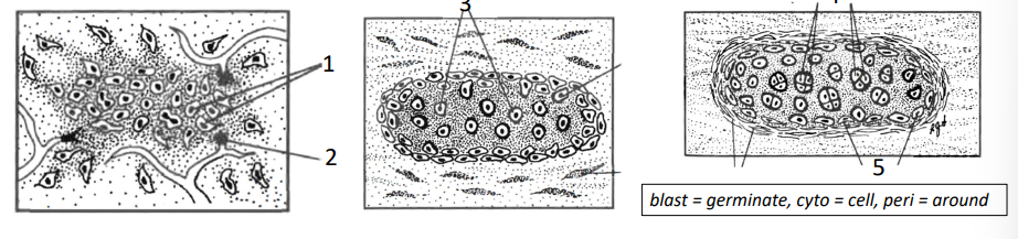
Describe the development of cartilage
1. Cartilage stem cells (chondroblasts) condense, proliferate & differentiate into cartilage cells (chondrocytes) at the centre of chondrification in week 5.
2. Blood vessels regress (cartilage = avascular; limits size of cartilage)
3. Chondrocytes secrete a gelatinous matrix around themselves separating the cells
4. Chondrocytes divide, creating nests surrounded by matrix
5. Cartilage grows by adding cells from its external surface (perichondrium)
Describe bone development
Osteoblasts condense at the centre of ossification
Osteoblasts differentiate into osteocytes and secrete calcified extracellular matrix (either endochondral or intramembranous ossification)
Why is the clavicle the first bone to ossify?
Intramembranous ossification
2 membranous primary ossification centres appear by 6 week
Fuse 1 week later
Endochondral ossification
Cartilage at both ends then develops (~ 7 weeks)
What are the 3 stages of vertebral development?
mesenchymal stage
cartilaginous stage
osseus stage
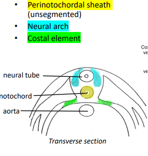
Mesenchymal Stage of Vertebral Development
Sclerotome cells migrate into 3 areas: perinotochordal sheath, neural arch and costal element
Body wall vessels from aorta form between somites (less dense)
Perinotochordal cells near vessels have better nutrition, growing large, and form the vertebral bodies,
Notochord squeexed between developing vertebral bodies become intervertebral discs
Cartilaginous Stage of Vertebral Development
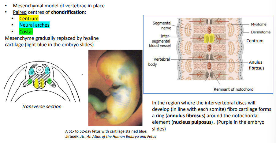
Mesenchymal model of vertebrae is in place
3 paired centres of chondrification form: centrum, neural arches and costal.
Mesenchyme is gradually replaced by hyaline cartilage
In the region where the intervertebral discs will develop, fibro cartilage forms a ring (annulus fibrosus) around the notochordal element (nucleus pulposus) .
Osseus Stage of Vertebral Development
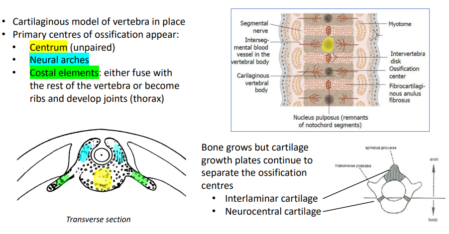
cartilaginous model of vertebrae in place
primary centres of ossification appear: centrum (unpaired), neural arches and costal elements (fuse with rest of vertebrae or become ribs and develop joints)
bone grows but cartilage growth plates continue to separate the ossification centres (Interlaminar cartilage and Neurocentral cartilage)
How is the intervertebral disc formed?
When the sclerotome splits, somitocele cells & sclerotome cells remaining in the plane of division coalesce form the annulus fibrosus (fibrocartilage ring) of the disc.

How is the nucleus pulposus formed?
Notochordal cells enclosed by the annulus fibrosus differentiate to form the nucleus pulposus of the disc. The regions of the notochord enclosed by the developing vertebral bodies degenerate and disappear.
Cartilaginous stage anomalies
As cartilage centres are paired, if one side fails to form and the other keeps growing, there is vertebral asymmetry (hemivertebrae) that cause scoliosis.
Osseus stage anomalies
If there are too many notochordal cells, as these cells inhibit ossification, centrum ossification can be inhibited and cause butterfly vertebrae. If too few of these cells, ossification can obliterate the discs and cause block vertebrae.
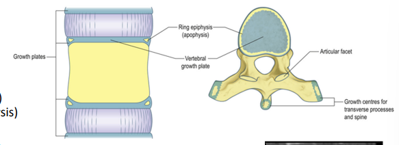
What are the 3 ossification centres?
centrum
neural arch
costal
What vertebral element does the centrum ossification centre form?
central portion of vertebral body
What 4 vertebral elements does the neural arch ossification centre form?
lateral portion of vertebral body
dorsal arch
zygapophyseal joints
mammillary processes
What 2 vertebral elements does the costal ossification centre form?
ribs in the thorax
other regions = transverse process
Describe growth of the vertebral column at birth.
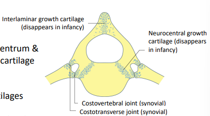
Primary ossification centres (centrum & neural arch) still separated by cartilage growth plates:
• Interlaminar cartilage
• Paired neurocentral cartilages

Describe growth of the vertebral column by 6-8 years.
• the growth plates close (ossified)
• but the vertebral body endplates are still unossified
• Vertebrae can’t increase diameter but can increase height.
Describe growth of the vertebral column at puberty.
5 secondary centres of ossification appear:
• Tip of spinous process
• Tip of transverse process
• Annular epiphysis (ring epiphysis) > annular apophysis (ring apophysis)
Processes continue to grow longer
Additional growth in height is possible due to ring epiphysis (vertebral bodies grow taller)
Describe growth of the vertebral column at adulthood.
• All epiphyses close
• Bone growth can only continue by surface remodelling
What causes adolescent idiopathic scoliosis?
Asymmetrical ossification of the epiphyseal ring.
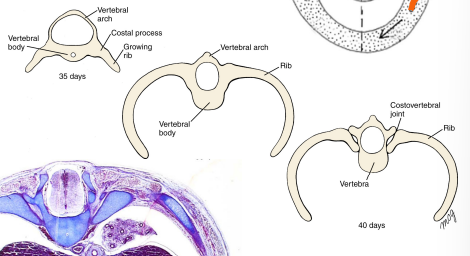
How do ribs develop?
During week 5 costal processes of thoracic vertebrae begin to elongate.
Grow around the curvature of the body wall toward the ventral body surface within the lateral plate mesoderm layer.
Costovertebral (synovial) joints form and separate ribs from vertebrae.
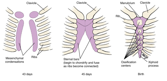
How does the sternum develop?
In week 6, paired mesenchymal condensations (sternal bars) form within the ventral body wall.
Sternal bars fuse together at their cranial ends.
Distal ends of the first 7 pairs of ribs connect with the lateral sternal edges.
Ribs join to sternum by costal cartilages.
Sternal bars fuse across the midline in a cranial‐to‐caudal direction.
Sternal ossification centers appear (60 days)
Xiphoid process does not ossify until birth
What ribs are true, false and floating?
true = 1-7
false = 8-12
floating = 11-12
How many pairs of vertebrae are there (and in what segments?)?
cervical = 7
thorax = 12
lumbar = 5
sacral = 5 fused together
coccyx = 3-4
Describe the curvature of each vertebral segment.
cervical = forward
thoracic = backward
lumbar = forward
sacrum = backward
Describe the cervical vertebrae.
spinous process is short and ends in a double point
upper articular facets face upward and inward
lower articular facets face downward and forward
transverse foramen present where the vertebral artery passes
How does C7 differ from the other cervical vertebrae?
It has a long spinous process ending in a single point.
Why are the cervical vertebrae the most mobile part of the spine?
curved shape of bodies (flexion/extension)
shallow slope of articular process (lateral flexion)
What movements are permitted by cervical vertebrae?
forward flexion
extension
lateral flexion
rotation (atlas and axis)
What is the joint between the atlas and occipital bone called?
atlantooccipital joint
Describe the thoracic vertebrae.
bodies become larger from above-down
articulate with pair of ribs at the end of the transverse process and where the pinnacle meets the body
transverse process arch sideways
spinous process points downwards
articular process are vertical (upper = backwards, lower = forwards)
What movements are permitted by thoracic vertebrae?
small amounts of forward flexion
lateral flexion
rotation
Describe the lumbar vertebrae.
massive body
transverse processes are small
spinous process is broad and points straight backwards
upper articular process face inward, lower face outward (= no rotation)
What movements are permitted by lumbar vertebrae?
forward flexion
extension
lateral flexion
Where does the vertebral canal end?
Sacral hiatus
Describe the structure of intervertebral discs
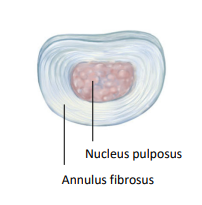
A tough outer rim of fibrocartilage (annulus fibrosus) and a soft centre called the nucleus pulposus. There is a thin layer of hyaline cartilage on the superior and inferior surfaces of each disc.
Interspinous ligaments
Run from the lower edge of one spinous process to the upper edge of the next.
Supraspinal ligament
Merges with the interspinous ligaments and runs the entire length of the vertebral column, connecting the tips of the spinous processes.
What is the function of the supraspinous and interspinous ligaments?
Limit flexion of the spine
Ligamentum flavum
A series of short ligaments that hold the vertebrae together. They lie on the front of the laminae and is made of yellow fibroelastic tissue. They limit flexion.
What ligaments hold the vertebral bodies together? Describe them.
anterior (thick and stronger, attached the upper and lower edges of the bodies) and posterior (runs along back of bodies, narrow at body, wide at disc) longitudinal ligaments that cover the front and sides of the vertebral bodies and the run the length of the vertebral column.
What is the function of the anterior longitudinal ligament?
Limits extension of the spine and prevents backward and forward movement of the vertebral bodies relative to each other.
What is the function of the posterior longitudinal ligament?
Limit flexion of spine.
Describe the posterior joints of the vertebrae.
surrounded by capsular ligaments which is loose enough to permit movement
articular processes are strong because the upper face forward and the lower backward. This prevents vertebrae above slipping forward.
What bones are involved in support and movement of the head?
thoracic and cervical vertebrae
upper ribs
clavicles
occipital bone
What 2 segments is the skull made of?
neurocranium
viscerocranium (facial skeleton)
What bones form the cranium?
occipital bone (behind + below)
2 parietal bones (side)
2 temporal bones (side)
sphenoid bone
frontal bone
ethmoid bone
Function of the occipital condyles.
Joint surfaces that articulate with the atlas (C1) to form the atlantooccipital joints.
What movement occurs at the atlantooccipital joints?
forward flexion
extension
lateral flexion
What movement occurs at the atlantoaxial joints?
rotation
Describe the atlas (C1).
no vertebral body
anterior arch in front that matches the posterior arch
lateral masses
large vertebral foramen that contains spinal cord and odontoid process of axis (C2)
upper articular surfaces are cup-shaped to match the occipital condyles
Describe the axis (C2).
body is prolonged by odontoid process (represents missing body of atlas and is the pivot where the head and atlas rotates
upper articular surfaces are in front of the lower, and are in straight line with odontoid process
odontoid process surrounded in front and at the sides by bone and is held in place behind by the transverse ligament of the axis. it is held in place from above by alar ligaments.