Pathology - Miscellaneous
1/113
Earn XP
Description and Tags
Name | Mastery | Learn | Test | Matching | Spaced | Call with Kai |
|---|
No analytics yet
Send a link to your students to track their progress
114 Terms
Massive adrenal gland enlargement, hepatomegaly and splenomegaly are common findings in what fatal autosomal recessive disorder?
A. Marfan syndrome
B. Beckwith-Wiedemann syndrome
C. Wolman disease
D. Wilson disease
C. Wolman disease
Wolman Disease is an autosomal recessive disorder that causes excessive lipid storage in the liver, spleen and adrenal glands. Most patients die in first 6 months of life. Sonographic findings include hepatosplenomegaly and massive adrenal gland enlargement.
Beckwith-Wiedemann syndrome is an autosomal dominant disorder that causes excessive growth. These patients are at an increased risk of developing several types of cancerous and noncancerous tumors, such as nephroblastoma or hepatoblastoma. Life expectancy is normal.
A hematoma of the rectus abdominis muscle that occurs below the arcuate line can extend into:
A. space of Retzius
B. pouch of Douglas
C. Morison pouch
D. the umbilicus
A. space of Retzius
An acute hematoma will be anechoic or mildly hypochoic to surrounding tissues. The linea alba stops the blood from crossing the midline, above the level of the arcuate line. Blood is not confined within the sheath below the arcuate line which allows the blood to extend into the pelvis (space of Retzius)
What type of hernia is a delayed complication of upper abdominal surgery?
A. direct inguinal hernia
B. indirect inguinal hernia
C. incisional hernia
D. amyand hernia
C. incisional hernia
Incisional Hernia is a delayed complication of abdominal surgery. It usually occurs in first few months after surgery and is more common with vertical incisions than with transverse incisions. Elderly, obese and malnourished patients have increased risk of an incisional hernia.
______ refers to a collection of serous fluid in the popliteal fossa.
A. arthroma
B. Baker cyst
C. ganglion cyst
D. Thompson cyst
B. Baker cyst
A Baker Cyst refers to a cyst that forms in the medial popliteal fossa and can extend into the calf muscle. Cyst formation is related to arthritis and it contains serous fluid. The cyst causes extrinsic compression of the popliteal vein, decreases venous outflow, causes swelling and pain in the lower leg. These are similar symptoms to DVT. It can rupture causing fluid to enter the calf causing symptoms similar to thrombophlebitis.
Pyloric stenosis is diagnosed when the muscle wall thickness exceeds:
A. 6mm
B. 8mm
C. 4mm
D. 2mm
C. 4mm
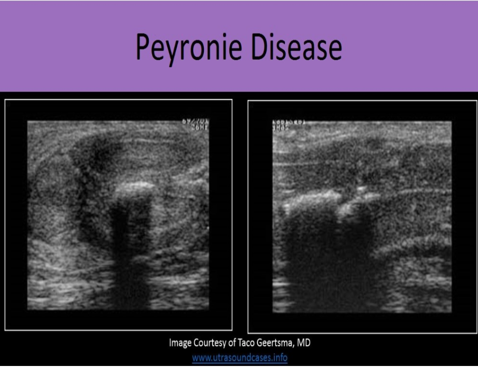
The development of scar tissue and fibrous plaque formation involving the tunica albuginea of the penis describes:
A. Peyronie disease
B. Penile fracture
C. Priapism
D. Squamous cell carcinoma of the penis
A. Peyronie disease
Peyronie Disease is the development of scar tissue and fibrous plaque formation that usually involves the tunica albuginea. It causes restriction and curvature of the affected side of the penis during erection and can be very painful. 2D imaging demonstrates hyperechoic areas along the outer margins of the corpus cavernosa.
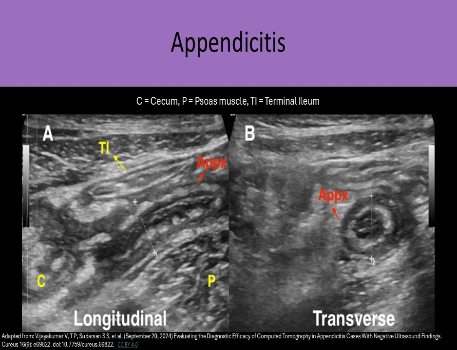
A patient presents with moderate RLQ pain and a low grade fever. An US exam of the area demonstrates a 7mm thick aperistaltic tube that is noncompressible. Which of the following best describes the findings?
A. appendicitis
B. intussusception
C. Crohn disease
D. varicocele
A. appendicitis
A thickened aperistaltic tube that is noncompressible is a hallmark sign of appendicitis. Crohn disease refers to inflammatory disease of the bowel. Bowel segments can be thickened, aperistaltic and noncompressible but will not appear a s a blind ended tube.
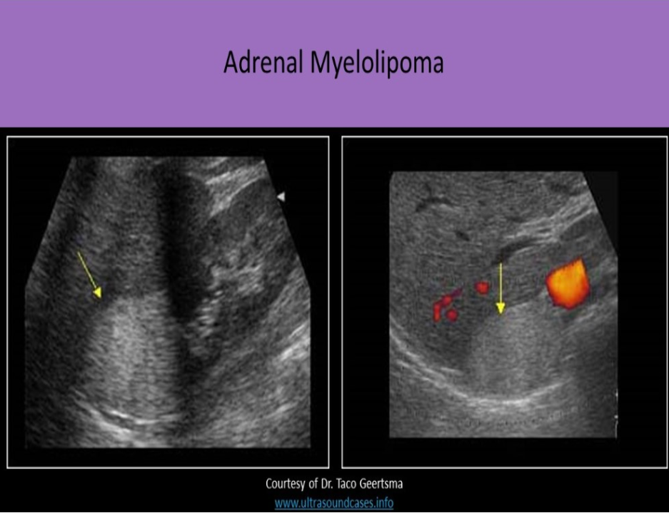
Which of the following is not an expected finding with adrenal myelolipoma?
A. attenuation artifacts
B. comet tail artifacts
C. propagation speed artifacts
D. increased echogenicity compared to adrenal and renal cortical tissues
B. comet tail artifacts
Adrenal myelolipomas are composed of fatty and bony elements that cause the mass to be echogenic on ultrasound evaluation. Fatty tissue causes increased attenuation of the ultrasound beam. Diaphragm disruption due to propagation speed artifact may be identified. The decrease in the speed of sound in fatty tissue can cause the portion of the diaphragm that is posterior to the mass to be placed more posterior on the image so the diaphragm looks "broken". Comet tail artifacts are caused by repetitive reflections at a media boundary between very different tissues. They are usually associated with tissue/gas or tissue/fluid interfaces.
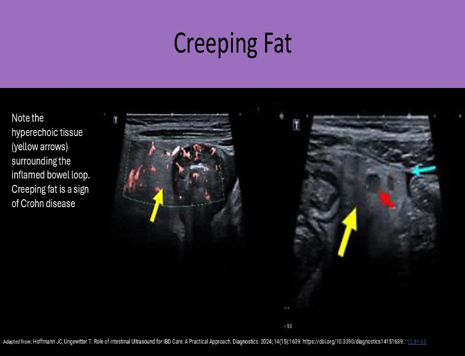
Creeping fat is a sign of :
A. Wilson disease
B. Mesenteric ischemia
C. Crohn disease
D. Pancreatic adenocarcinoma
C. Crohn disease
Creeping fat refers to fat creeping onto the margins of the involved gut segment. It creates an echogenic halo around the mesenteric border of the gut. It is associated with perienteric inflammation seen with Crohn disease.
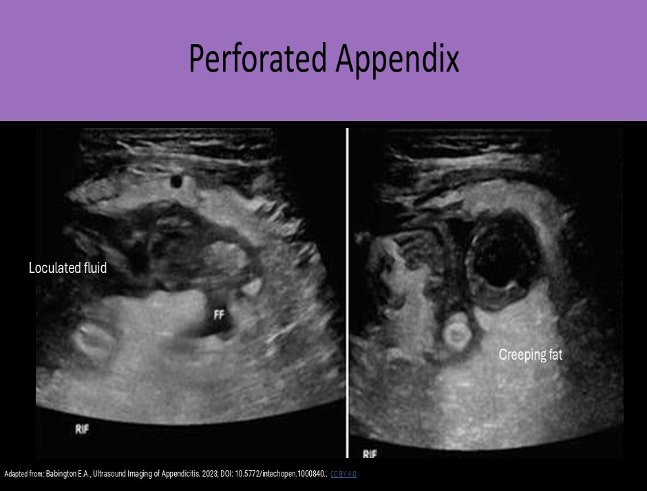
Phlegmon formation, abscess formation, prominent pericecal fat and loculated pericecal fluid collections are sonographic findings that are most suggestive of:
A. mononucleosis
B. appendiceal perforation
C. acute pancreatitis
D. Addison disease
B. appendiceal perforation
Appendiceal perforation is associated with loculated pericecal fluid, phlegmon formation, abscess formation, and prominent inflamed pericecal fat.
Pancreatitis can be associated with similar findings of phlegmon, abscess, loculated fluid, but not normally found in the area of the cecum.
All of the following correctly describe a mechanical bowel obstruction, except:
A. adhesions are the most common cause in adults
B. best evaluated by US after patient drinks at least 32oz of water
C. must assess Gl tract caliber and content
D. physical impediment to the progression of the luminal contents
B. best evaluated by US after patient drinks at least 32oz of water
When a mechanical obstruction is suspected, the patient should NOT consume any food or water until they have been evaluated for the blockage. Adding more substances into the Gl tract will only exacerbate their symptoms. A mechanical obstruction indicates that there is a physical impediment blocking the GI tract.
Sonographic evaluation includes:
Describe location of gut loops
Evaluate the caliber of the segment and size of obstruction
Describe the contents of the segment; solid, fluid, gas
Assess peristalsis
A 55yr old female presents with a fever, leukocytosis and LLQ pain. She states she suffers from Crohn disease. The exam demonstrates a 2.7cm ill-defined hypoechoic mass in the LLQ that is surrounded by a small amount of ascites. There is no flow within the mass and no movement with peristalsis. These findings are most suggestive of:
A. Retroperitoneal lymphadenopathy
B. Intussusception
C. Appendicitis
D. Abscess
D. Abscess
Crohn disease can be associated with intraperitoneal abscess formation. An ill-defined mass with no internal vascularity that is surrounded by fluid should causes suspicion of an abscess. Intussusception, appendicitis and lymphadenopathy would demonstrate vascularity and/or motion. In most patients the appendix is in the RLQ.
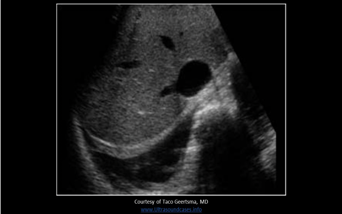
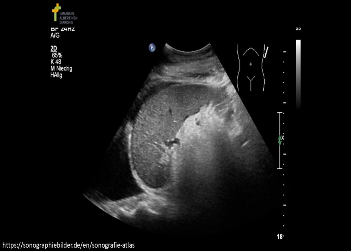
Where is free fluid documented on the image?
A. pleural space
B. subphrenic space
C. subhepatic space
D. paracolic gutter
A. pleural space
The image demonstrates a fluid above the diaphragm in the RUQ. There is moderate pleural effusion present in the pleural space.
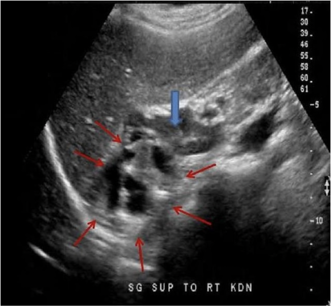
The blue arrow indicates the adrenal gland in a 30 yr old male with a palpable flank mass and no other related history. Which of the following best describes the findings on the image demonstrated by the red arrows?
A. complex adrenal mass located superior to the gland, most likely cortical carcinoma
B. complex renal mass extending superior to the adrenal gland, most likely medullary sponge kidney
C. dilated bowel loops posterior to the adrenal gland, most likely an intestinal hernia
D. complex adrenal mass, most likely a lipoma
A. complex adrenal mass located superior to the gland, most likely cortical carcinoma
Adrenal cortical carcinoma usually presents as a complex adrenal mass. The solid and cystic components cause a very irregular appearance.
Which of the following is the most common cancer of the Gl tract?
A. Kaposi sarcoma
B. Krukenberg tumor
C. Adenocarcinoma
D. Gastric sarcoma
C. Adenocarcinoma
A doctor refers an infant for a chest ultrasound exam due to a history of a chest mass seen in the left lower lobe on an x-ray. A well defined hypoechoic mass is identified in the area of interest. Color Doppler demonstrates arterial supply to the mass from a branch of the aorta. These findings are most suggestive of:
A. atelectasis
B. pulmonary sequestration
C. hemothorax
D. pneumonia
B. pulmonary sequestration
Pulmonary sequestration is a congenital anomaly that occurs when a mass of nonfunctional lung tissue forms that does not communicate with the bronchial tree. It has separate blood supply from the aorta, instead of the pulmonary arteries. Ultrasound may demonstrate a hypochoic chest mass. Color Doppler is used to demonstrate a systemic artery (not pulmonary) supplying the hypoechoic mass in both types of sequestration
Concentric wall thickening of the terminal ileum, hyperemia and mesenteric fatty proliferation are signs of what Gl tract abnormality?
A. Crohn disease
B. colitis
C. diverticulitis
D. appendicitis
A. Crohn disease
Crohn disease typically affects the terminal ileum. Colitis affects the sigmoid colon and rectum. The primary Sonographic signs of Crohn disease include concentric wall thickening (> 3 mm), hyperemia and mesenteric fatty proliferation (creeping fat) with reactive inflammatory fat.
Which of the following correctly describes myelolipoma?
A. originate in the adrenal medulla
B. hypoechoic mass with extensive posterior enhancement
C. may see varied levels of attenuation lateral to the mass
D. adrenal tumor that is composed of fat and bony elements
D. adrenal tumor that is composed of fat and bony elements
An adrenal myelolipoma usually originates in the adrenal cortex and is composed of fat and bony elements. The mass is hyperechoic and causes varied levels of sound attenuation, posterior shadowing and propagation speed artifact. Large masses can be associated with acute retroperitoneal hemorrhage. These masses can be associated with Cushing syndrome, Conn syndrome and adrenal hyperplasia.
The diaphragm sign, displaced crus sign, and bare area sign are indicative of what abnormality?
A. pneumonia
B. diaphragmatic hernia
C. ascites
D. pleural effusion
D. pleural effusion
Diaphragm Sign - when using an abdominal approach to view the fluid, fluid below the diaphragm and more centrally located = ascites; fluid above the diaphragm and more peripherally located = pleural effusion.
Displaced Crus Sign - if the fluid is displacing the crus away from the spine, it is located in the chest cavity
Bare Area Sign - pleural fluid will extend behind the liver at the level of the bare area, ascites cannot reach this area
What is the most accurate Sonographic predictor of pyloric stenosis?
A. Cross-sectional diameter
B. Gastric volume
C. Channel length
D. Muscle wall thickness
D. Muscle wall thickness
Excessive muscle wall thickness is the most accurate predictor of pyloric stenosis.
A patient with a history of Conn disease presents for an abdomen ultrasound exam. Two 1cm round hypoechoic tumors are identified in the left adrenal gland. These findings are most suggestive of:
A. aldosteronomas
B. adenomas
C. myelolipomas
D. abscess formation
A. aldosteronomas
Hyperaldosteronism (Conn disease/syndrome) - increased aldosterone levels, can lead to multiple small round hypochoic masses in the adrenal cortex (aldosteronomas).
Acute pain in the RLQ is commonly associated with _______ acute pain in the LLQ is commonly associated with _______
A. appendicitis, irritable bowel syndrome
B. appendicitis, cecal colitis
C. diverticulitis, irritable bowel syndrome
D. appendicitis, diverticulitis
D. appendicitis, diverticulitis
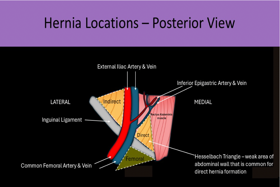
Which of the following statements is correct regarding hernias?
A. Varicose vein formation is the most significant complication of a hernia
B. Most hernias are closed using a skin graft taken from the opposite groin
C. Herniorrhaphy is the most common surgical procedure performed on post-partum females
D. Patients with one hernia should be evaluated for other types of ipsilateral and contralateral groin or anterior abdominal hernias
D. Patients with one hernia should be evaluated for other types of ipsilateral and contralateral groin or anterior abdominal hernias
Patients with one hernia, usually have multiple and they should be evaluated for other types of ipsilateral and contralateral groin or anterior abdominal hernias .
Strangulation is the most significant complication of a hernia. Herniorrhaphy is one of the most common surgical procedures performed in the US. Most patients that undergo repair are males less than 1yr of age. Surgical mesh is used to cover/close the hernia in most procedures.
Most malignant tumors will demonstrate evaluation.
A. high resistance
B. low resistance
C. absent
D. increased diastolic flow reversal
B. low resistance
Most malignancies are constantly growing and "invading" so this requires constant flow throughout the cardiac cycle. Doppler will demonstrate increased velocity flow with increased diastolic flow consistent with a low resistance waveform.
All of the following are signs of pyloric stenosis, except:
A. cervix
B. sandwich
C. olive
D. doughnut
B. sandwich
Olive sign - enlarged pylorus is palpable in the infant abdomen; feels like an olive, in size and shape
Doughnut sign - used to describe cross sectional appearance of the pylorus on ultrasound
Cervix sign - used to describe the longitudinal sectional appearance of the pylorus on ultrasound
McBurney point is an area in the right lower quadrant that is associated with the appendix.
Which of the following describes a giant cell tumor of the hand?
A. moves with the adjacent tendon with flexion and extension
B. does not move with the adjacent tendon with flexion and extension
C. hypochoic structure that demonstrates posterior enhancement
D. anechoic structure that demonstrates posterior shadowing
B. does not move with the adjacent tendon with flexion and extension
Giant Cell Tumor of the hand is a benign soft tissue mass that arises from the palmar tendons. It is the 2nd most common tumor of the hand. The mass does not move with flexion or extension of adjacent tendons. Sonographic characteristics include homogeneously hypoechoic mass with NO enhancement and most will have some internal vascularity.
Which of the following describes the best way to image the patellar tendon with ultrasound?
A. due to the proximity of the tendon to bone, the patellar tendon cannot be evaluated with US
B. evaluate the patellar attachments from an anterior approach
C. patient must be evaluated with the leg fully extended
D. the central portion of the tendon is best imaged from the posterior approach
B. evaluate the patellar attachments from an anterior approach
The patellar tendon connects the patella to the tibial tuberosity. Imaging is performed from the anterior approach. Tears are associated with sports that involve jumping and it most commonly detaches from the distal patella. When detached, the patient is unable to completely extend their leg and patella is displaced superiorly. A posterior approach would not be beneficial in viewing the central portion of the tendon due to the bony structures between the transducer and the tendon.
A right sided pheochromocytoma will displace the ipsilateral kidney
A. anteriorly
B. medially
C. inferiorly
D. superiorly
C. inferiorly
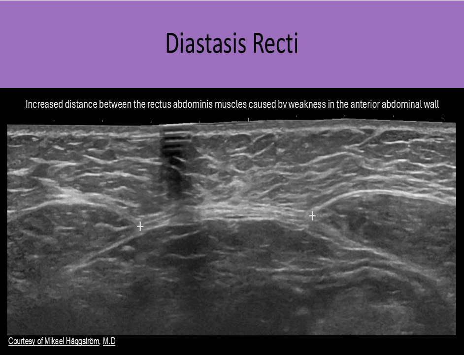
A patient with diastasis recti will have an increased:
A. distance between the bladder and the rectum
B. risk of developing hepatocellular carcinoma
C. distance between the rectus abdominis muscles
D. risk of developing carpal tunnel syndrome
C. distance between the rectus abdominis muscles
Diastasis Recti is diagnosed when there is an increased distance between the rectus abdominis muscles caused by weakness in the anterior abdominal wall.
The linea alba is normally a narrow fibrous band in the midline abdomen that separates the two rectus abdominis muscles. Muscle weakness and thinning/stretching of the linea alba cause diastasis recti. This abnormality is seen with pregnancy, obesity and in newborns. It can be differentiated from a ventral hernia because there is no fascial defect. It presents as a midline bulge that increases with increased abdominal pressure.
Bacterial infection, internal bleeding and viral infection typically share what abdominal ultrasound finding?
A. abscess formation
B. retroperitoneal fibrosis
C. free air in the peritoneal cavity
D. ascites with debis/septations
D. ascites with debis/septations
Ascites is a common finding with abdominal viral or bacterial infections.
Bleeding of the intraperitoneal organs also causes ascites. Ascites may appear complex and contain debris/septations if there is associated bacterial infection, internal bleeding, or viral infection. Abscess formation may be seen with the infections, but not typically associated with internal bleeding.
All of the following correctly describe a scalp hematoma, except:
A. caput succedaneum hematoma is associated with vacuum assisted delivery
B. described by their location related to the galea aponeurosis and skull periosteum
C. most commonly caused by motor vehicle trauma
D. sonographic appearance varies with age of the hematoma
C. most commonly caused by motor vehicle trauma
The scalp is composed of 5 layers of tissue
S - skin
C - connective tissue
A - (galea) aponeurosis
L - loose connective tissue
P - periosteum
Hematoma formation usually occurs due to birth trauma, although they are also seen with head trauma
Most resolve on their own within a few days
Subdivided by their location related to the galea aponeurosis and skull periosteum caput succedaneum - subcutaneous hematoma, most commonly caused by vacuum assisted delivery
subgaleal hematoma - hematoma within the potential space between the galea aponeurosis and the skull periosteum cephalohematoma - subperiosteal layer
Sonographic appearance varies with age of the hematoma
Loculated fluid in the abdomen usually ______, while free fluid in the abdomen will ______
A. indicates a benign process, indicate a malignant process
B. displaces adjacent structures, fills spaces around structures
C. fills spaces around structures, displaces adjacent structures
D. is resorbed over time by the body, require a paracentesis to remove the fluid
B. displaces adjacent structures, fills spaces around structures
Free fluid in the abdomen tends to fill spaces around the organs, while loculated fluid tends to displace surrounding structures.
A patient presents for a groin ultrasound with a history of right inguinal hernia repair 3 years ago. He's currently having right groin pain. What should you do next for this patient?
A. call the referring physician and get an order for an MRI because ultrasound cannot demonstrate recurrent hernias
B. because the patient had prior hernia repair in the right groin, no maneuvers are performed during imaging
C. assess the edges of the mesh in the groin while the patient performs the Valsalva maneuver
D. because the highly reflective mesh will severely limit the US exam of the right groin, no maneuvers are performed during imaging
C. assess the edges of the mesh in the groin while the patient performs the Valsalva maneuver
Hernia repair is usually performed using a mesh material. Most recurrent hernias form from the edges of the mesh. The edges of the mesh should be evaluated with dynamic maneuvers.
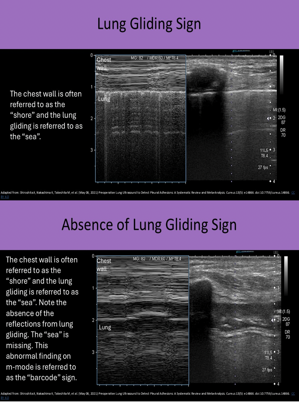
Which of the following correctly describes a pneumothorax?
A. fluid accumulation between the visceral and parietal pleura
B. the lung-gliding sign is not present with free air in the chest
C. There are two types of pneumothorax, transudate and exudate
D. ultrasound is not able to demonstrate a pneumothorax
B. the lung-gliding sign is not present with free air in the chest
Ultrasound can be used to evaluate lung motion and the presence of certain artifacts can indicate lung disease. Lung-gliding sign - visceral pleura and underlying aerated lung can be seen gliding across the parietal pleura in the normal lung.
Pneumothorax: Ultrasound most helpful in evaluating patients that cannot sit upright for a chest x-ray. Free air in the chest can be a complication caused by thoracentesis. The free air can be seen between the visceral and parietal pleura and the lung-gliding sign is lost.
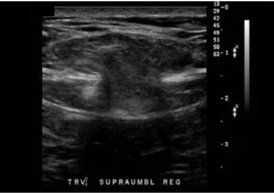
A 50yr old male presents with paraumbilical pain and a palpable mass. The findings on the image are most suggestive of:
A. A large rectus sheath lipoma
B. A large rectus sheath hematoma
C. Abdominal wall hernia with no bowel protrusion
D. Abdominal wall hernia with bowel protrusion
D. Abdominal wall hernia with bowel protrusion
The image demonstrates an opening in the peritoneum and muscular fascia of the abdominal wall with a "mushroom cloud" of intestine extending anteriorly through the herniation. The Valsalva maneuver is helpful in evaluating the presence/absence of bowel protruding through a herniation in the abdominal wall. The increase in intra-abdominal pressure should force the bowel through the opening and peristalsis should occur upon release.
What is the primary reason a retroperitoneal lymph node dissection (RLND) is performed?
A. to evaluate the lymph tissue for functionality
B. to determine the extent of a systemic infection
C. to prevent lymphocele formation on an upcoming renal transplant
D. to determine if systemic chemotherapy is needed
D. to determine if systemic chemotherapy is needed
Retroperitoneal Lymph Node Dissection (RLND):
Retroperitoneal lymph nodes are removed for evaluation for signs of metastasis
Surgical technique used to the remove the lymph nodes
Testicular cancer usually spreads to these lymph nodes first
Patients with cancer may require an RLND to determine whether chemotherapy is needed
Lymphocele formation is a common post-procedure complication
A hernia with a narrow neck:
A. should not be evaluated with the Valsalva maneuver
B. can be closed with compression therapy instead of surgery
C. is usually completely reducible with compression
D. is at an increased risk for incarceration and strangulation
D. is at an increased risk for incarceration and strangulation
A hernia with a narrow neck is at an increased risk for incarceration and strangulation. All hernias should be evaluated with dynamic maneuvers. The reducibility should be assessed with compression. A narrow neck significantly limits reducibility and increases the risk for incarceration and strangulation. Hernia repair is required to close the orifice.
Ascites is usually associated with all of the following, except:
A. renal failure
B. congestive heart failure
C. hepatocellular carcinoma
D. oncocytoma
D. oncocytoma
Oncocytoma is a benign renal mass that is not typically related to ascites. CHF involves poor cardiac function and decreased circulation which can lead to fluid accumulation in the body, including the abdomen. Renal and liver failure leads to decreased filtration of fluid and waste from the blood which also leads to fluid accumulation in the body. Liver malignancy leads to the formation of ascites because the tumor formation increases internal liver pressure. Fluids are forced into the abdomen due to the increased pressure.
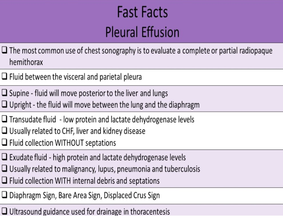
Which of the following is a common cause of exudate pleural effusion?
A. congestive heart failure
B. kidney failure
C. cirrhosis
D. malignancy
D. malignancy
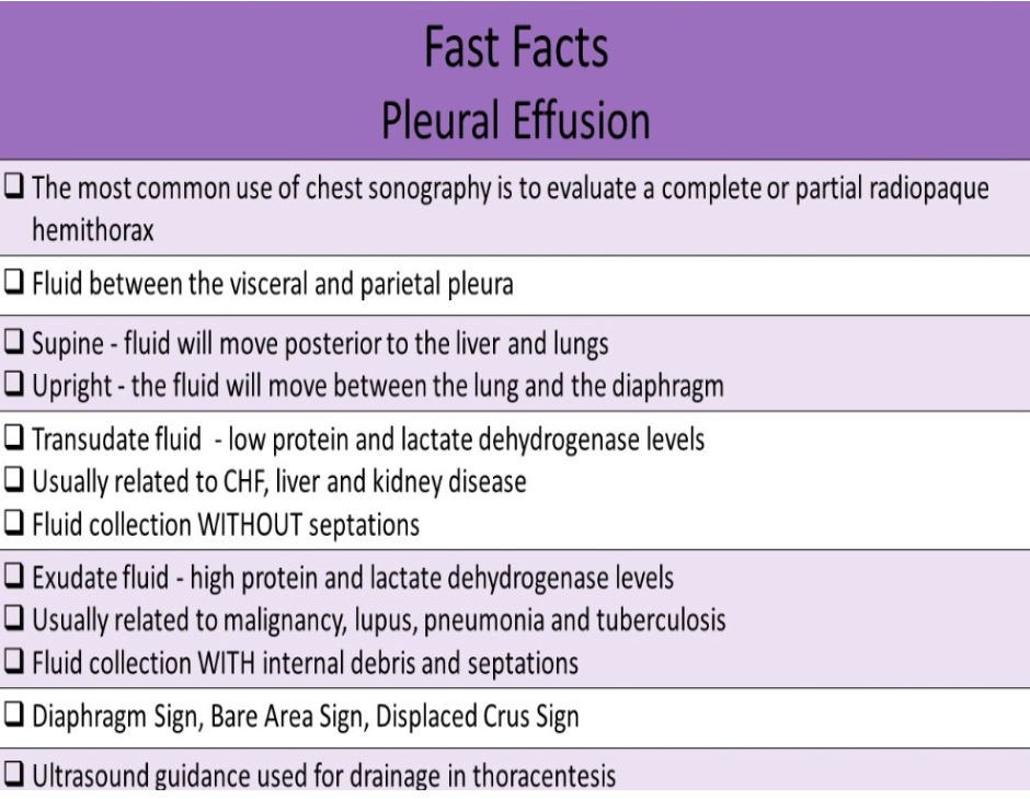
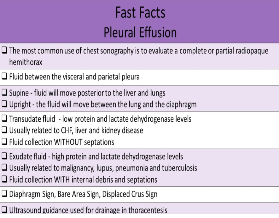
Transudate fluid has low protein and lactate dehydrogenase concentrations. Free fluid is pushed outside the capillary bed due to high pressure in the capillary beds. It is usually related to CHF, liver and kidney disease.
Exudate fluid has high protein and lactate dehydrogenase concentration. It is usually related to malignancy, lupus, pneumonia and tuberculosis. Free fluid leaks outside the capillary cells due to inflammation.
Which of the following describes how to differentiate a ventral hernia from diastasis recti?
A. ventral hernias never contain bowels loops, diastasis recti always contain bowel loops
B. ventral hernias require an increase in abdominal pressure for visualization, diastasis recti is best visualized with the patient relaxed
C. ventral hernias have an associated fascial defect, but there is no defect with diastasis recti
D. ventral hernias are a weakening of the anterior abdominal wall, diastasis recti is a weakening of the lateral abdominal wall
C. ventral hernias have an associated fascial defect, but there is no defect with diastasis recti
Diastasis Recti is diagnosed when there is an increased distance between the rectus abdominis muscles caused by weakness in the anterior abdominal wall. The linea alba is normally a narrow fibrous band in the midline abdomen that separates the two rectus abdominis muscles. Muscle weakness and thinning/stretching of the linea alba cause diastasis recti. This abnormality is seen with pregnancy, obesity and in newborns. It can be differentiated from a ventral hernia because there is no fascial defect. It presents as a midline bulge that increases with increased abdominal pressure.
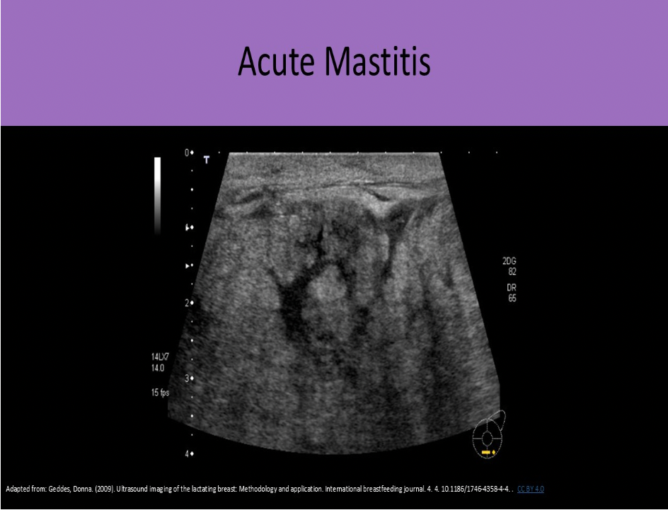
What changes occur on the US image when scanning breast tissue affected by acute mastitis?
A. improved visualization of the ductal system due to tissue edema
B. posterior enhancement due to tissue edema
C. decreased penetration due to tissue edema
D. dilated ductal system within normal breast tissues
C. decreased penetration due to tissue edema
Fluid in the breast tissues will cause an increase in the sound attenuation. This attenuation will reduce penetration and resolution.
Which of the following is a malignant mass of the retroperitoneum that is composed of muscle tissue?
A. Fibrosarcoma
B. Mesothelioma
C. Liposarcoma
D. Rhabdomyosarcoma
D. Rhabdomyosarcoma
Rhabdomyosarcoma - composed of malignant striated muscle tissue, hyperechoic with areas of necrosis
Liposarcoma - malignant fatty mass, most common retroperitoneal neoplasm, hyperechoic with areas of necrosis
Fibrosarcoma - composed of malignant connective tissue, hypochoic, infiltrative mass
Mesothelioma - composed of benign epithelial cells, echogenic mass with irregular margins
You identify free fluid above the right diaphragm that contains internal debris and septations. The fluid collection has a honeycomb appearance.
These findings are most suggestive of?
A. exudative pleural effusion
B. pneumothorax
C. hemothorax
D. transudative pleural effusion
A. exudative pleural effusion
Exudative pleural effusions are usually complex collections with internal debris and septations. They may be multiloculated with a honeycomb appearance.
Transudative pleural effusions are anechoic or hypochoic fluid collection without septations. They are usually seen immediately deep to the chest wall.
What congenital defect presents as a blind-ended, fluid filled bowel loop connected to a normal peristalsing small bowel loop?
A. appendicitis
B. pyloric stenosis
C. Meckel diverticulum
D. epiploic appendagitis
C. Meckel diverticulum
Meckel Diverticulum is a small pouch in the wall of the intestine that develops due to the failure of the vitelline duct to obliterate during fetal development. It is the most common congenital anomaly of the gastrointestinal tract. Most patients are asymptomatic. Gastrointestinal hemorrhage is the most common symptom. Ultrasound may demonstrate a blind-ended, fluid filled bowel loop connected to the normal, peristalsing small bowel segment in the RLQ.
Which of the following correctly describes a mechanical bowel obstruction?
A. best evaluated by US after patient drinks at least 32oz of water
B. adhesions are the most common cause of mechanical obstruction in adults
C. paralysis of the wall muscles inhibit peristalsis and the progression of the luminal contents
D. functional obstruction is much more common than mechanical obstruction
B. adhesions are the most common cause of mechanical obstruction in adults
A functional instruction is caused when paralysis of the wall muscles inhibits peristalsis and the progression of the luminal contents. Mechanical obstruction by luminal contents is much more common than functional. When a mechanical obstruction is suspected, the patient should NOT consume any food or water until they have been evaluated for the blockage. Adding more substances into the Gl tract will only exacerbate their symptoms. A mechanical obstruction indicates that there is a physical impediment blocking the Gl tract.
Sonographic evaluation includes:
Describe location of gut loops
Evaluate the caliber of the segment and size of obstruction
Describe the contents of the segment; solid, fluid, gas
Assess peristalsis
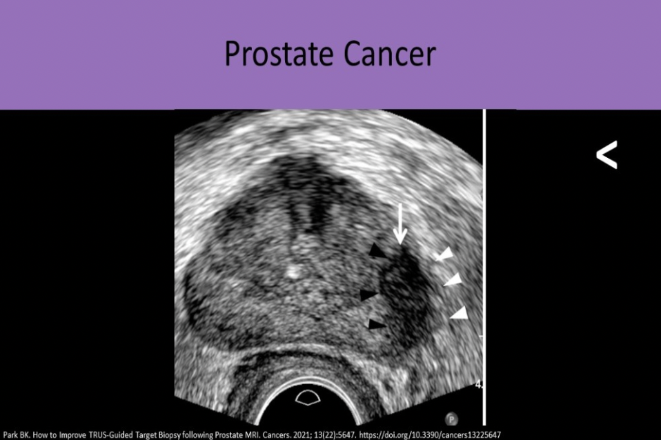
Which zone of the prostate has the highest risk for carcinoma development?
A. peripheral
B. central
C. stromal
D. transitional
A. peripheral
Prostate Cancer Location: 70% peripheral zone, 20% transitional zone, 10% central zone
Bacterial infection, internal bleeding and viral infection typically share what ultrasound finding?
A. retroperitoneal fibrosis
B. free air in the peritoneal cavity
C. abscess formation
D. ascites with debis/septations
D. ascites with debis/septations
Ascites is a common finding with abdominal viral or bacterial infections.
Bleeding of the intraperitoneal organs also causes ascites. Ascites may appear complex and contain debris/septations if there is associated bacterial infection, internal bleeding, or viral infection. Abscess formation may be seen with the infections, but is not typically associated with internal bleeding.
Which of the following statements is true regarding rectus sheath hematomas?
A. An acute hematoma will appear hyperechoic compared to surrounding tissues
B. Linea alba stops the blood from spreading into the pelvis
C. Coumadin therapy is a common cause for hematoma formation
D. Arcuate line stops the blood from extending across the midline
C. Coumadin therapy is a common cause for hematoma formation
An acute hematoma will be anechoic or mildly hypochoic to surrounding tissues. The linea alba stops the blood from crossing the midline, above the level of the arcuate line. Blood is not confined within the sheath below the arcuate line which allows the blood to extend into the pelvis (space of Retzius).
Acute hematomas usually form due to trauma but can be related to coagulotherapy (Coumadin).
Which of the following is NOT required when reporting a suspected hernia?
A. reducibility
B. contents of the hernia sac
C. tenderness at the area of interest
D. suspected age of the hernia
D. suspected age of the hernia
When reporting any type of suspected hernia, you must include the following:
Dynamic maneuvers used in the examination
Presence or absence of a hernia
Hernia size
Hernia contents
Reducibility of the hernia
Tender versus nontender during the exam
How can you differentiate a ganglion cyst from a giant cell tumor of the hand?
A. ganglion cyst will demonstrate posterior enhancement, giant cell tumor will demonstrate posterior shadowing
B. ganglion cyst will demonstrate posterior enhancement, giant cell tumor will not demonstrate enhancement
C. giant cell tumors will move along with the adjacent tendon, ganglion cyst will not move with the adjacent tendon
D. giant cell tumor will demonstrate posterior enhancement, ganglion cyst will not demonstrate enhancement
B. ganglion cyst will demonstrate posterior enhancement, giant cell tumor will not demonstrate enhancement
Because giant cell tumors are soft tissue masses, they will not demonstrate posterior enhancement. Ganglion cysts have the classic signs of a simple cyst on ultrasound, including enhancement. Giant cell tumors do not move with the adjacent tendon but ganglion cysts do move with the adjacent tendon.
Which of the following is true regarding pyloric stenosis?
A. abnormal channel length > 1mm
B. presents as a short thick pyloric canal
C. abnormal muscle thickness >4mm
D. more common in females
C. abnormal muscle thickness >4mm
Pyloric Stenosis most commonly affects males 2-10 weeks of age. Symptoms include projectile vomiting and dehydration. Excessive hypertrophy of the pyloric muscle controlling digestive flow out of stomach can cause obstruction to normal digestive flow.
Results are abnormal when:
Muscle thickness > 4mm
Channel length > 1.2cm
Cross Section > 1.5cm
What penile structure is usually fractured with penile trauma?
A. glans penis
B. corpus spongiosum
C. pampiniform plexus
D. corpus cavernosum
D. corpus cavernosum
The corpus cavernosum on one or both sides can be fractured causing subcutaneous bleeding and significant pain.
Which of the following is true regarding the image of the RUQ of a neonate?
A. The pylorus is thickened and the stomach is demonstrated medially.
B. The pylorus is thickened with an adjacent solid mass.
C. The pylorus is not thickened but there is a fluid collection seen in the stomach.
D. The pylorus and the stomach are both demonstrated normally on the image.
A. The pylorus is thickened and the stomach is demonstrated medially.
Note the excessive thickness of the pyloric walls. The stomach is distended with fluid and food material.
Most patients with adrenal cortical cancer have associated:
A. Conn Disease
B. Budd Chiari Syndrome
C. Cushing Syndrome
D. Addison Disease
C. Cushing Syndrome
Most patients with adrenal cortical cancer have associated Cushing Syndrome.
Malignant ascites is described as:
A. anechoic fluid with free-floating bowel loops and septations
B. complex fluid collection with matted bowel loops
C. anechoic fluid with free-floating bowel loops
D. hyperechoic fluid with septations
B. complex fluid collection with matted bowel loops
Benign ascites - anechoic fluid with free-floating bowel
Malignant ascites - complex fluid collection with matted bowel loops
Which of the following is a common cause of transudate pleural effusion?
A. malignancy
B. pneumonia
C. congestive heart failure
D. tuberculosis
C. congestive heart failure
Transudate fluid has low protein and lactate dehydrogenase concentrations.
Free fluid is pushed outside the capillary bed due to high pressure in the capillary beds. It is usually related to CHF, liver and kidney disease.
Exudate fluid has high protein and lactate dehydrogenase concentration. It is usually related to malignancy, lupus, pneumonia and tuberculosis. Free fluid leaks outside the capillary cells due to inflammation.
The most common benign breast mass is the _____ and the most common malignant breast mass is _____
A. ductal carcinoma in situ, invasive ductal carcinoma
B. acute mastitis, chronic mastitis
C. fibroadenoma, invasive ductal carcinoma
D. lipoma, invasive lobular carcinoma
C. fibroadenoma, invasive ductal carcinoma
A patient presents with right groin pain. You identify a hernia sac medial to the common femoral vein. What type of hernia is present?
A. spigelian
B. direct inguinal
C. umbilical
D. femoral
D. femoral
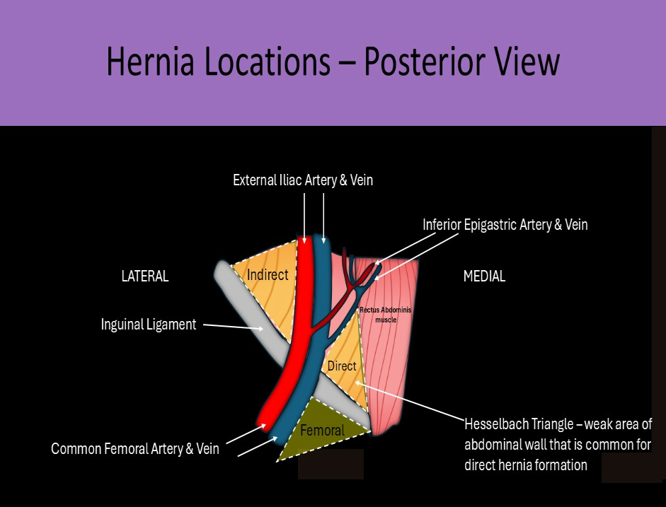
In a femoral hernia, the hernia sac usually lies medial to the common femoral vein. (below the inguinal ligament) Umbilical and spigelian hernias are abdominal hernias. A direct inguinal hernia occurs superior to the inguinal ligament and inferior and medial to the inferior epigastric artery origin
The findings on the image are most suggestive of what penile abnormality?
A. Priapism
B. Penile cancer
C. Peyronie disease
D. Penile fracture
C. Peyronie disease
Peyronie Disease causes the development of scar tissue and fibrous plaque formation that usually involves the tunica albuginea. It causes restriction and curvature of the affected side of the penis during erection and can be very painful. 2D imaging demonstrates hyperechoic areas along the outer margins of the corpus cavernosa.
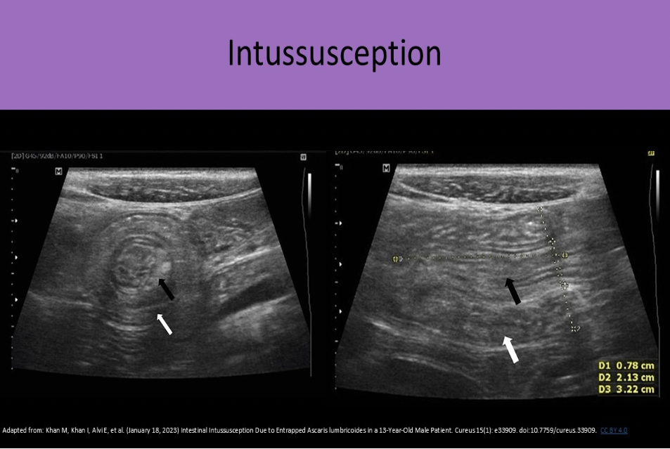
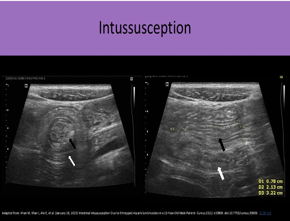
A 2yr old presents with a swollen abdomen and pain for 3 days. The doctor suspects an intussusception is present. How will this appear on the exam?
A. fluid filled colon with multiple intramural masses
B. prominent focal area of concentric rings of bowel
C. fluid filled colon with inflamed walls
D. thickened pylorus muscle
B. prominent focal area of concentric rings of bowel
An intussusception refers to a segment of bowel that involutes into itself. You will see a focal area of concentric rings of bowel that do not persitalse or change shape.
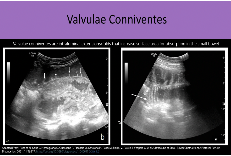
Which portion of the Gl tract contains numerous valvulae conniventes that cause a feathery appearance of the structure on ultrasound?
A. jejunum
B. ilium
C. appendix
D. cecum
A. jejunum
The jejunum is the second portion of small bowel. It contains valvulae conniventes that are intraluminal extensions/folds that increase surface area for absorption. They are abundant in proximal small bowel, but decreased in number in distal small bowel loops. The jejunum has a feathery appearance on ultrasound due to the valvulae conniventes. Valvulae conniventes are more abundant in jejunum than in distal ileum
The image demonstrates a coronal view of the left upper quadrant. Where is free fluid documented on the image?
A. subhepatic space and pleural space
B. subphrenic space and pleural space
C. subphrenic space and splenorenal space
D. subphrenic space and paracolic gutter
B. subphrenic space and pleural space
The image demonstrates a fluid above and below the diaphragm in the LUQ. There is moderate pleural effusion and mild ascites in the subphrenic space.
What is the key anatomic landmark in differentiating direct, indirect and spigelian hernias?
A. rectus abdominis muscles
B. inferior epigastric artery
C. linea alba
D. internal iliac artery
B. inferior epigastric artery
The inferior epigastric artery is the key anatomic landmark in differentiating direct, indirect and spigelian hernias.
Direct inguinal - herniated structures medial to the lEA Indirect Inguinal - herniated structures lateral to the lEA
Spigelian - herniated structures located just lateral to where the spigelian fascia is penetrated by the lEA
Demonstration of gastrohepatic and retroperitoneal lymphadenopathy in a patient is most suggestive of:
A. renal cell carcinoma
B. gallbladder carcinoma
C. irritable bowel syndrome
D. lymphoma
D. lymphoma
Lymphoma is a cancer of the lymphatic system, Finding lymphadenopathy in multiple location in the body is suggestive of lymphoma, instead of a single primary cause
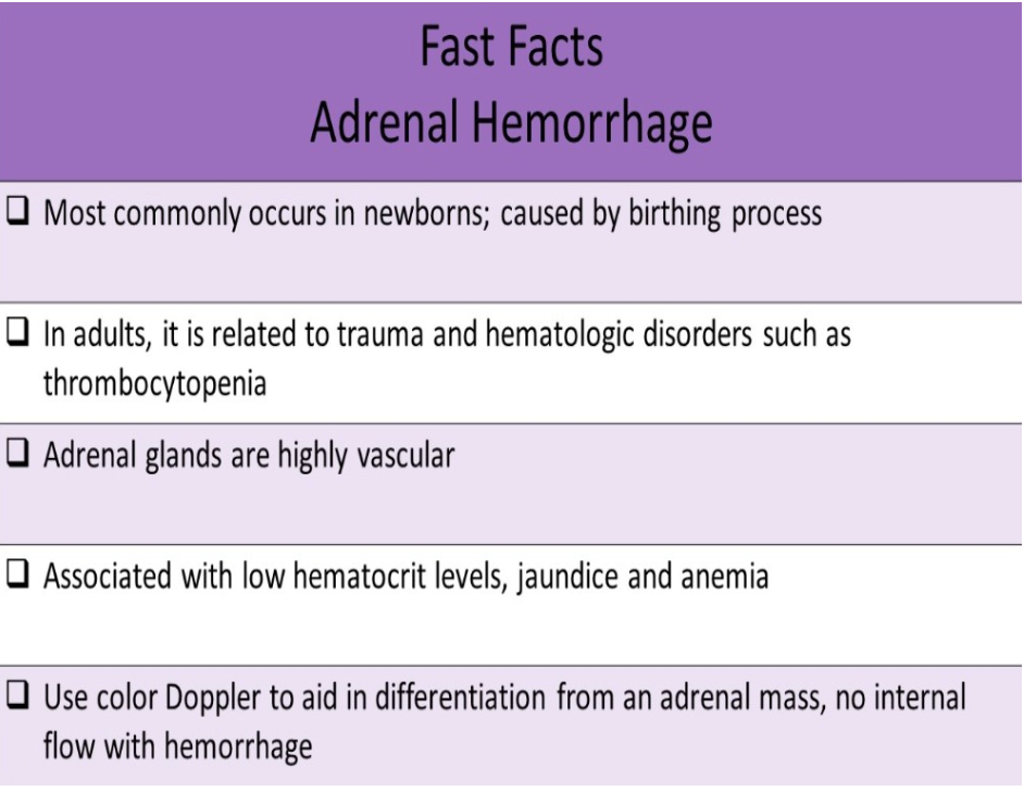
Adrenal gland hemorrhage:
A. in adults is usually related to trauma or hematologic disorders, such as thrombocytopenia
B. is most commonly seen in patients over 70yrs of age
C. is associated with very high hematocrit levels
D. is very rare because the adrenal glands are hypovascular fatty organs
A. in adults is usually related to trauma or hematologic disorders, such as thrombocytopenia
The adrenal glands are highly vascular organs. Adrenal Hemorrhage most commonly occurs in newborns and is caused by the birthing process. In adults, it is related to trauma and hematologic disorders, such as thrombocytopenia. Lab testing demonstrates decreased hematocrit (indicates internal bleeding). The Sonographic characteristics vary with the age of the thrombus.
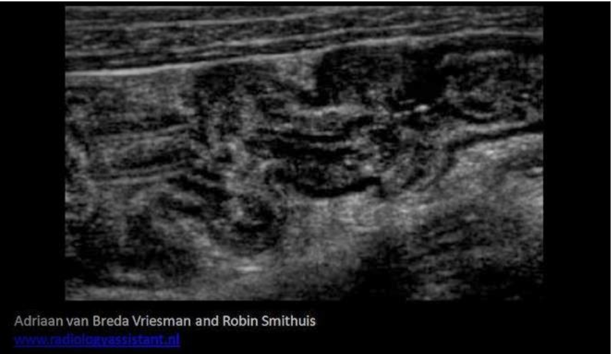
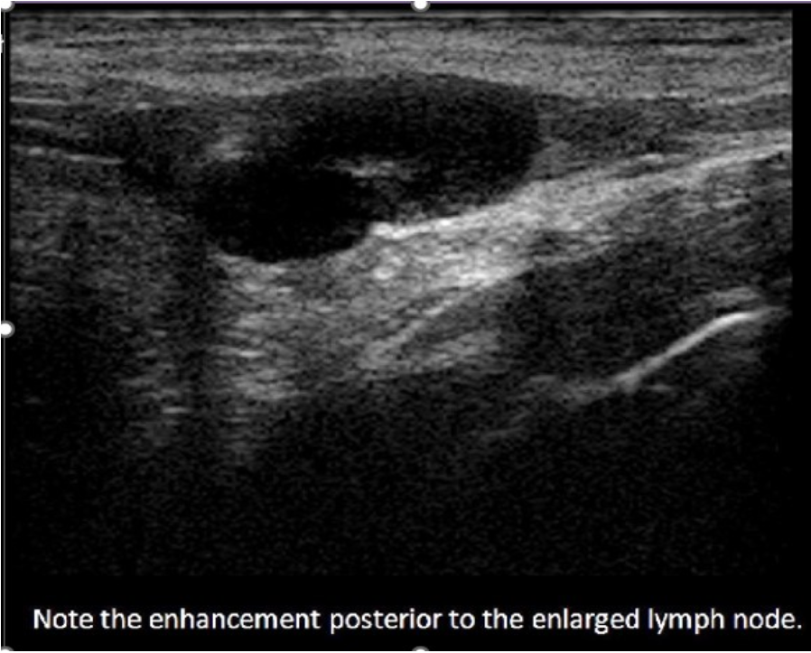
A 50yr old male presents with RLQ pain, watery diarrhea and a low grade fever. The ultrasound exam demonstrates a prominent lymph node in the area of pain and the image below was also obtained at the area of pain. These findings are most consistent with?
A. Colitis
B. Irritable bowel syndrome
C. Intussusception
D. Appendicitis
A. Colitis
The patient symptoms are related to an infection, most likely in the RLQ area of pain. Colitis will cause the thickening of the bowel wall seen on the image. The haustra are visible which indicates a colon segment and not the appendix. Irritable bowel syndrome (IBS) is a diffuse functional disorder of the colon that would not cause a fever or enlarged nodes.
Which of the following is most likely to be confused with a breast carcinoma?
A. sebaceous cyst
B. multiple fibroadenomas in both breast
C. fat necrosis after breast surgery
D. single 5 cm fibroadenoma in the breast
C. fat necrosis after breast surgery
A malignant breast mass demonstrates irregular shape and borders, indistinct margins, hypochoic, hetergeneous, posterior shadowing. Scar tissue can present with similar findings. Fibroadenomas are well-defined oval masses that are hypoechoic and homogeneous. They also demonstrate posterior enhancement.
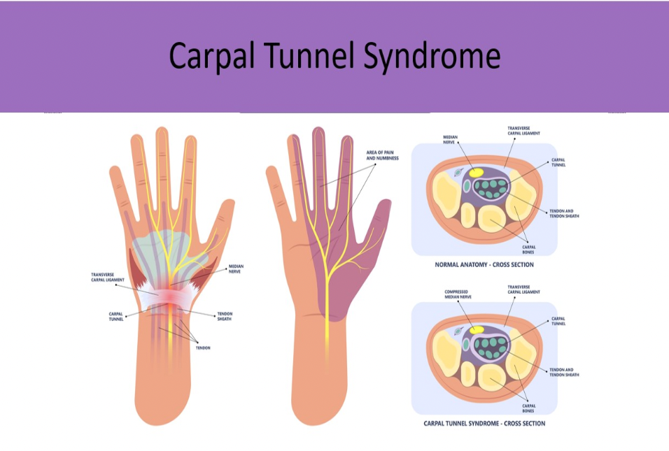
Which of the following describes carpal tunnel syndrome?
A. Compression of the blood vessels as they enter/exit the wrist causing thrombus formation and emboli
B. Damage to the cartilage between the carpal bones of the wrist causing swelling and pain with motion
C. Compression of the median nerve by the carpal sheath causing numbness, weakness and pain
D. Damage to the median nerve by chronic compression between the metacarpal bones of the palm
C. Compression of the median nerve by the carpal sheath causing numbness, weakness and pain
Carpal tunnel syndrome refers to compression of the median nerve by the carpal sheath causing numbness, weakness and pain in the wrist/hand.
_______ is the most common malignant neoplasm found in the prostate.
A. Adenocarcinoma
B. Central zone sarcoma
C. Transitional zone sarcoma
D. Metastasis
A. Adenocarcinoma
Priapism is an abnormality of:
A. the testicles
B. the urinary bladder
C. the penis
D. the prostate
C. the penis
Priapism is an unwanted, persistent, painful erection that lasts more than 4 hours. It can be spontaneous or caused by certain medications.
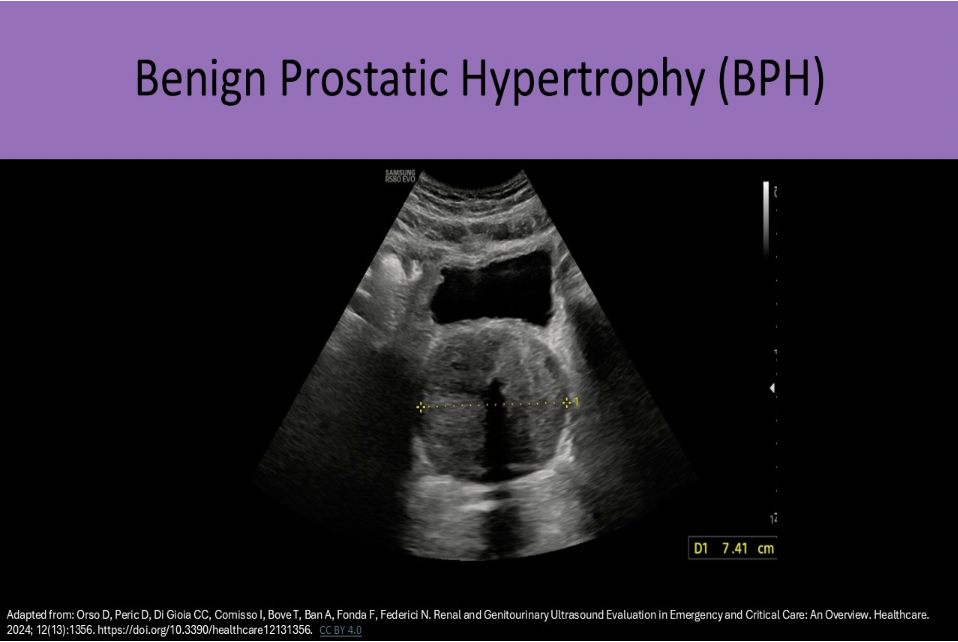
A 70yr old male presents for transrectal ultrasound due to PSA level of 7ng/ml, urinary frequency and hematuria. The US exam demonstrates a 40cc prostate volume with heterogeneity and calcifications centrally. The peripheral zone appears normal. These findings are most consistent with:
A. normal prostate
B. benign prostatic hypertrophy
C. prostatitis
D. carcinoma of the prostate
B. benign prostatic hypertrophy
BPH occurs in the transitional zone. Carcinoma and prostatitis most commonly occur in the peripheral zone. BPH causes increased levels of PSA in the blood. Carcinoma also causes increased PSA (>10ng/ml) but usually much more significantly than BPH.
What is the most common retroperitoneal neoplasm?
A. Мухоma
B. Liposarcoma
C. Rhabdomyosarcoma
D. Fibroma
B. Liposarcoma
Liposarcoma is a malignant fatty mass of the retroperitoneum, It is the most common retroperitoneal neoplasm, Sonographic presentation is a hyperechoic mass with areas of necrosis.
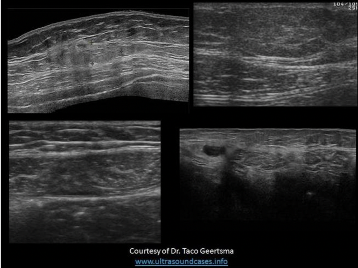
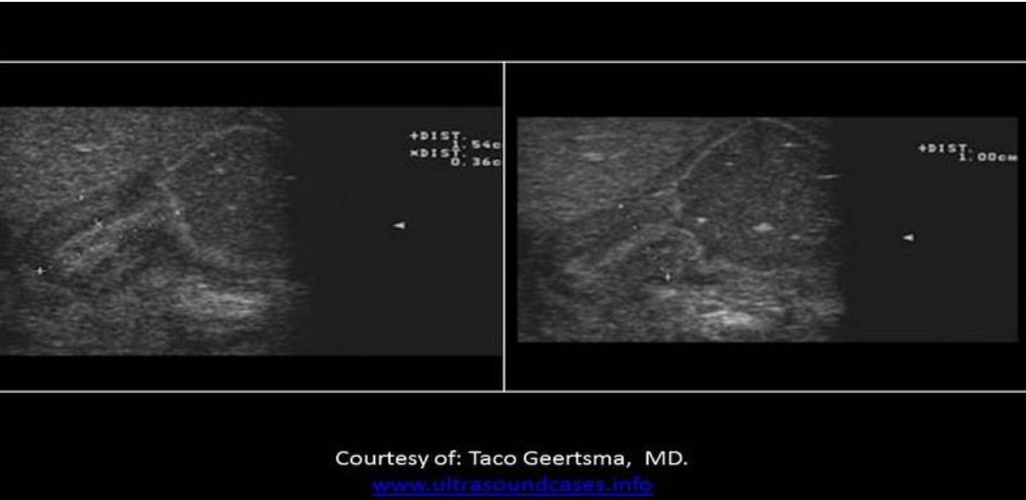
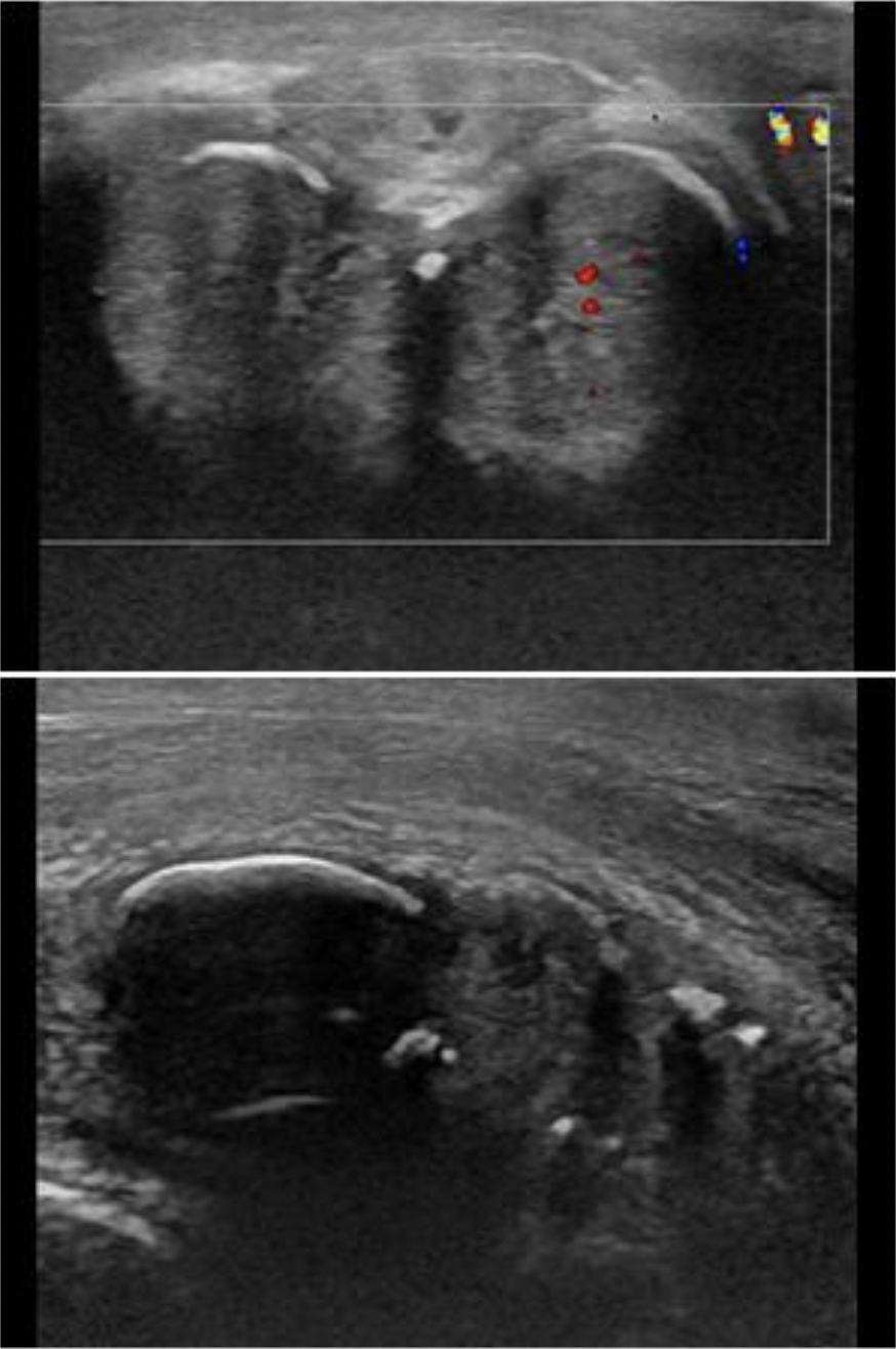
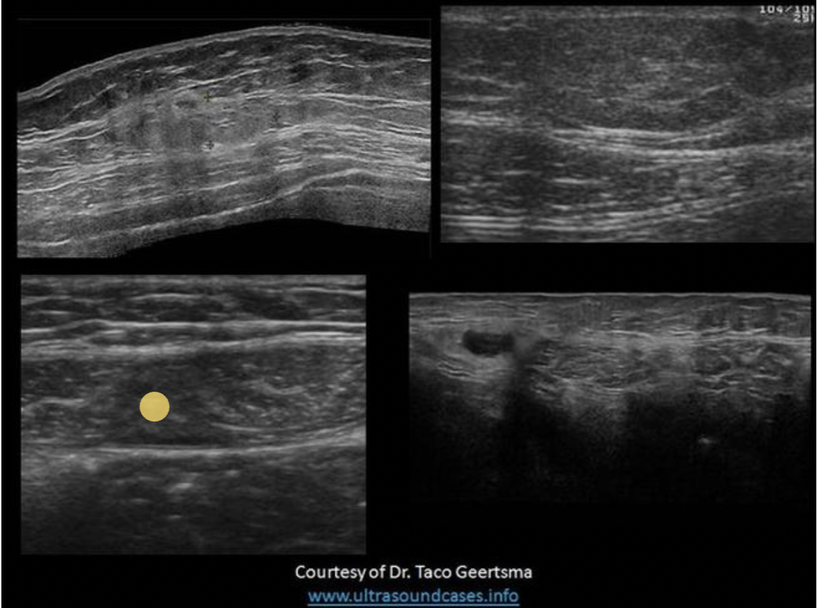
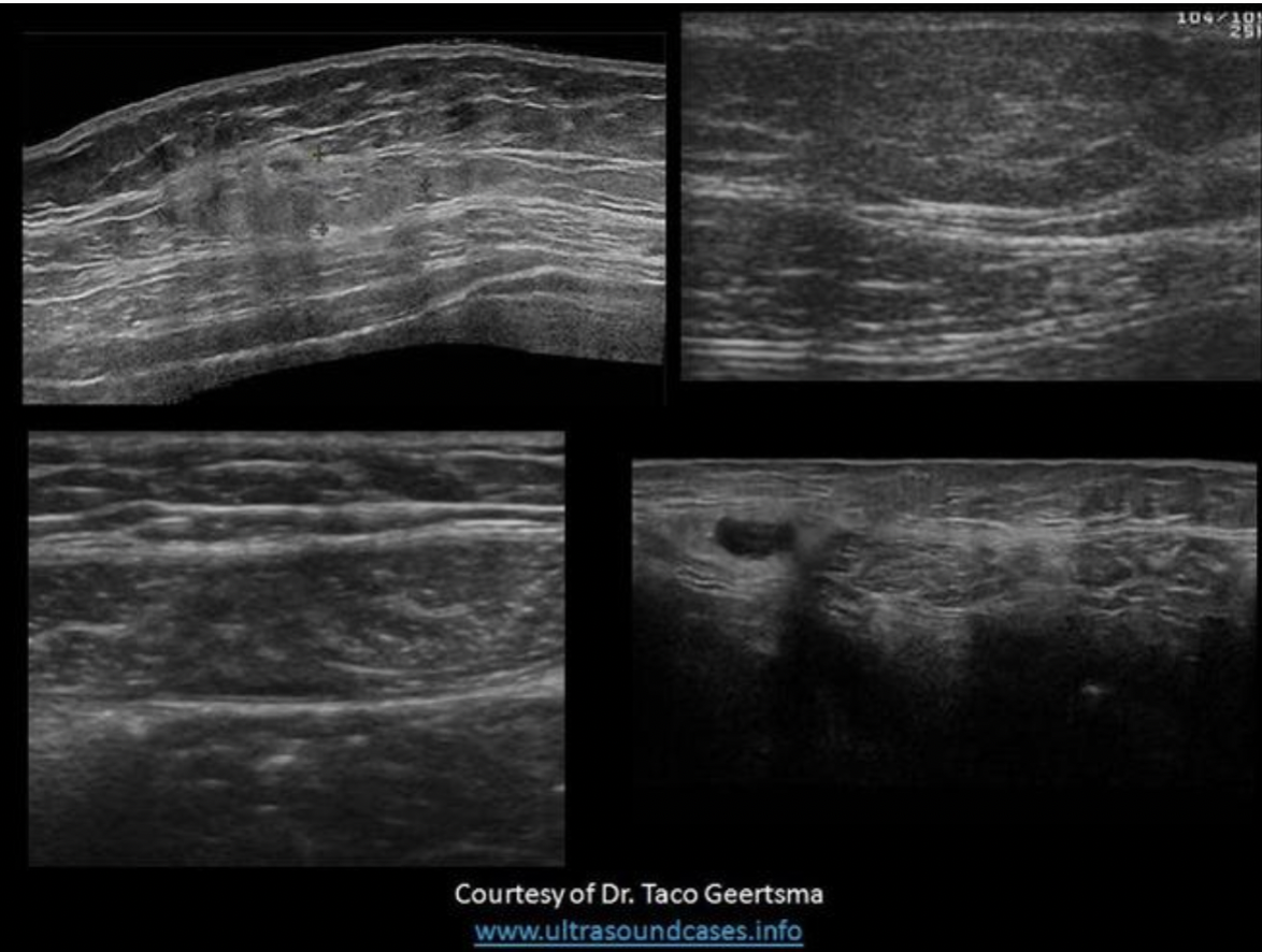
Find the rectus sheath hematoma
Top left is a benign mass with lobulated borders and the top right is a lipoma. Both are located in the subcutaneous tissues/fat between the skin and the anterior rectus sheath. The lipoma has smooth borders and an echogenicity similar to the surrounding fat.
The bottom right image demonstrates a spigelian hernia, between the right rectus abdominis and right oblique muscles.
The bottom left image demonstrates a hematoma in the rectus abdominis muscle, between the anterior and posterior rectus sheaths.
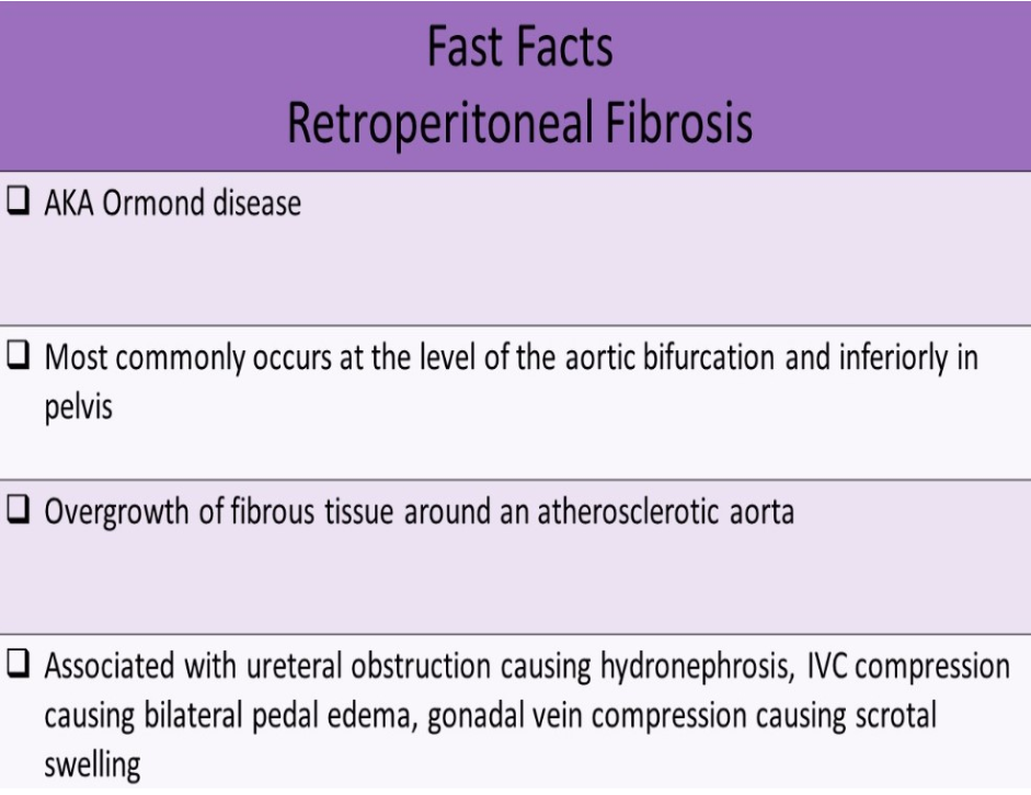
Bilateral ureteral obstruction can be caused by?
A. splenic varices
B. retroperitoneal fibrosis
C. liver mass in the posterior right lobe
D. renal cell carcinoma of the left kidney
B. retroperitoneal fibrosis
Retroperitoneal fibrosis typically occurs at the level of the aortic bifurcation in the pelvis area. The ureters pass through this space to reach the bladder. If both ureters are impinged, bilateral hydronephrosis will occur, The other choices listed would only cause unilateral hydronephrosis.
What causes symptoms in a patient with carpal tunnel syndrome?
A. impingement of the palmar arch
B. impingement of the radial nerve
C. impingement of the median nerve
D. impingement of the ulnar nerve
C. impingement of the median nerve
Carpal tunnel syndrome is caused by impingement of the medial nerve. Patients are evaluated due to symptoms of "pins and needles" in the wrist. The Tinel sign is the tingling or prickling sensation elicited by the percussion of an injured nerve. A positive Tinel sign indicates carpal tunnel syndrome is present at the wrist
Which of the following is a common finding in hepatitis, cirrhosis and hepatic abscess formation?
A. Hepatoma
B. Hydatid cyst
C. Portal HTN
D. Ascites
D. Ascites
Hepatitis and abscess formation are related to infections in the abdomen. Fluid accumulation is a common inflammatory response. Cirrhosis causes decreased liver function and filtration of the Gl tract blood. The excess fluids and waste from the portal system may accumulate within the peritoneal cavity in the form of ascites.
Hepatoma and portal HTN are associated with cirrhosis, but not abscess formation.
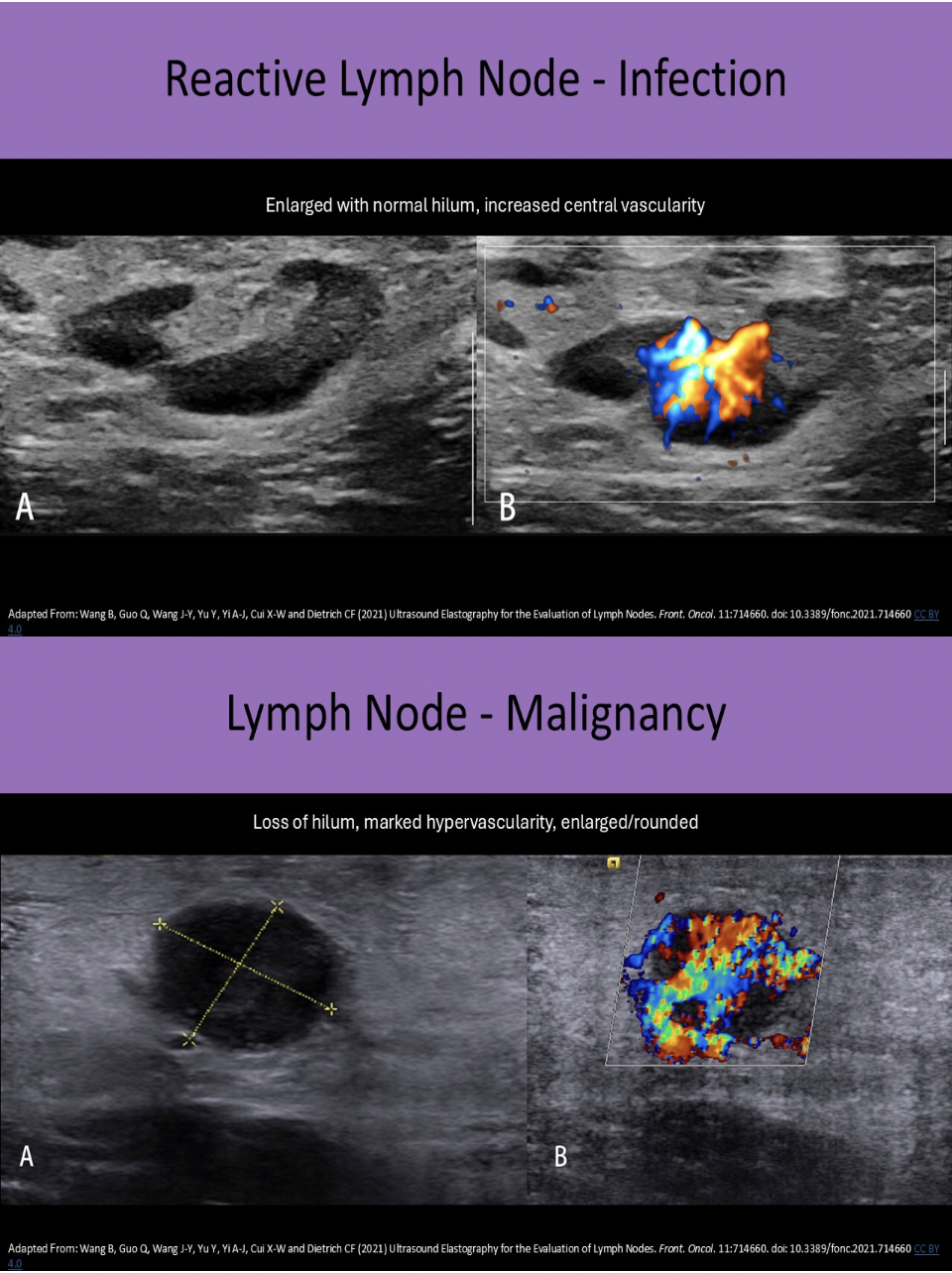
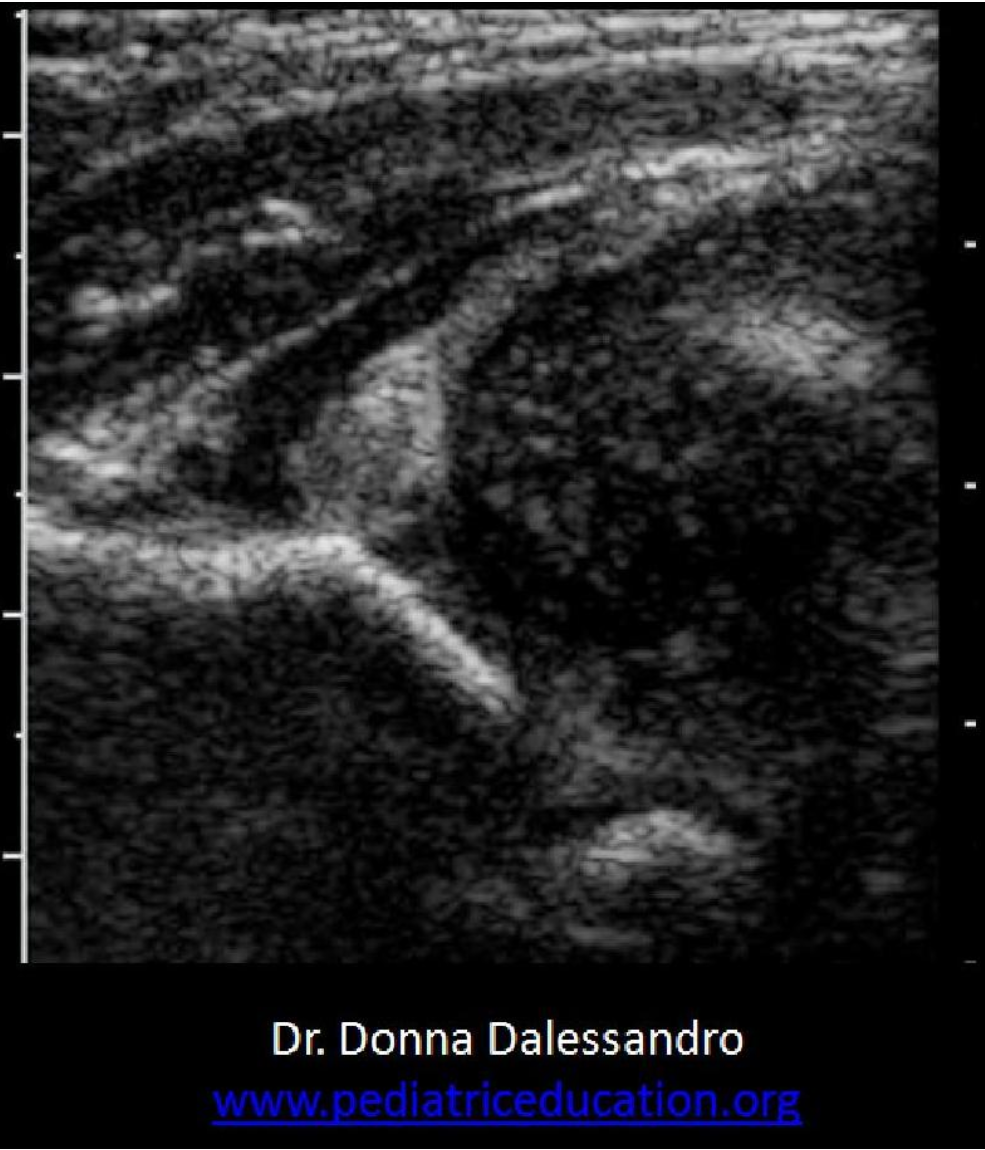
An enlarged, rounded lymph node with loss of hilar definition is most suggestive of associated _____, while an enlarged, oval lymph node with normal hilar characteristics is most suggestive of associated _____
A. viral infection, bacterial infection
B. infection, malignancy
C. malignancy, infection
D. bacterial infection, viral infection
C. malignancy, infection
An enlarged, rounded lymph node with loss of hilar definition = suspicious for underlying malignancy
An enlarged, oval lymph node with normal hilar characteristics = suspicious for underlying infection
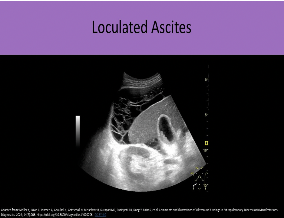
Loculated ascites with echogenic debris and matted bowel loops are most suggestive of:
A. congestive heart failure
B. Budd Chiari syndrome
C. peritoneal metastasis
D. portal hypertension
C. peritoneal metastasis
CHF and portal HTN are associated with transudative ascites. The fluid has no cellular debris or proteins and appears anechoic. Free floating bowel loops are a common finding.
Exudative ascites is associated with infection and malignancy. The fluid has echogenic cellular debris and proteins. Loculations and matted bowel loops are common findings.
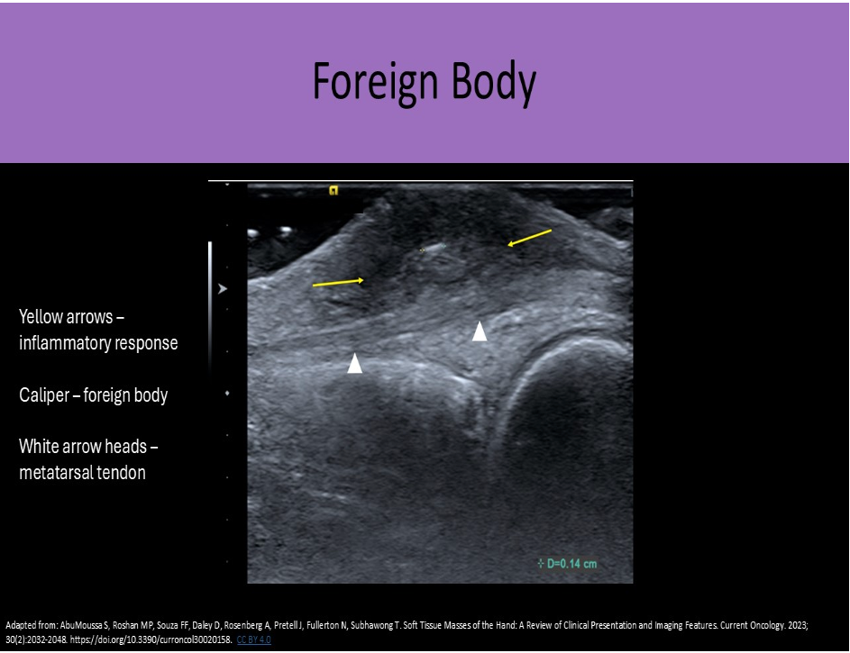
A patient presents for a superficial ultrasound to rule out a foreign body in their foot. Which of the following statements is true regarding the evaluation of a this patient?
A. The radiologist should be consulted because MRI is preferred over Sonography for foreign body detection.
B. Most foreign bodies are echogenic with significant posterior enhancement.
C. Inflammation usually leads to a hypochoic ring surrounding the foreign body.
D. Improved visualization of the foreign body occurs as the US beam becomes more parallel to the structure.
C. Inflammation usually leads to a hypochoic ring surrounding the foreign body.
MRI is contraindicated for some types of foreign bodies (metallic). Most foreign bodies present as an echogenic structure with some degree of posterior shadowing. Inflammation usually leads to a hypochoic ring surrounding the foreign body. Improved visualization of the foreign body occurs as the US beam becomes more perpendicular to the structure.
Adrenal hemorrhage is most commonly seen in ______
A. adults over 50yrs
B. newborns
C. juvenile patients with chromosomal defects
D. adults over 70yrs
B. newborns
Adrenal Hemorrhage usually presents as jaundice and anemia. It most commonly occurs in newborns and is caused by the birthing process. In adults, it is related to trauma and hematologic disorders such as thrombocytopenia. Adrenal glands are highly vascular organs and hemorrhage results in decreased hematocrit (indicates internal bleeding). Sonographic Appearance: Mass with varied echogenicity with age of thrombus
Which statement is true regarding the image displayed?
A. The alpha angle will significantly exceed 60 degrees which indicates dislocation.
B. The infant hip appears normal with normal femoral head location within the acetabulum.
C. The alpha angle appears less than 43 degrees and the femoral head coverage is less than 50% which indicates dislocation.
D. The beta angle appears less than 43 degrees and the femoral head coverage is less than 50% which indicates dislocation.
C. The alpha angle appears less than 43 degrees and the femoral head coverage is less than 50% which indicates dislocation.
Note the position of the femoral head in relationship to the iliac bone. The Graf classification system is used to assess potential dislocation. The femoral head is visibly more than 50% outside of the acetabulum, therefore the femoral head coverage is LESS than 50%. The alpha angle will be less than 43 degrees with dislocation.
Which of the following is caused by a pituitary tumor?
A. Cushing disease
B. Adrenal hemorrhage
C. Myelolipoma
D. Conn syndrome
A. Cushing disease
Cushing Disease is caused by excessive secretion of ACTH by the pituitary gland.
It is related to a pituitary tumor.
Lab Testing: hypersecretion of ACTH by pituitary gland causes increased cortisol production by the adrenal gland; excess cortisol causes increased glucose production (hyperglycemia)
Cushing syndrome is associated with excess cortisol, no matter what the cause.
Steroid use is the most common cause of Cushing syndrome, but adrenal tumors can also be related. Cushing syndrome is characterized by truncal obesity, hirsutism, amenorrhea, HTN, fatigue, hyperglycemia. It is usually caused by excessive cortisol secretion from an adrenal mass.
The term used to describe the involution of the small bowel upon itself is:
A. intussusception
B. colitis
C. Crohn syndrome
D. irritable bowel syndrome
A. intussusception
An intussusception refers to a segment of bowel that involutes into itself. You will see a focal area of concentric rings of bowel that do not persitalse or change shape.
The most common cause of small bowel obstruction in adults is ______. The most common cause of small bowel obstruction in an infant is ______
A. bezoars, pyloric stenosis
B. extrinsic compression by pelvic mass, intussusception
C. adhesions, intussusception
D. diverticulitis, pyloric stenosis
C. adhesions, intussusception
Which of the following statements is true regarding retroperitoneal lymph nodes?
A. Both normal and abnormal lymph nodes exhibit mild posterior shadowing.
B. Normal lymph nodes exhibit posterior enhancement.
C. Abnormal lymph nodes exhibit posterior enhancement.
D. A lymph node is considered abnormally enlarged once it reaches 3cm in length.
C. Abnormal lymph nodes exhibit posterior enhancement.
Normal lymph nodes do not exhibit posterior enhancement. They are composed of mainly fatty tissue and lymphatic tissue which absorb and scatter sound. Abnormal lymph nodes do exhibit posterior enhancement due to the increased fluid within the inflamed tissues. The increased fluid within the tissues allows more sound to be transmitted through the node instead of reflected by the tissues. This leads to the enhancement of the reflection from the tissues posterior to the enlarged node. Lymph nodes larger than 1cm are considered suspicious and those over 2cm are considered abnormally enlarged.
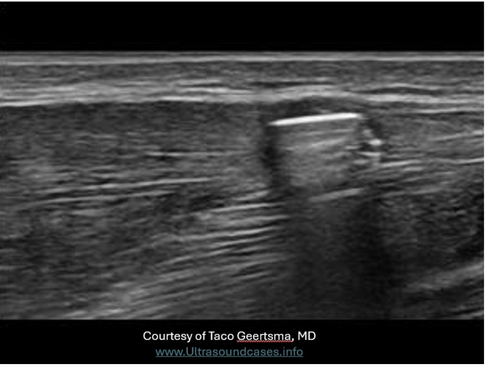
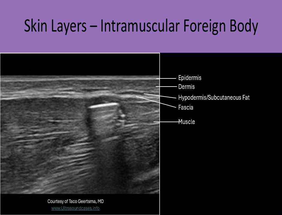
The foreign body on the image is located in what tissue layer of the upper arm?
A. hypodermis
B. dermis
C. muscle
D. epidermis
C. muscle
The ______ zone is the prostate zone most commonly affected by BPH.
A. paraprostatic
B. peripheral
C. transitional
D. central
C. transitional
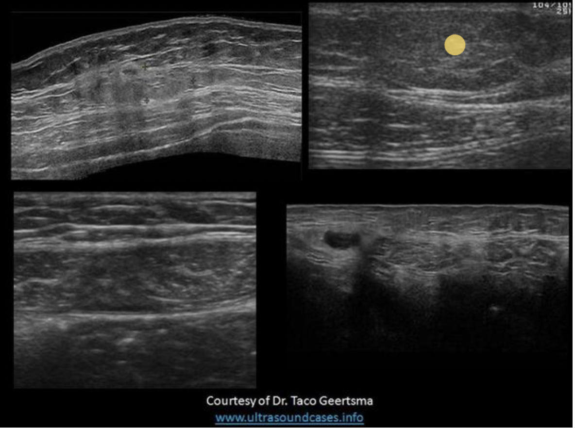
Find the lipoma
The top two images demonstrate solid masses that are superficial to the rectus abdominis muscle. Top left is a benign mass with lobulated borders and the top right is a lipoma. Both are located in the subcutaneous tissues/fat. The lipoma has smooth borders and an echogenicity similar to the surrounding fat.
The bottom right image demonstrates a spigelian hernia, between the right rectus abdominis and right oblique muscles.
The bottom left image demonstrates a hematoma in the rectus abdominis muscle, between the anterior and posterior rectus sheaths.
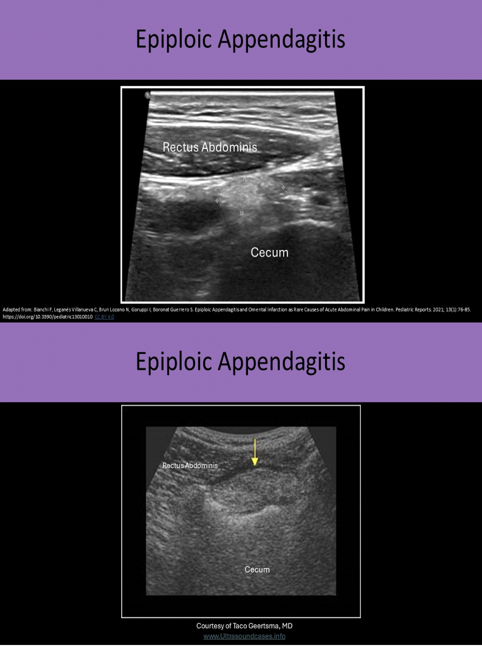
What is a epiploic appendagitis?
A. another term to describe Crohn disease
B. perforated appendix with pericolic fluid
C. a fatal type of diverticulitis
D. inflammation of the thick fatty strands that attach to the serosal surface of the colon
D. inflammation of the thick fatty strands that attach to the serosal surface of the colon
Epiploic appendages are thick fatty strands that attach to the serosal surface of the colon. Torsion or thrombosis of these strands can cause ischemia or infarction. This leads to localized inflammation and pain. On US it appears as an echogenic finger-like projection from the colon wall. The surrounding pericolic fat becomes thickened and echogenic. It can simulate appendicitis and it is very important to differentiate the two because the treatments are very different.
Primary sclerosing cholangitis is usually seen in patients with:
A. pancreatic head mass
B. ulcerative colitis
C. biliary ascariasis
D. HIV infection
B. ulcerative colitis
Bacterial cholangitis is nearly always associated with ductal obstruction (stone or tumor). Bile is infected by gram negative bacteria. Parasitic cholangitis involves liver flukes or ascariasis. May see worms within ducts as echogenic tortuous strands in the lumen. HIV cholangitis occurs in patients with advanced HIV infection. Most patients with primary sclerosing cholangitis have inflammatory bowel disease or ulcerative colitis. It causes chronic inflammation and fibrosis of the biliary ducts.
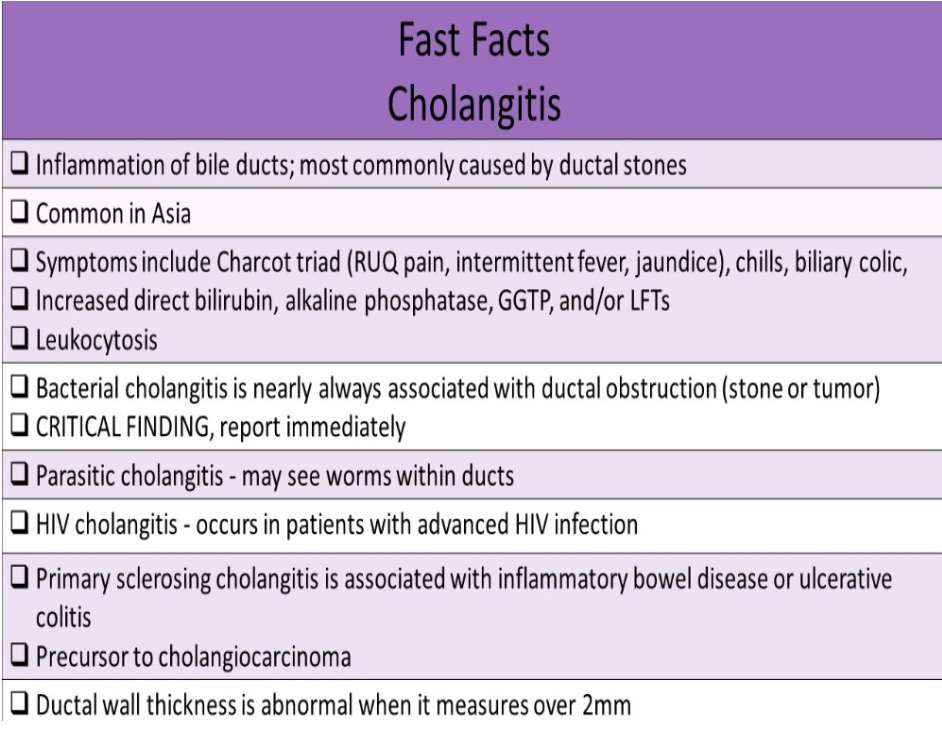
A 3yr old is scanned due to vomiting and a palpable lump in the abdomen.
These findings are most suggestive of:
A. Pyloric stenosis
B. Biliary ascaris
C. Intussusception
D. Appendicitis
C. Intussusception
The image demonstrates intussusception with the the inversion of one portion of the intestine within another. Note the body marker indicates the area of interest is on in the mid to lower quadrant on the left side. The aorta is also visualized on the image. Pyloric stenosis is diagnosed in early infancy and would be located in the epigastric/RUQ area. The aorta would not be demonstrated in the area of the appendix.
What is one of the most common findings on a transrectal ultrasound in prostatitis?
A. heterogeneous gland with hypovascularity
B. homogeneous gland with hypovascularity
C. multiple macrocalcifications
D. hypochoic halo at the periurethral area
D. hypochoic halo at the periurethral area
Prostatitis demonstrates a heterogeneous peripheral gland with a hypoechoic halo at the periurethral area. Abscess formation can occur later. There is marked hypervascularity in tissues affected.
Which of the following correctly describes penile cancer?
A. usually found on the proximal shaft in circumcised men
B. most commonly demonstrates a complex cystic mass
C. associated with inguinal lymphadenopathy
D. most common type of penile cancer is transitional cell carcinoma
C. associated with inguinal lymphadenopathy
Penile Cancer is usually identified on the glans penis on circumcised men or on the foreskin in uncircumcised men. Symptoms include: Focal area of skin thickening or discoloration, Palpable lump, An ulcer that may bleed, A reddish, velvety rash under the foreskin, Small, crusty bumps, Flat, bluish-brown growths, Odorous discharge or bleeding under the foreskin.
Penile cancer is associated with inguinal lymphadenopathy.
The most common cancer to affect the penis is squamous cell carcinoma. If a focal mass is present, it is usually solid and hypochoic with irregular borders and internal vascularity
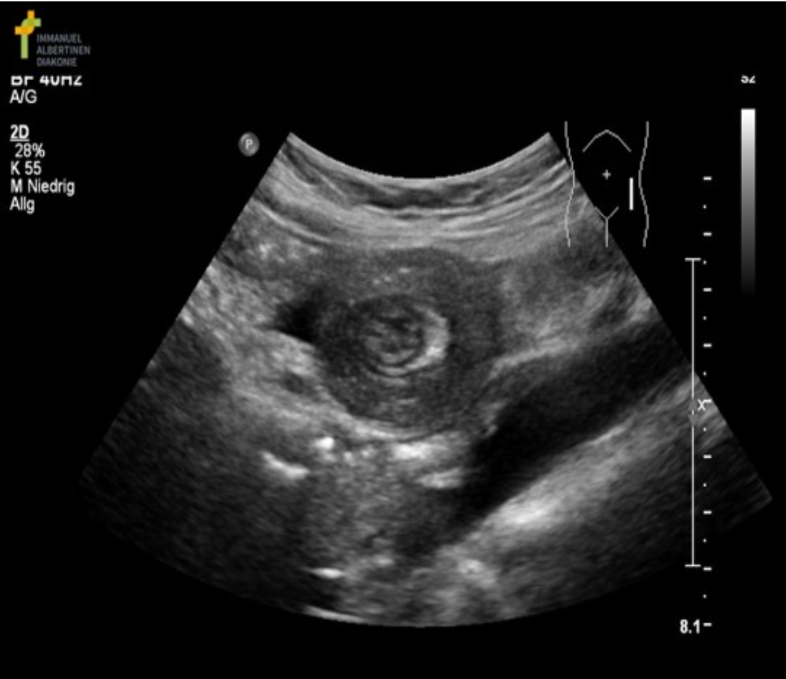
The SMA is identified to the right of the SMV in a patient with acute abdomen symptoms. What should you suspect?
A. Pyloric stenosis
B. Normal anatomy
C. Intussusception
D. Midgut malrotation
D. Midgut malrotation
Midgut Malrotation refers to any variation in the normal rotation and fixation of the Gl tract during development. It is usually associated with malposition of the SMA and SMV. Varices are usually present and the left gastric vein is the most common portosystemic collateral seen with malrotation. Doppler is used to evaluate flow in the SMA, SMV and collaterals. Flow reversal occurs in the left gastric vein. Color Doppler can be used to assess loss of flow to bowel wall.
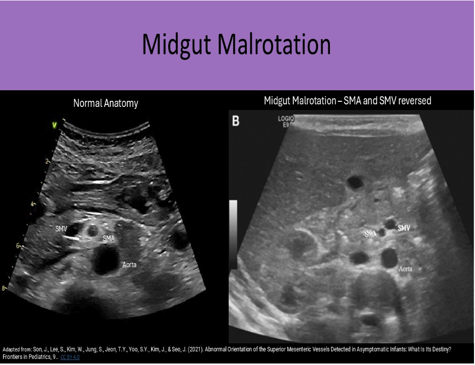
The image demonstrates a transverse view of the right and left side of the abdominal wall, about 5cm lateral to the umbilicus. Which of the following statements is true regarding the findings on the image?
A. There is a heterogeneous, rounded mass located within the left rectus sheath, within the muscle layers and anterior to the peritoneal lining.
B. There is a homogeneous, rounded mass located within the left rectus sheath, anterior to the muscle layers and the peritoneal lining.
C. There is a heterogeneous, rounded mass located within the muscle layers and posterior to the peritoneal lining on the left side.
D. The image demonstrates the normal appearance of the muscle structures of the anterior abdominal wall bilaterally.
A. There is a heterogeneous, rounded mass located within the left rectus sheath, within the muscle layers and anterior to the peritoneal lining.
The image demonstrates a large hematoma within the rectus sheath of the abdominal wall.
Which of the following describes the Sonographic appearance of adrenal hyperplasia?
A. multiple small echogenic nodules in the affected gland
B. bilateral, diffuse gland enlargement
C. complex cystic mass formation in both glands
D. thickened, echogenic cortex and loss of differentiation with the medulla
B. bilateral, diffuse gland enlargement
Adrenal hyperplasia presents as bilateral, diffuse gland enlargement due to the hyperfunction.
Which of the following is not usually treated by surgical intervention?
A. spigelian hernia
B. pyloric stenosis
C. appendicitis
D. intussusception
D. intussusception
Intussusception is treated by nonsurgical reduction. Air or contrast is administered through the rectum (enema) to push the involuted bowel back out of the colon. The other choices all require surgical treatment.
The Achilles' tendon most commonly ruptures:
A. at the proximal insertion point
B. mid-tendon
C. about 4 cm from the distal insertion point
D. at the distal insertion point
C. about 4 cm from the distal insertion point
The Achilles' tendon most commonly ruptures about 4 cm from the distal insertion point at the calcaneus.
_____ is acute symptomatic inflammation of a tendon, while ______ refers to asymptomatic degenerative changes in the tendon
A. Sprain, Tear
B. Tear, Sprain
C. Tendinitis, Tendinosis
D. Tendinosis, Tendinitis
C. Tendinitis, Tendinosis
If an inguinal hernia does not increase in size with Valsalva strain, ______ should be suspected.
A. malignancy
B. spontaneous closure
C. strangulation
D. infection
C. strangulation
If the hernia does not increase in size with strain, incarceration/strangulation should be suspected.