Complete collection of IB Biology Topic 1 information as prescribed by the for the May 2024 Exam Syllabus. Majority adopted straight from Old-Bio Ninja, includes majority of the "Extra Content" prescribed by Old-Bio Ninja as well.
Functions of Life (MR SHENG)
Metabolism
Undertakes essential chemical reactions
Reproduction
Produces offspring sexually or asexually
Sensitivity
Responsive to internal and external stimuli
Homeostasis
Maintains stable inner environment
Excretion
Able to remove toxic waste products
Nutrition
Exchanges material with the environment
Growth/movement
Changes shape/size/position
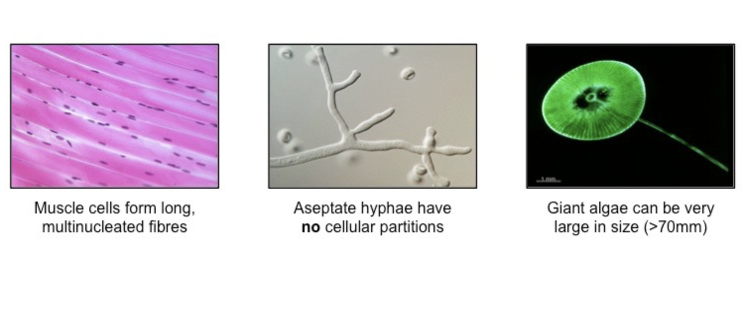
Cell Theory Exceptions
Striated Muscle
Individual cells fuse to form long multi-nucleated fibres
Challenges idea that cells always function as autonomous units
Giant Algae
Certain species can be very large
Challenges idea that larger organisms are always made up of many microscopic cells
Aseptate Fungal Hypae
Hyphae may be connected by a continuous cytoplasm
Challenges idea that living structures are composed of discrete cells
Cell Theory (3 points)
Living organisms are composed of cells (or cell products)
The cell is the smallest unit of independent life
Cells can only arise from pre-existing cells (mitosis/meioses)
Functions of life in Autotroph (Scenedesmus)
Chlorophyll pigments allow organic molecules to be produced via photosynthesis
Metabolism
Daughter cells form as non-motile autospores via the internal asexual division of the parent cell
Reproduction
Scenedesmus may exist as unicells or form colonies for protection
Sensitivity / Responsiveness
Scenedesmus exchange gases and other essential materials via diffusion
Nutrition and Excretion
Functions of life in Heterotroph (Paramecium)
Paramecium (Heterotroph)
Food particles are enclosed within small vacuoles that contain enzymes for digestion
Metabolism
Paramecia divide asexually through fission, however horizontal gene transfer can occur via conjugation
Reproduction
Surrounded by small hairs (cilia) which allow it to move
Sensitivity/Responsiveness
Essential gases enter (Oxygen) exit (Carbon Dioxide) the cell to keep balance via diffusion
Homeostasis
Solid wastes are removed via an anal pore, while liquid wastes are pumped out via contractile vacuoles
Excretion
Paramecia engulf food via a specialised membranous feeding groove called a cytosome
Nutrition
Autotroph
An organism that can produce its own food using light, carbon dioxide, or other chemicals.
Heterotroph
An organism that eats other plants or animals for energy and nutrients.
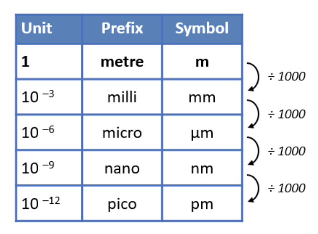
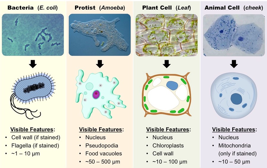
Cell Scale
Cells and their components are measured according to the metric system
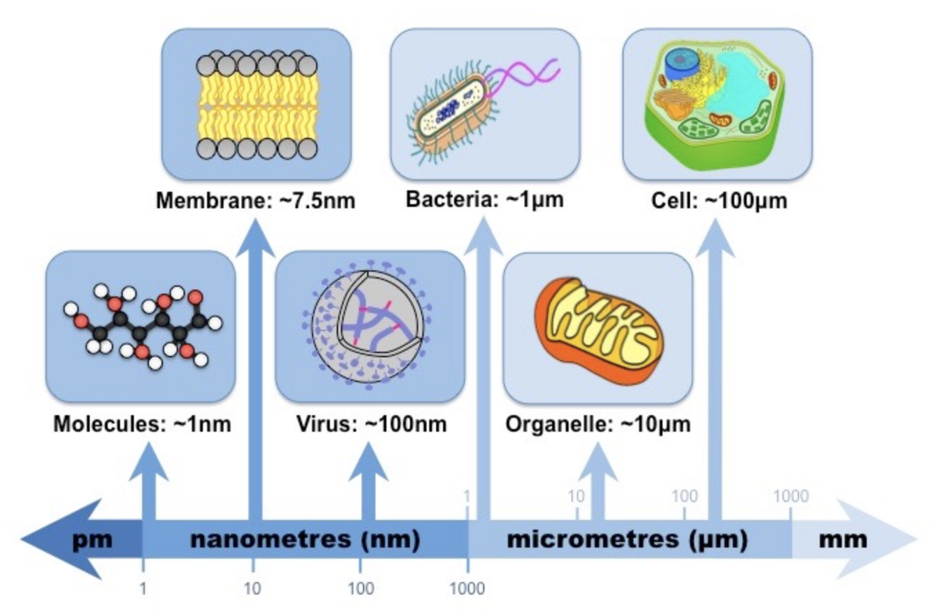
Relative Sizes of Biological Material
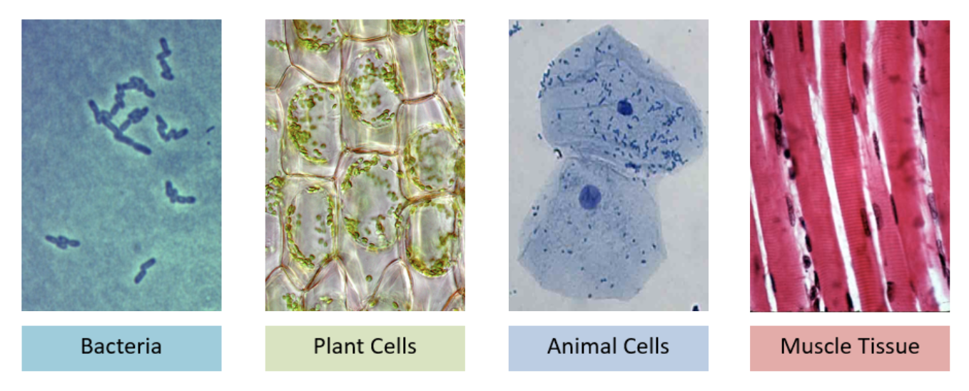
Microscopes Function
Objects that are too small for the naked eye may be visualised with microscopes
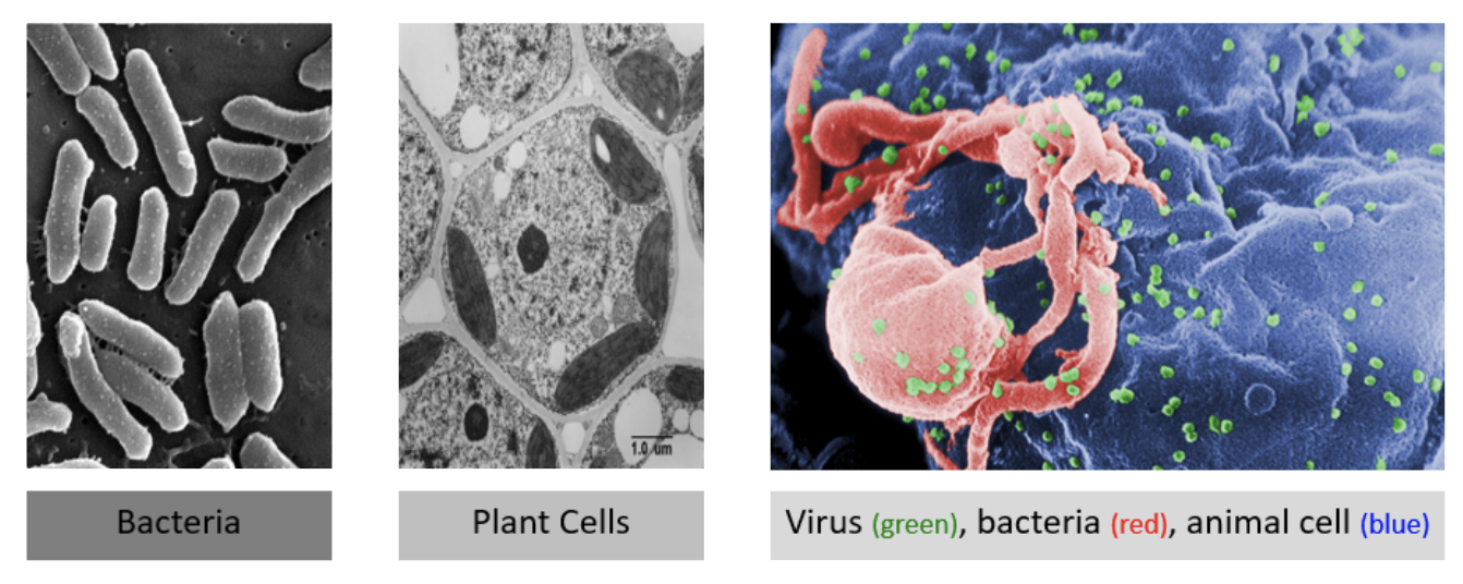
Types of Microscopy: Light v.s. Electron
Light Microscopy
Views living specimens in natural colour
Lower resolution and magnification
Electron Microscopy
Views dead specimens in monochrome
Has a higher resolution and magnification
Cell Structures under Light Microscopy
Cell Structures under Electron Microscopy
Magnification Formula
MIA
Magnification = Imagine Size/Actual Size
M = I / A
A = I / M
Function of Cell’s Surface Area vs Volume
Volume / Mass
The rate of metabolism of a cell is a function of its mass (larger cell needs more energy)
Surface Area
The rate of material exchange is a function of its surface area (large membrane surface equates to more material movement)
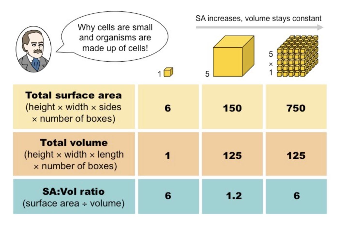
SA:Vol Ratio and Cell Growth
As a cell grows volume increases faster than surface area, leading to decreased SA:Vol ratio
If metabolic rate > rate of vital material exchange cell dies
Hence growing cells tend to divide and remain small to maintain high SA:Vol ratio
This way Volume stays constant with growth and SA can increase
High SA:Vol ratio is important to what functions
Cells and tissues specialised for gas exchange will increase surface area to optimise material transfer
Villi of intestinal tissue
To maximise nutrient exchange from digestive tract to blood stream
Microvilli in alveolar cells
To maximise gas exchange in lungs for consistent oxygen sypply
Drawing Microscopic Structures: Conventions of Diagram
A title should be included to identify the specimen (e.g. name of organism, tissue or cell)
A magnification or scale should be included to indicate relative size
Identifiable structures should be clearly labelled (drawings should only reflect what is seen, not idealised versions)
Emergent Properties
Arises when the interaction of individual components produce new functions.
Help living organisms better adapt to their environments and increase their chances of survival.
“Emergent Property” is a characteristic an entity gains when it becomes part of a bigger system.
Multicellular Organisms: Function / Purpose
Multicellullar organisms are capable of completing functions that unicellular organisms could not undertake due to the collective actions of individual cells combining to create new synergistic effects
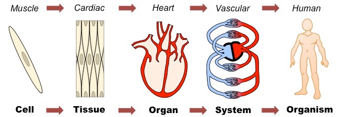
Multicellular Organisms: Organisation
Cells may be grouped together to form tissues
Organs are then formed from the functional grouping of multiple tissues
Organs that interact may form organ systems capable of carrying out specific body function
Organ systems collectively carry out the life functions of the complete organism
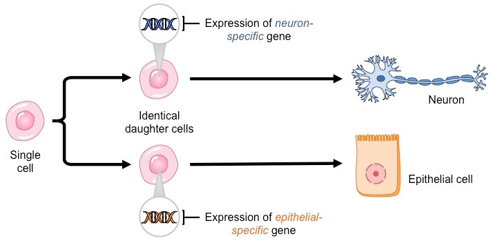
Cell Differtiation: Definition
Differentiation is the process during development whereby newly formed cells become more specialised and distinct from one another as they mature.
Cell Differentiation: Process
All cells in a multicellular organism share an identical genome
Each cell contains entire set of genetic instructions for that organism
The activation of specific genes within a cell will cause it to differentiate
Activation occurs by chemical signals
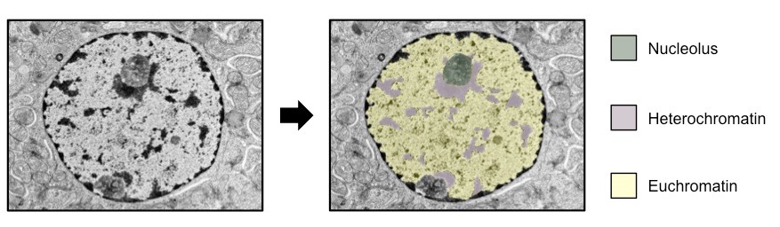
Gene Packaging
Inside nucleus of eukaryotic cells, DNA is packaged with proteins to form chromatin
Active genes - Packaged in expanded form called euchromatin
Inactive genes - Packaged in more condensed form called heterochromatin (saves space, not transcribed)
Differentiated cells will have different regions of DNA packaged as euchromatin and heterochromatin according to their specific function.
Stem Cells: Definition and Key Qualities
Stem cells are unspecialised cells that have two key qualities:
Self Renewal - They can continuously divide and replicate
Potency - They have the capacity to differentiate into specialised cell types
Necessary for embryonic development as they are an undifferentiated cell source from which all other cell types may be derive
Types of Stem Cells and Non-Stem Cells
Totipotent - Can form any cell type including extra-embryonic tissue (eg. zygote)
Pluripotent - Can form any cell type (eg. embryonic stem cells)
Multipotent - Can differentiate into a number of closely related cell types (eg haematopoietic adult stem cells)
Unipotent - Cannot differentiate, but are capable of self-renewal (eg. muscle stem cells)
Non-Stem Cells - Cells not capable of self-renewal (eg. amitotic nerve tissues). Cannot regenerate or replace, therefore damage often calls for stem cell therapy
Stem Cell Therapy: Process
Use of biochemical solutions to trigger differentiation of stem cells into desired cell type
Surgical implantation of cells into the patient’s own tissue
Suppression of host immune system to prevent rejection of cells (if stem cells are from foreign source)
Careful monitoring of new cells to ensure they do not become cancerous
Stem Cell Therapy Example: Stargardt’s Disease
What is Stargardt’s Disease
Inherited form of juvenile macular degeneration (early vision loss)
Causes progression vision loss to point of blindness
Caused by a gene mutation that impairs energy transport in retinal photoreceptor cells, causing them to degenerate
Stem Cell Treatment
Treated by replacing dead cells in retina with functioning ones derived from stem cells
Stem Cell Therapy Example: Parkinson’s Disease
What is Parkinson’s Disease
A degenerative disorder of the central nervous system caused by the death of dopamine-secreting cells in the midbrain
Dopamine is a neurotransmitter responsible for transmitting signals involved in the production of smooth, purposeful movements
Consequently, individuals with Parkinson’s disease typically exhibit tremors, rigidity, slowness of movement and postural instability.
Stem Cell Treatment
Treated by replacing dead nerve cells with living, dopamine producing ones.
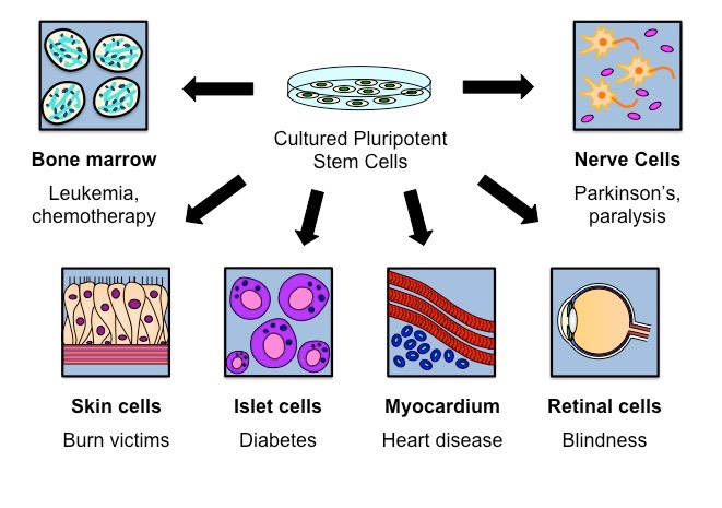
Stem Cell Therapy Examples: Others
Leukemia: Bone marrow transplants for cancer patients who are immunocompromised as a result of chemotherapy
Paraplegia: Repair damage caused by spinal injuries to enable paralysed victims to regain movement
Diabetes: Replace non-functioning islet cells with those capable of producing insulin in type I diabetics
Burn victims: Graft new skin cells to replace damaged tissue
Stem Cells: Sources
Embryos - may be specifically created by therapeutic cloning
Umbilical cord blood or placenta of new-born
Certain adult tissues like bone marrow (not pluripotent)
Reasons for the therapeutic use of Stem Cells
Unspecialized/undifferentiated stem cells can divide / differentiate along different pathways; (Qualities of Stem Cells)
Stem cells are accessible as they come from embryos/bone marrow/umbilical cord blood/adult tissue; (Accessible Sources)
Stem cells can regenerate/repair diseased/damaged tissues in people;
valid specific example; (Qualities of Stem Cells)
drugs can be tested on stem cells (in laboratories to see if they are harmful); (Reduced Human Risk)
Stem Cell Therapy: Ethical Considerations
Limited scope of application - Multipotent adult tissue only effective for certain conditions
Availability and Access - Stem cells derived from umbilical cord blood need to be stored and preserved at cost (expensive and limited access)
Embryo Destruction - Embryos hold the greatest yield of pluripotent stem cells, but their use means the destruction of a potential living organism
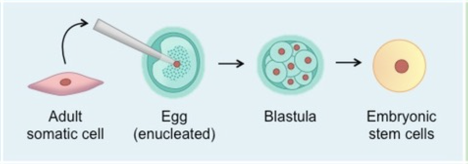
Artificial Stem Cell Techniques: Somatic Cell Nuclear Transfer (SCNT)
Involves the creation of embryonic clones by fusing a diploid nucleus with an enucleated egg cell (therapeutic cloning)
Advantages:
Indistinguishable from embryo-derived cells
Meaning Totipotent Stem Cells can be derived
Disadvantages:
Involves ex vivo (out of body) creation of embryos
More embryos are created than needed, raising ethical concerns about the exigency of excess embryos
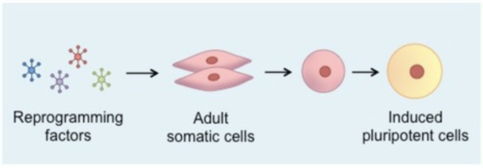
Artificial Stem Cell Techniques: Nuclear Reprogramming
Induce a change in the gene expression profile of a cell in order to transform it into a different cell type (transdifferentiation)
Advantages:
Stem cells are autologous to adult doner (derived from same person needing them) so no risk of incompatibility
Disadvantages:
Involves the use of oncogenic retroviruses and transgenes, increasing the risk of health consequences (ie. cancer)
Prokaryotic Cells: Definition, How they divide, and Classification
Prokaryotes are organisms whose cells lack a nucleus, they divide by Binary Fission. They typically do not possess any membrane-bound organelles.
Kingdom: Monera
Domains:
Archaebacteria - found in extreme environments like high temperatures, salt concentrations or pH (ie. extremophiles)
Eubacteria - traditional bacteria including most known pathogenic forms (ie. E. coli, S. aureus, etc.)
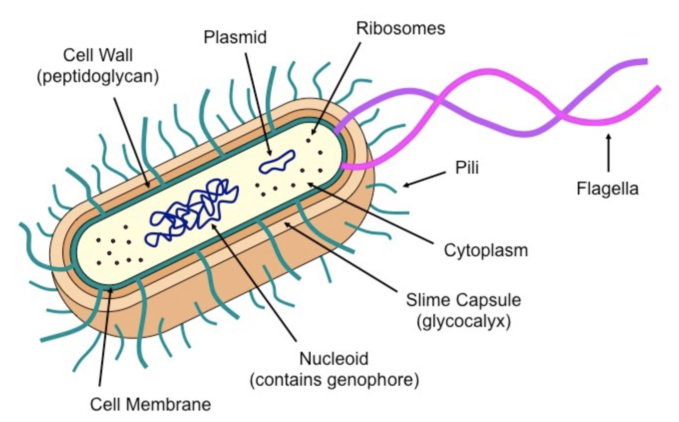
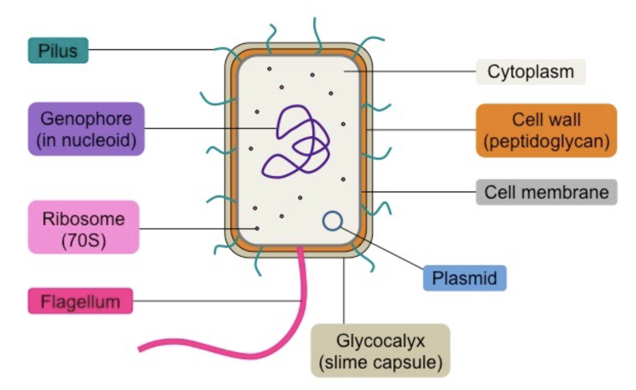
Prokaryotic Cells: Features
Cytoplasm: Internal fluid component of the cell
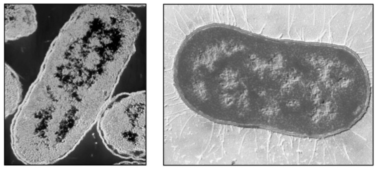
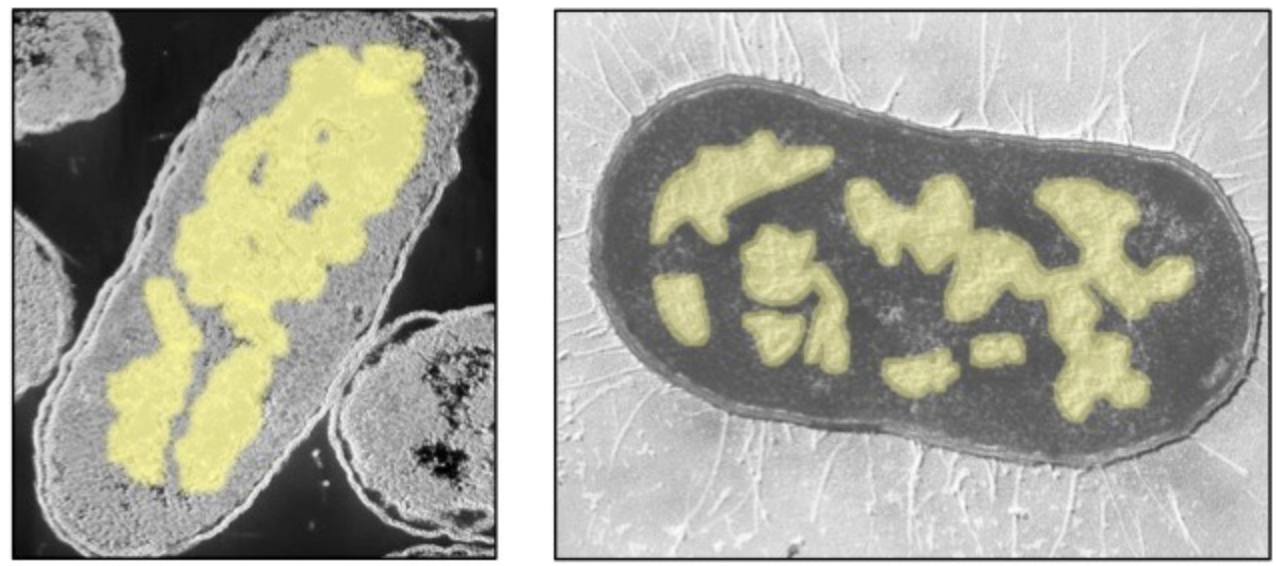
Nucleoid: region of the cytoplasm where the DNA is located (DNA is circular and called “Genophore”)
Plasmids: autonomous circular DNA molecules that may be transferred between bacteria (horizontal gene transfer)
Ribosomes: complexes of RNA and protein that are responsible for polypeptide synthesis (prokaryote ribosome = 70S)
Cell membrane: Semi-permeable and selective barrier surrounding the cell
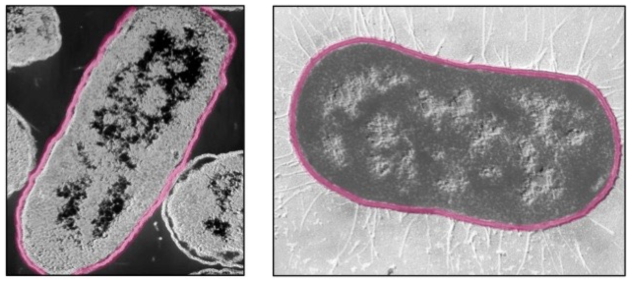
Cell wall: Rigid outer covering made of peptidoglycan; maintains shape and prevents bursting (lysis)
Slime capsule: a thick polysaccharide layer used for protection against desiccation (drying out) and phagocytosis
Flagella: Long, slender projections containing a motor protein that enables movement (singular: flagellum)
Pili: Hair-like extensions that enable adherence to surfaces (attachment pili) or mediate bacterial conjugation (sex pili)
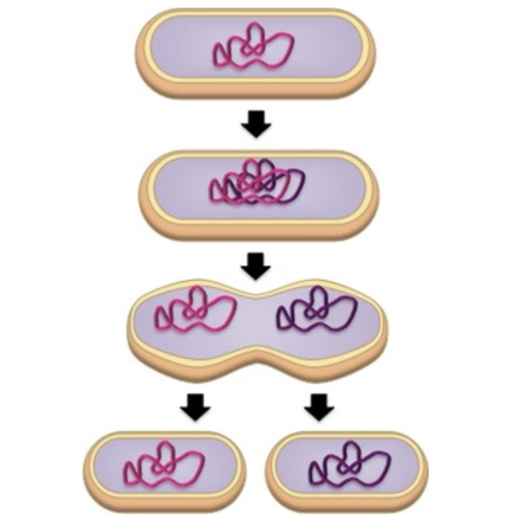
Binary Fission: Definition and Process
A form of asexual reproduction used by prokaryotic cells.
Process:
The circular DNA is copied in response to a replication signal
The two DNA loops attach to the membrane
The membrane elongates and pinches off (cytokinesis) forming two cells
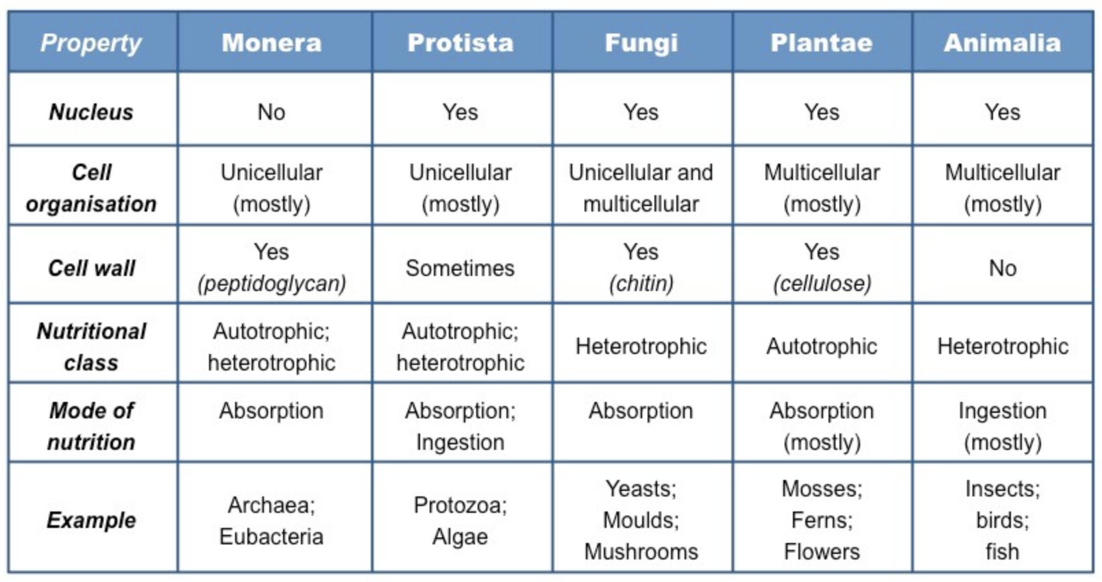
Eukaryotic Cells: Definition and Classification
Eukaryotes are organisms whose cells contain a nucleus and several membrane-bound organelles.
They have more complex structures and are believed to have evolved from prokaryotic cells.
Compartmentalised by membrane-bound structures that perform specific roles.
Kingdoms:
Protista: Unicellular organisms; or multicellular organisms without specialised tissue
Fungi: have a cell wall made of chitin and obtain nutrition via heterotrophic absorption
Plantae: have a cell wall made of cellulose and obtain nutrition autotrophically (via photosynthesis)
Animalia: no cell wall and obtain nutrition via heterotrophic ingestion
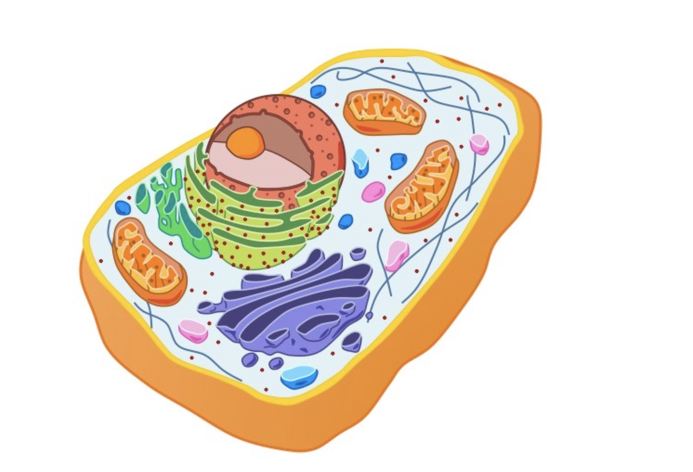
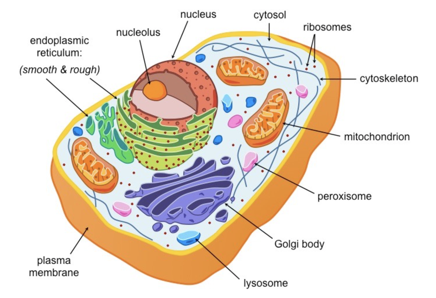
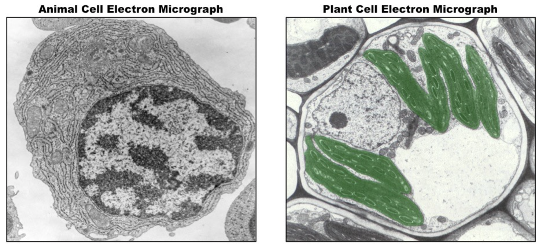
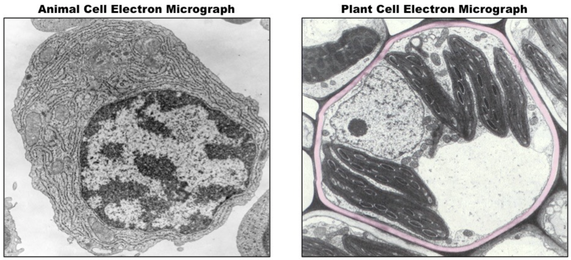
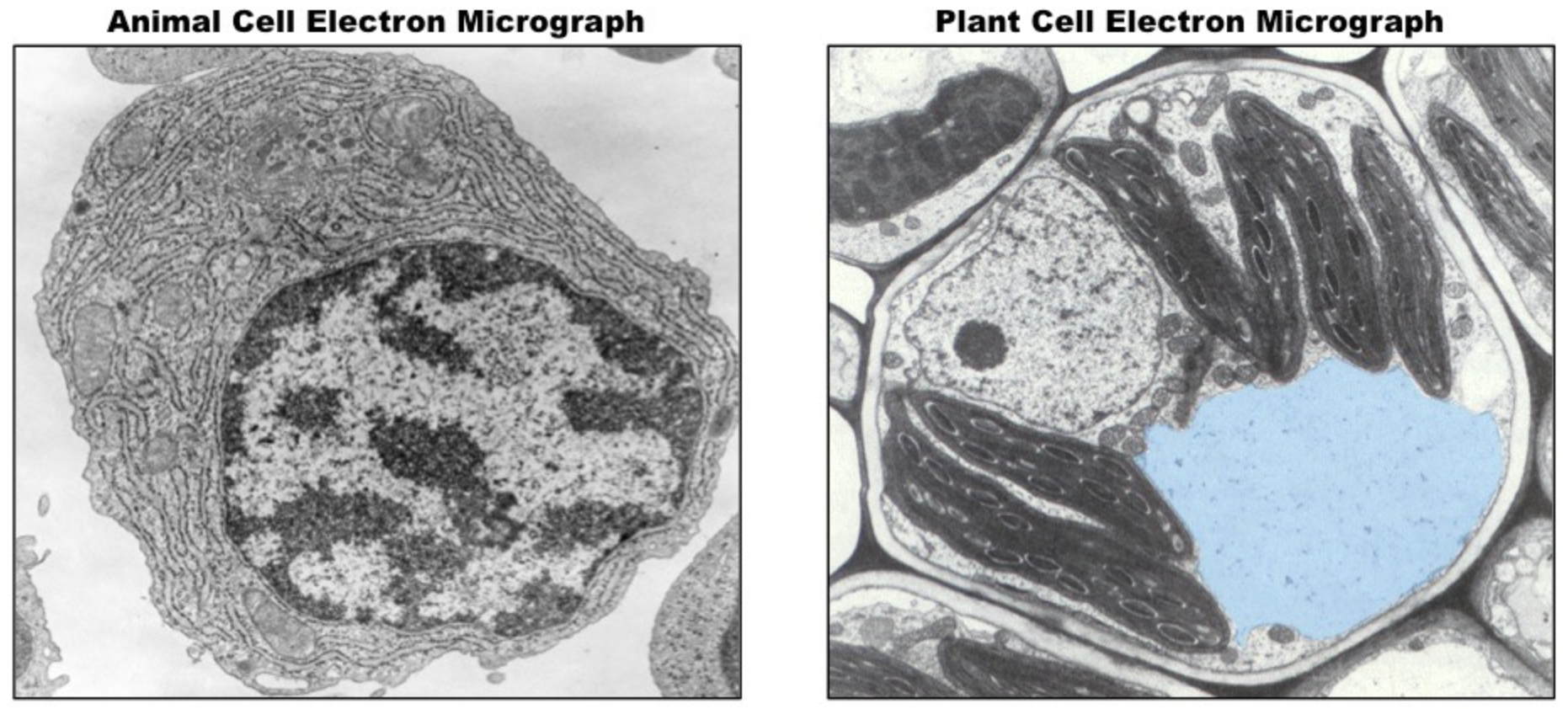
Animal Cell: Typical Structure and Features
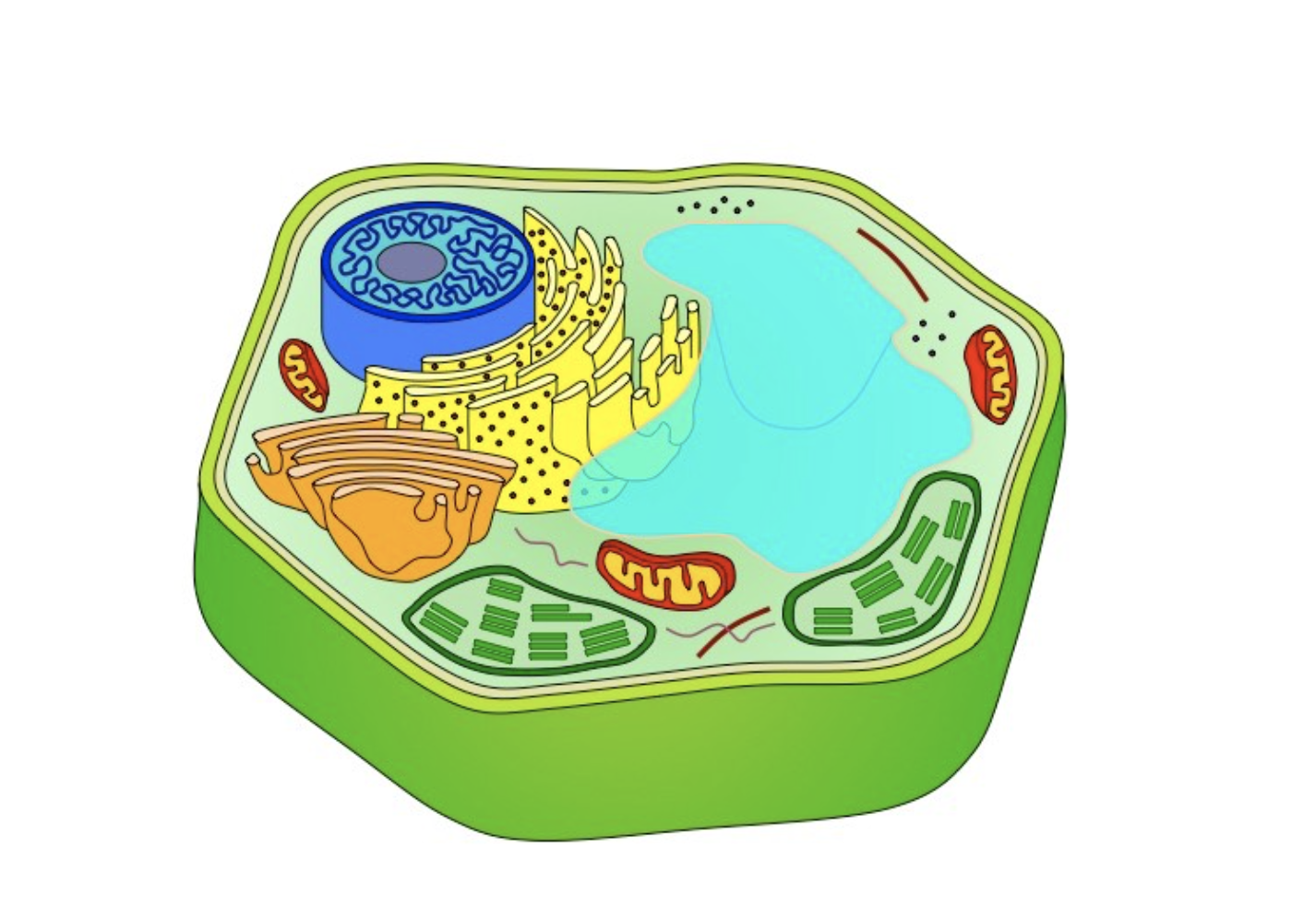
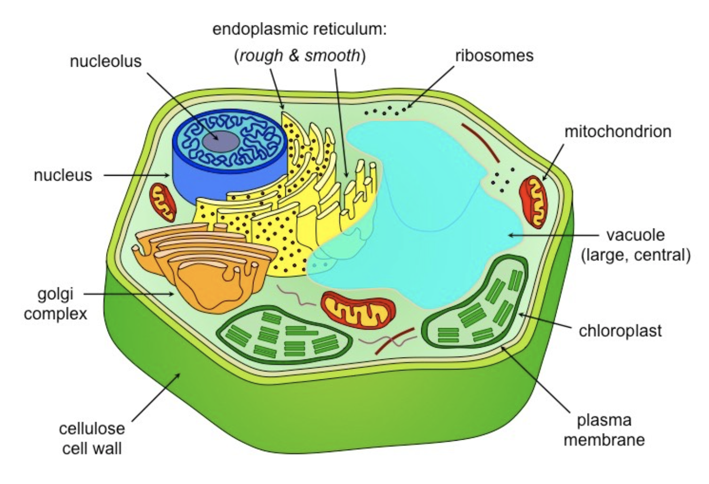
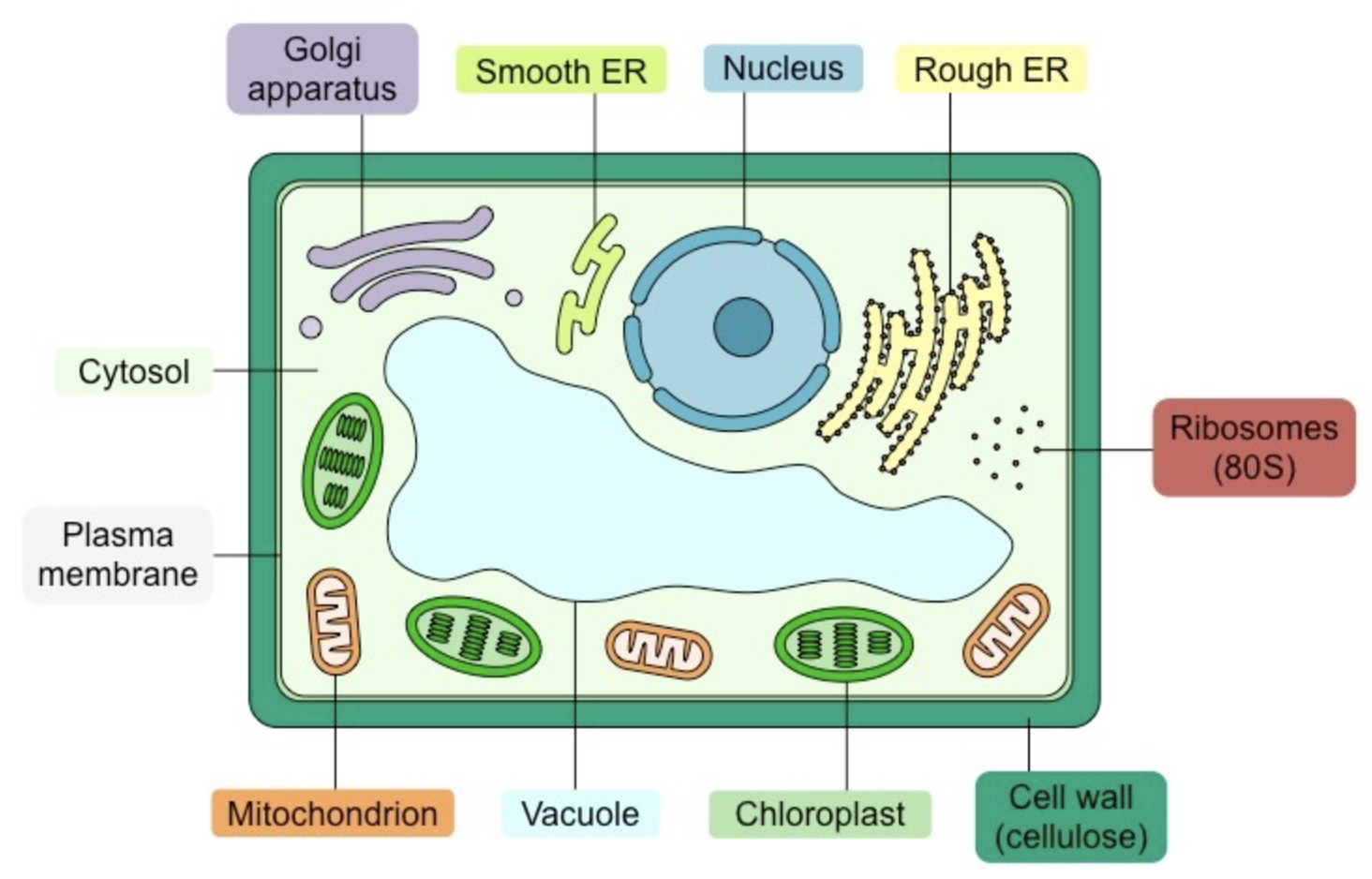
Plant Cell: Typical Structure and Features
Universal Organelles (prokaryote & eukaryote)
Ribosomes
Cytoskeleton
Plasma membrane
Ribosomes: Structure and Function
Structure:
Two subunits made of RNA and Protein
Larger in eukaryotes
Eukaryote: 80S
Prokaryote: 70S
Function:
Site of polypeptide synthesis (translation)
Cytoskeleton: Structure and Function
Structure:
A filamentous scaffolding within the cytoplasm (the fluid portion of the cytoplasm is the cytosol)
Function:
Provides internal structure and mediates intracellular transport (less developed in prokaryotes)
Plasma Membrane: Structure and Function
Not an organelle per se, but a vital structure.
Structure:
Phospholipid bilayer embedded with proteins
Function:
Semi-permeable and selective barrier surrounding the cell
Eukaryotic Organelles (animal and plant)
Nucleus
Endoplasmic Reticulum
Golgi Apparatus
Mitochondrion
Peroxisome
Centrosome
Nucleus: Structure and Function
Structure:
Double membrane structure with pores
Contains an inner region called a nucleolus
Function:
Stores genetic material (DNA) as chromatin
Nucleolus is site of ribosome assembly
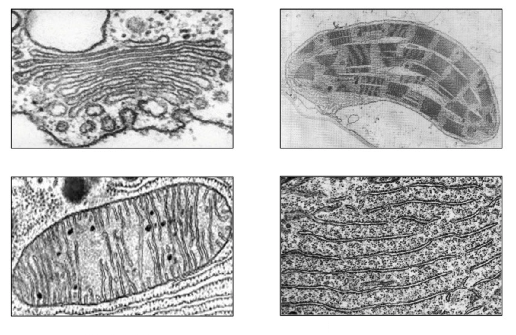
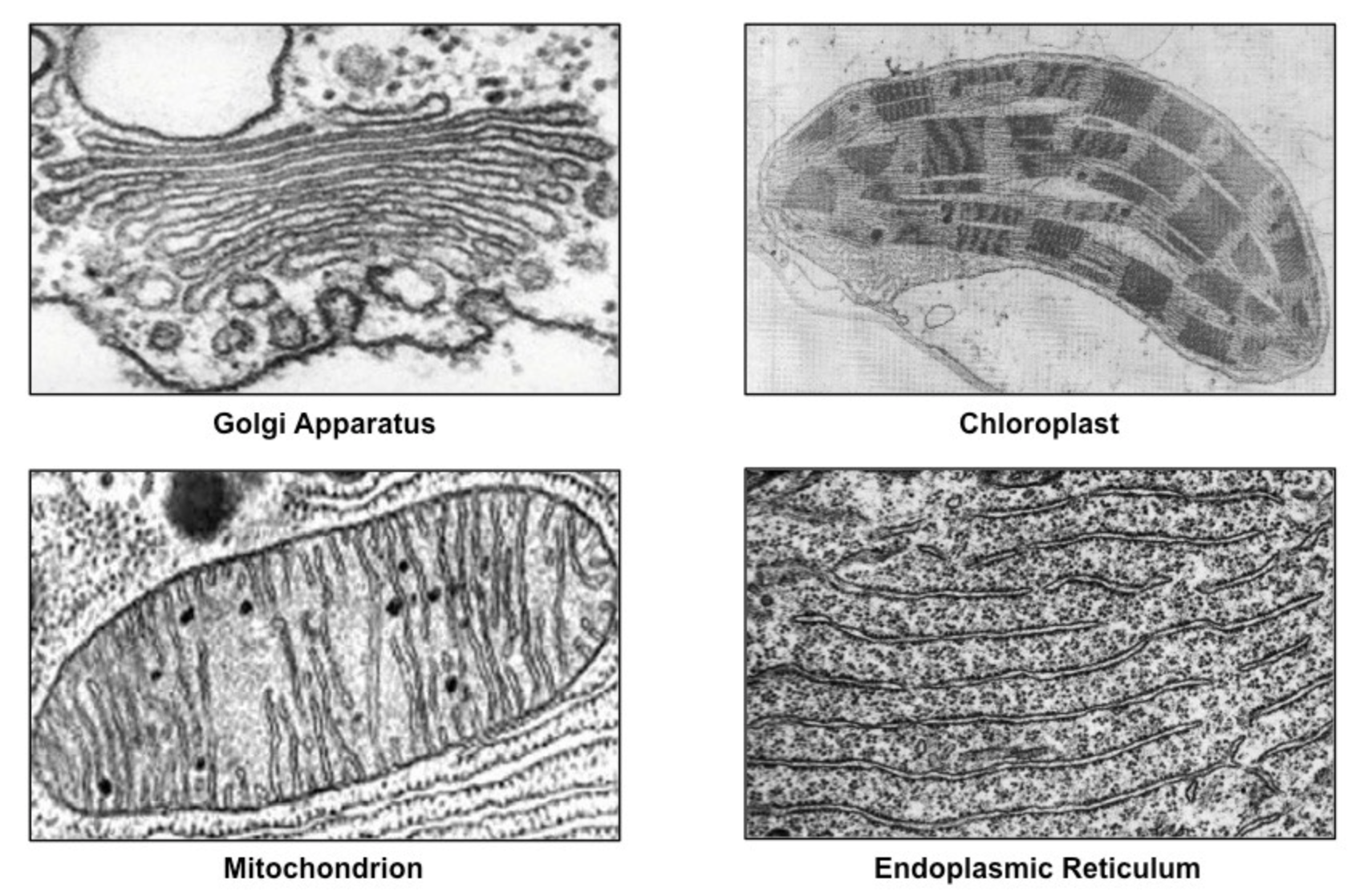
Endoplasmic Reticulum: Structure and Function
Structure:
Membrane network that may be bare (smooth ER) or studded with ribosomes (rough ER)
Function:
Transports materials between organelles
Smooth ER = lipids
Rough ER = proteins
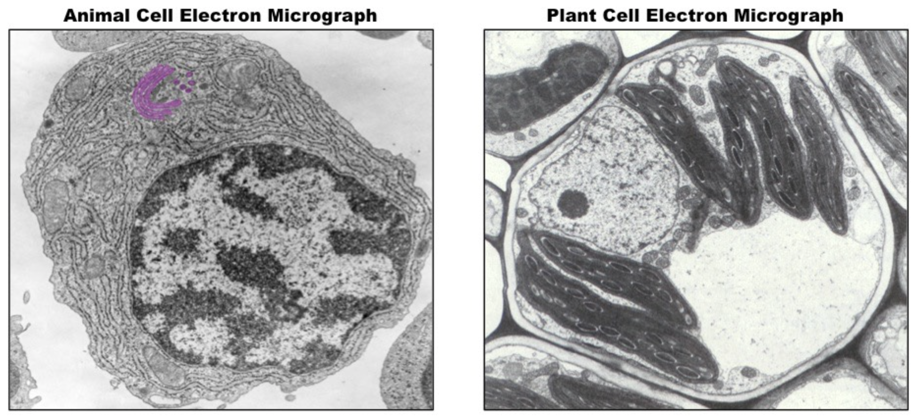
Golgi Apparatus: Structure and Function
Structure:
An assembly of vesicles and folded membranes located near the cell membrane
Function:
Involved in sorting, story, modification, and export of secretory products
Mitochondrion: Structure and Function
Structure:
Double membrane structure, inner membrane highly folded into internal cristae
Function:
Site of aerobic respiration (ATP production)
Peroxisome: Structure and Function
Structure:
Membranous sac containing a variety of catabolic enzymes
Function:
Catalyses breakdown of toxic substances and other metabolites
Centrosome: Structure and Function
Structure:
Microtubule organising centre (contains paired centrioles in animal cells but not plant cells)
Function:
Radiating microtubules form spindle fibers and contribute to cell division (mitosis/meiosis)
Eukaryotic Organelles (Plant Cells Only)
Chloroplast
Vacuole
Cell Wall
Chloroplast: Structure and Function
Structure:
Double membrane structure with internal stacks of membranous discs (thylakoids)
Function:
Site of photosynthesis - manufactured organic molecules are stored in various plastids (other plant cells, chloroplast is one type of plastid)
Vacuole: Structure and Function
Structure:
Fluid-filled internal cavity surrounded by a membrane (tonoplast)
Function:
Maintains hydrostatic pressure (animal cells may have small, temporary vacuoles)
Cell Wall: Structure and Function
Not an organelle per se, but a vital structure.
Structure:
External outer covering made of cellulose
Function:
Provides support and mechanical strength, prevents excess water uptake.
Lysosome: Structure and Function (Animal Cell Only)
Considered animal cell only, presence in plant cells is subject to debate.
Structure:
Membranous sacs filled with hydrolytic enzymes
Function:
Breakdown/hydrolysis of macromolecules
Electron Microscope: Definition, types, and advantages over light microscopes
Definition
Electron microscopes use electron beams focused by electromagnets to magnify and resolve microscopic specimens.
Types
Transmission Electron Microscopes (TEM)
Generate high-resolution cross-sections of objects
Scanning Electron Microscopes (SEM)
Display enhanced depth to map the surface of objects in 3D
Advantages (Over Light Microscopes)
Much higher range of magnification (can detect smaller structures)
Much higher resolution (provides clearer and more detailed images)
Disadvantage
Cannot display living specimens in natural colour
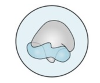
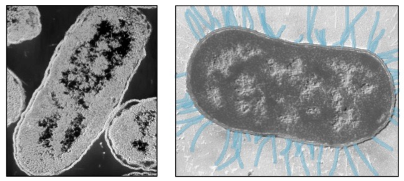
Prokaryote Micrograph: Nucleoid
Prokaryote Micrograph: Cell Wall
Prokaryote Micrograph: Pili
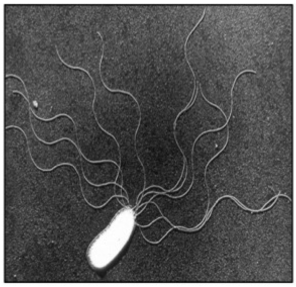
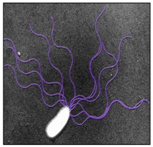
Prokaryote Micrograph: Flagella
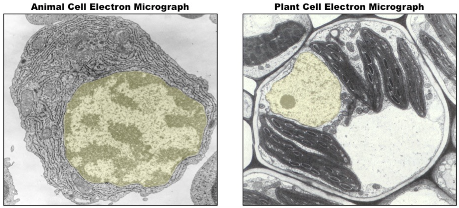
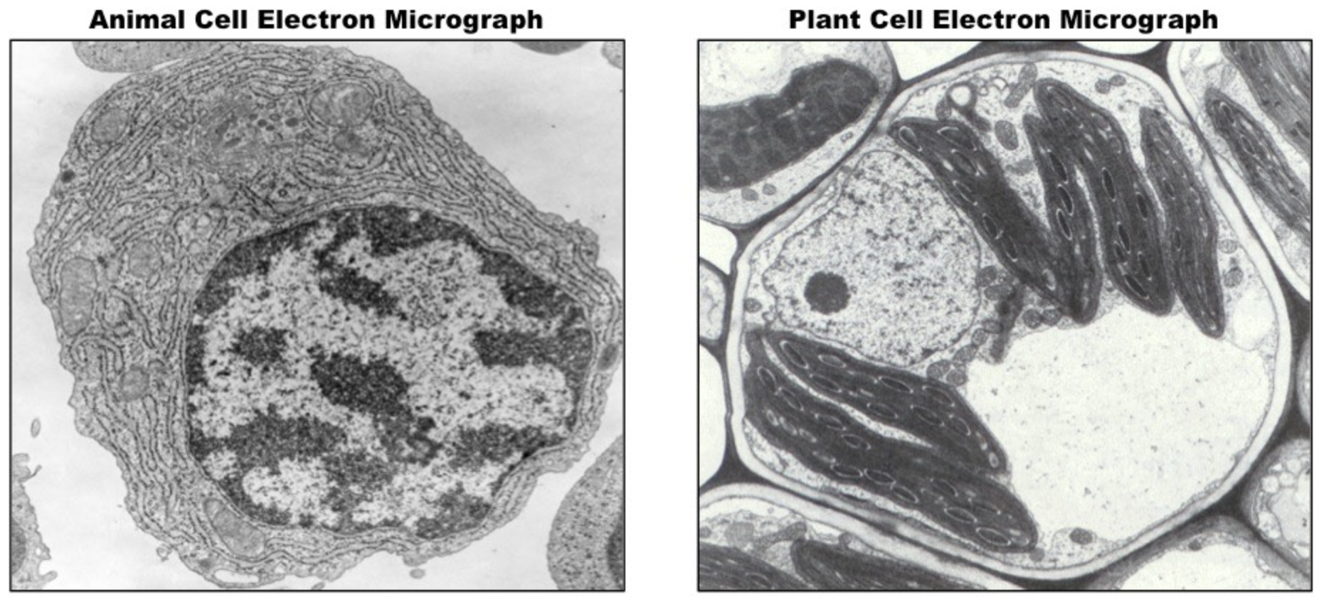
Eukaryote Micrograph: Nucleus
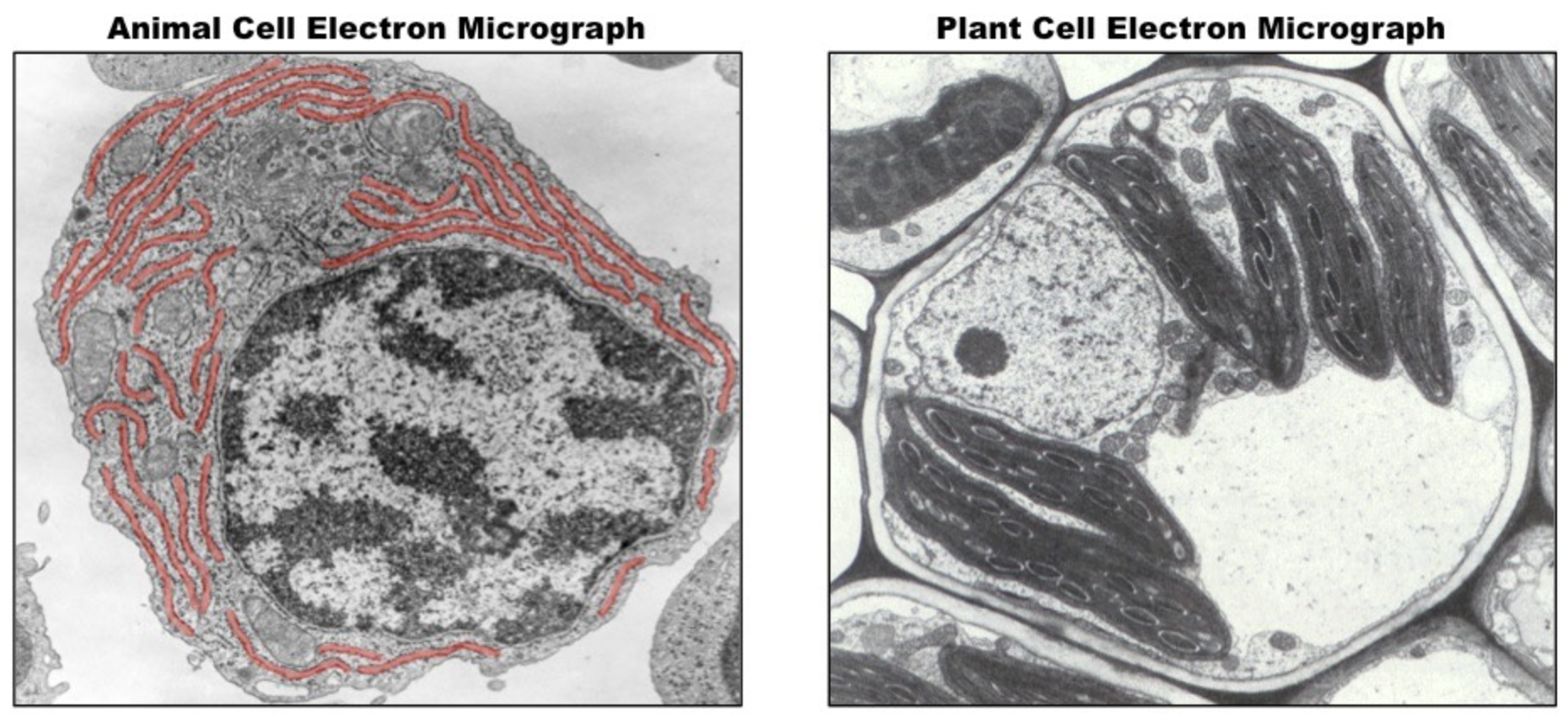
Eukaryote Micrograph: Endoplasmic Reticulum (ER)
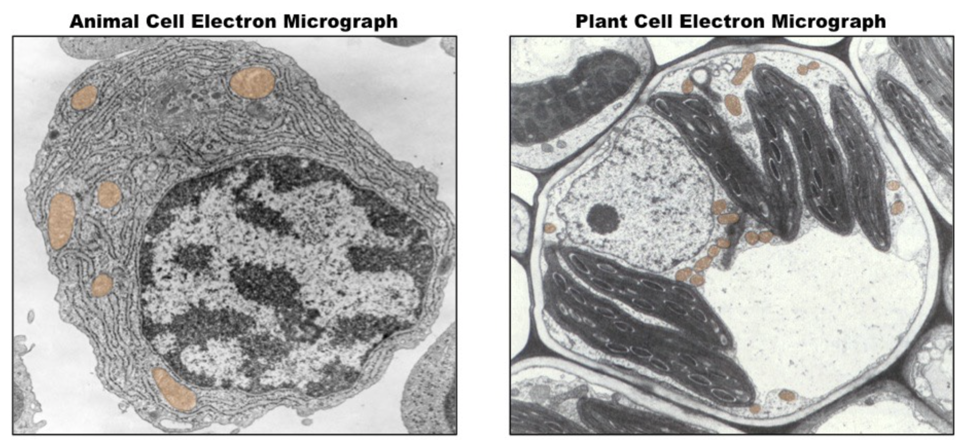
Eukaryote Micrograph: Mitochondria
Eukaryote Micrograph: Golgi Apparatus
Eukaryote Micrograph: Chloroplast
Eukaryote Micrograph: Cell Wall
Eukaryote Micrograph: Vacuole
Label the Organelles
Deduce Cell Function Based on Abundance:
Mitochondria
Endoplasmic Reticulum
Lysosomes
Chloroplasts
Mitochondria:
Cells with many mitochondria typically undertake energy-consuming processes (e.g. neurons, muscle cells)
ER:
Cells with extensive ER networks undertake secretory activities (e.g. plasma cells, exocrine gland cells)
Lysosomes:
Cells rich in lysosomes tend to undertake digestive processes (e.g. phagocytes)
Chloroplasts:
Cells with chloroplasts undergo photosynthesis (e.g. plant leaf tissue but not root tissue)
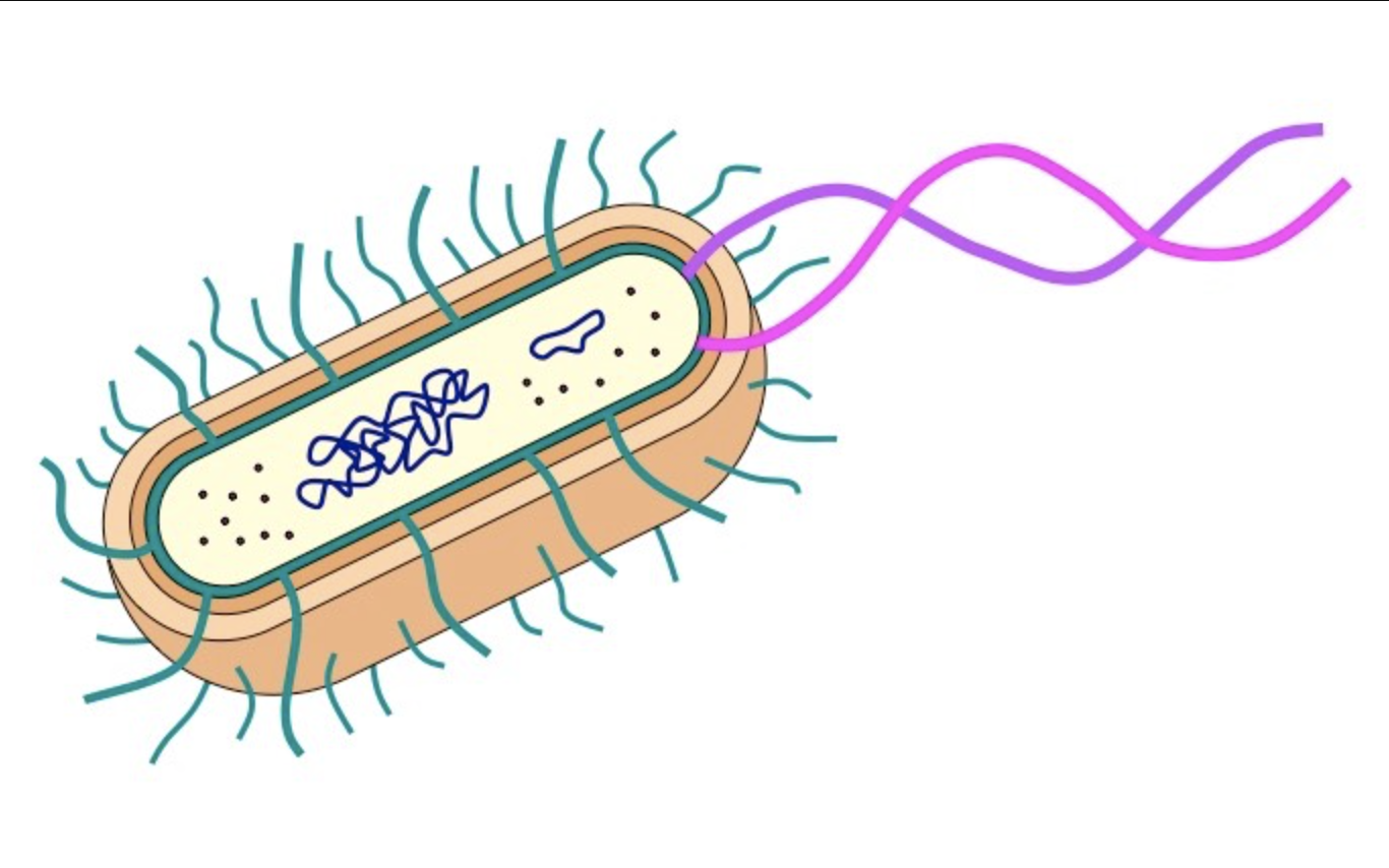
Prokaryotic Cell: Label and Draw
Key Features:
Pili – shown as single lines
Flagella – shown as thicker and significantly longer lines than the pili
Ribosomes – labelled as 70S
Cell wall – labelled as being composed of peptidoglycan; thicker than cell membrane
Shape – appropriate for bacteria chosen (e.g. E. coli is a rod-shaped bacillus)
Size – appropriate dimensions (e.g. length of cell twice the width)
Prokaryotic Animal Cell: Label and Draw
Key Features:
Nucleus – shown as double membrane structure with pores
Mitochondria – double membrane with inner one folded into cristae ; no larger than half the nucleus in size
Golgi apparatus – shown as a series of enclosed sacs (cisternae) with vesicles leading to and from
Endoplasmic reticulum – interconnected membranes shown as bare (smooth ER) and studded (rough ER)
Ribosomes – labelled as 80S
Cytosol – internal fluid labelled as cytosol (‘cytoplasm' is all internal contents minus the nucleus)
Prokaryotic Plant Cell: Label and Draw
Key Features:
Vacuole – large and occupying majority of central space (surrounded by tonoplast)
Chloroplasts – double membrane with internal stacks of membrane discs (only present in photosynthetic tissue)
Cell wall – labelled as being composed of cellulose ; thicker than cell membrane
Shape – brick-like shape with rounded corners
Domain/Kingdom Classification: cell characteristics
Prokaryotes:
Kingdom - Monera
Domains - Archaea and Eubacteria
Eukaryotes:
Kingdom - Eukarya
Domains - Protist, Plant, Fungi, and Animal
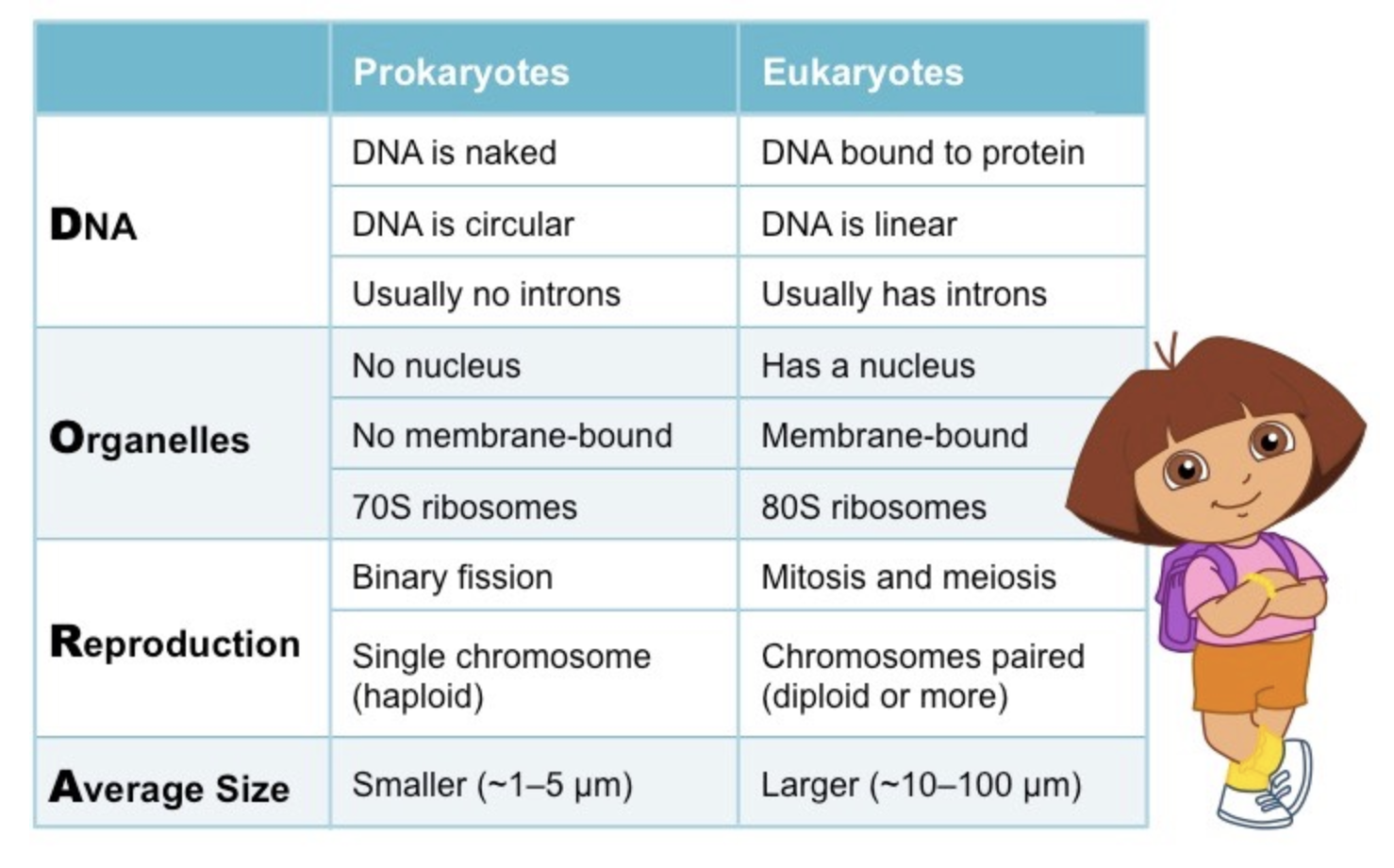
Points of Comparison: Prokaryotic vs Eukaryotic Cells (DORA)
DNA
Organelles
Reproduction
Average Size
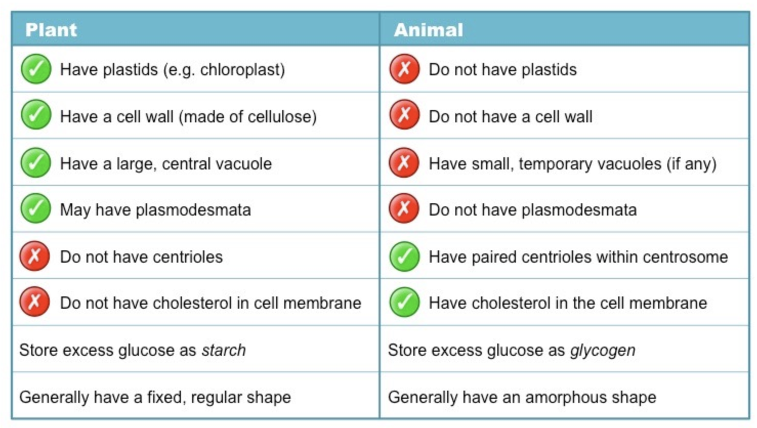
Points of Comparison: Animal vs Plant Cells
Both have:
DNA stored within a nucleus
Larger ribosomes (80S in size)
A variety of membrane-bound organelles (e.g. mitochondria, ER, golgi apparatus)
Differences:
Presence or absence of specific sub-cellular structures
Composition of the plasma membrane
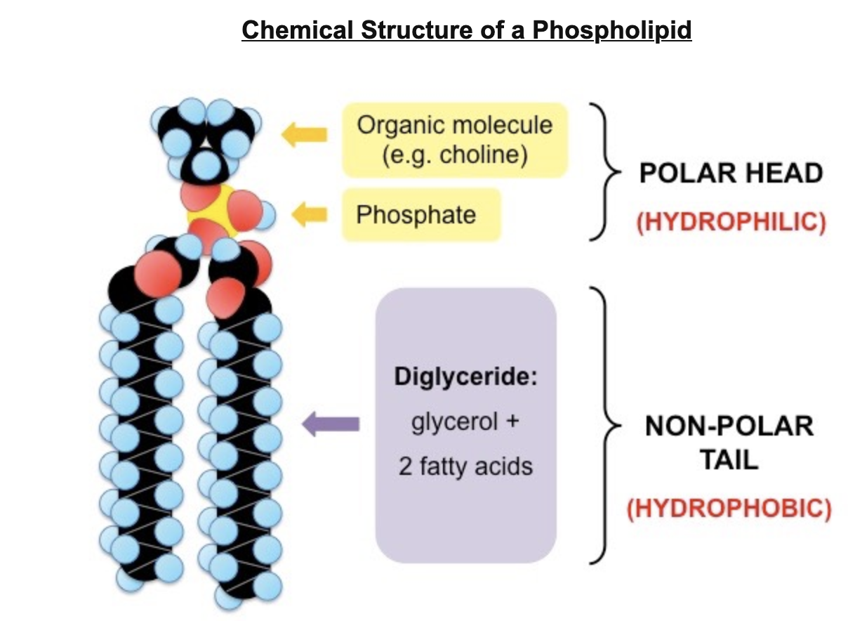
Structure of Phospholipids
Amphipathic molecules, meaning they have clearly discernible hydrophilic and hydrophobic regions
Phospholipids typically share a common basic structure that includes:
A polar organic molecule (e.g. choline, serine)
A phosphate group
A glycerol molecule (replaced by sphingosine in sphingomyelin)
Two fatty acid tails (may be saturated or unsaturated)
Main Parts
Polar head
Hydrophilic
Composed of a glycerol and a phosphate molecule
Two non-polar tails
Hydrophobic
Composed of fatty acid (hydrocarbon) chains
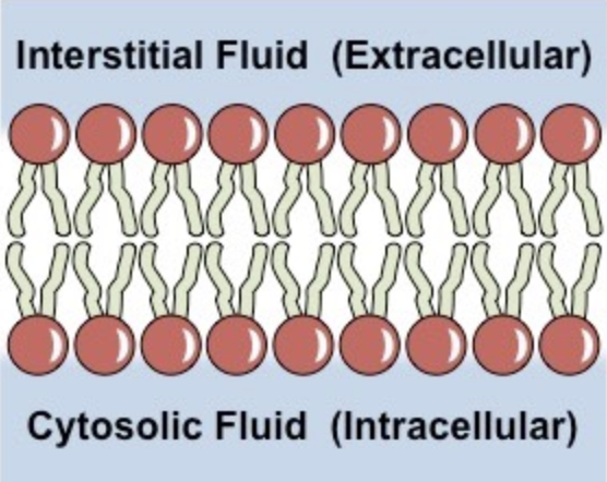
Phospholipid Arrangement in Membranes
Phospholipids spontaneously arrange into a bilayer
The hydrophobic tail regions face inwards
Thereby shielded from surrounding polar fluids
The two hydrophilic head regions associate with the extra/intracellular fluids
Hydrophilic property allows association
Interstitial Fluid (Extracellular)
Cyrosolic Fluid (Intracellular)
Properties of the Phospholipid Bilayer
Held together by weak hydrophobic interactions between the tails
Hydrophilic / Hydrophobic layers restrict the passage of many substances
Individual phospholipids can move within the bilayer, allowing for membrane fluidity and flexibility
Fluidity allows for the spontaneous breaking and reforming of membranes (endocytosis/exocytosis)
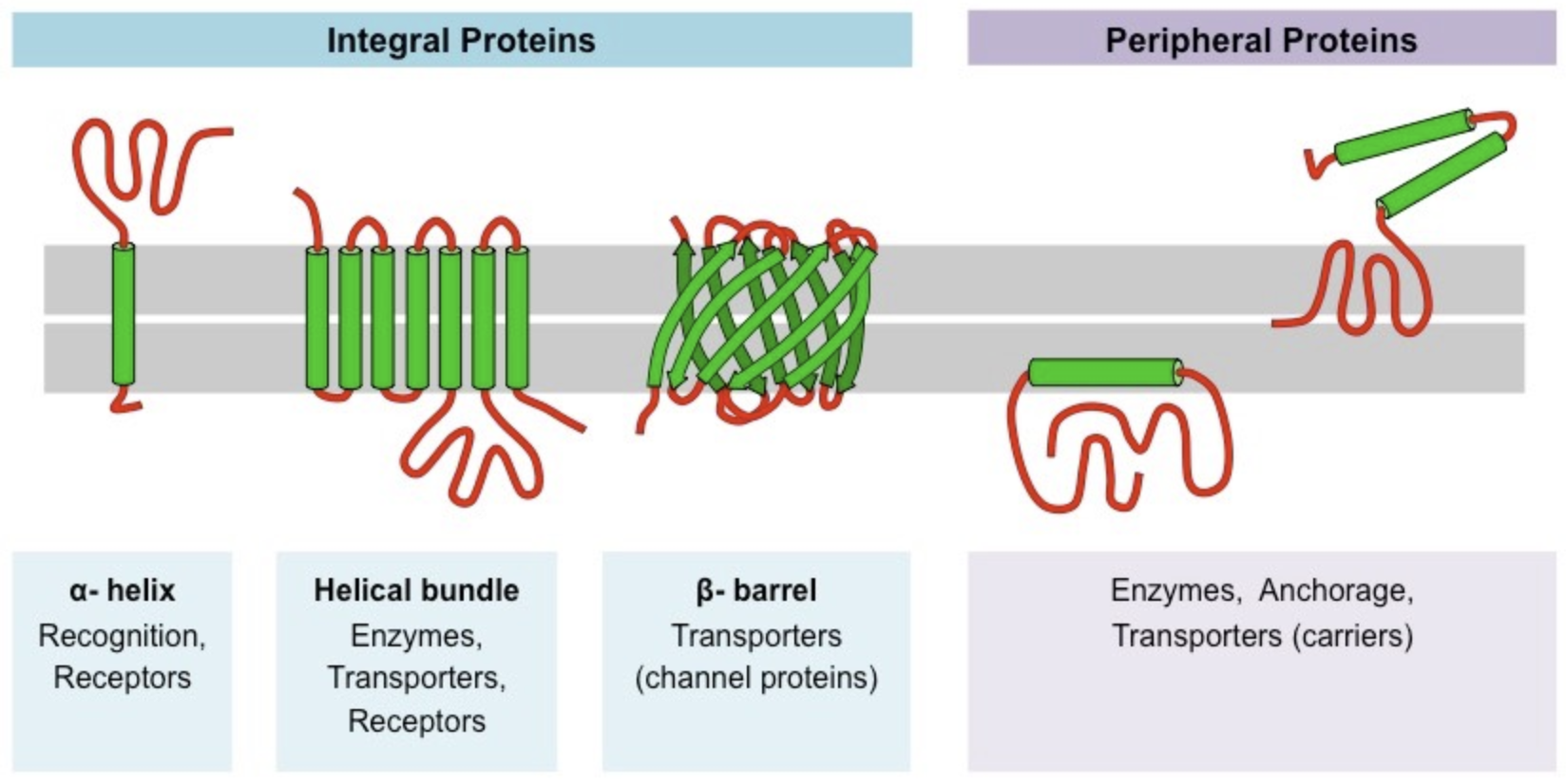
Membrane Proteins: What and Integral vs Peripheral
Phospholipids are embedded with membrane proteins which may be either permanently or temporarily attached to the membrane
Integral Proteins
Permanently attached to membrane
Typically transmembrane (span across bilayer)
Peripheral Proteins
Temporarily attached by non-covalent interactions
Associate with one surface of the membrane
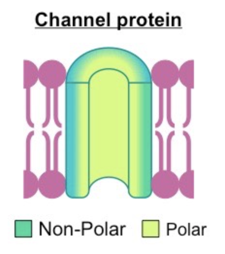
Structure of Membrane Proteins: Polarity
Aminos within the proteins are localised by polarity:
Non-Polar (hydrophobic)
Associate directly with lipid bilayer (makes up outter lining of the protein)
Polar (hydrophilic)
Located internally
Hydrophilic because face aqueous solutions passing through the membrane
Structure of Membrane Proteins: Tertiary Structures
Typically adopt one of two tertiary structures:
Single helices/helical bundles
Beta Barrels (common in channel proteins)
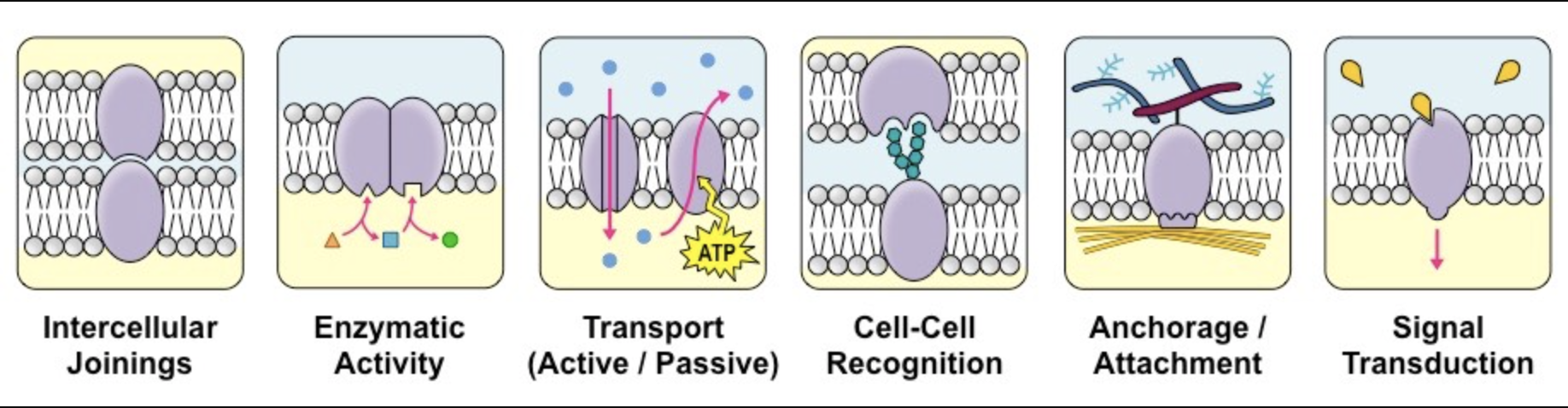
Functions of Membrane Proteins (JET RAT)
Junctions
Serve to connect and join two cells together
Enzymes
Fixing to membranes localised metabolic pathways
Transport
Responsible for facilitated diffusion and active transports (pumps)
Recognition
May function as markers for cellular identification
Anchorage
Attachment points for cytoskeleton and extracellular matrix
Transduction
Function as receptors for peptide hormones
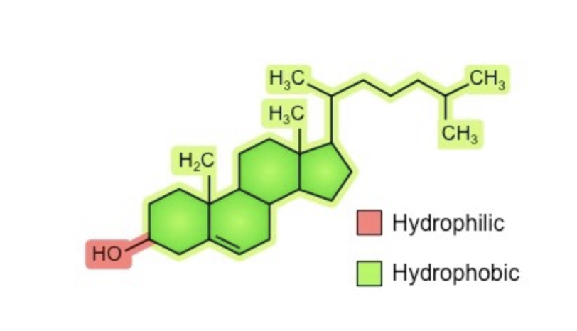
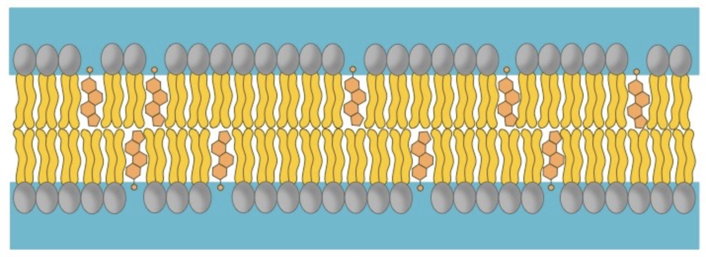
Cell Membrane Cholesterol: Structure and Placement
Amphipathic molecule (like phospholipids)
Hydroxyl (-OH) group is hydrophilic and alights towards the phorphate heads of phospholipids
Remainder is hydrophobic and associates with phospholipids tails
Cell Membrane Cholesterol: Functions
Without Cholesterol
Phospholipid bilayers are fluid and in constant movement relative to one another.
Main Function
Cholesterol functions to maintain integrity and mechanical stability, only in animal cells because plant cells have rigid cell walls of cellulose to maintain structure.
Specific Functions
Cholesterol immobilises the outer surface of the membrane, reducing fluidity
It makes the membrane less permeable to very small water-soluble molecules that would otherwise freely cross
It functions to separate phospholipid tails and so prevent crystallisation of the membrane
It helps secure peripheral proteins by forming high density lipid rafts capable of anchoring the protein
Regulation with Temperature
Cholesterol acts as a bi-directional regulator of membrane fluidity
At high temperatures it stabilises the membrane and raises the melting point
At low temperatures it intercalates between the phospholipids and prevents clustering
Membrane Fluidity
Cell membranes are fluid, meaning they are not fixed in position and can adopt amorphous shapes.
Membrane fluidity is enhanced at higher temperatures.
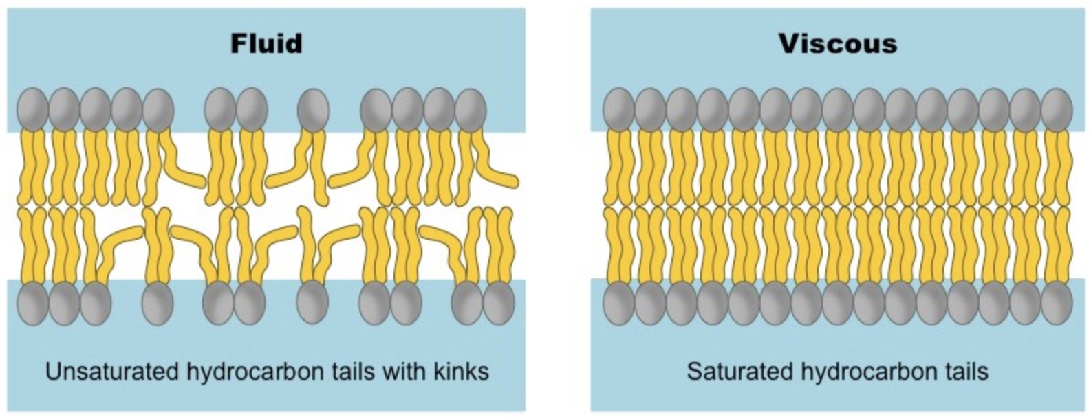
Phospholipid Structure and Fluidity
Phospholipids may vary in the length and relative saturation of the fatty acid tails
Shorter Fatty Acid Tails = more fluid
Less viscous and more susceptible. to changes in kinetic energy
Unsaturated Fatty Acid Tails = less fluid
Lipid chains with double bonds (unsaturated) have kinked hydrocarbon tails
Harder to pack tightly
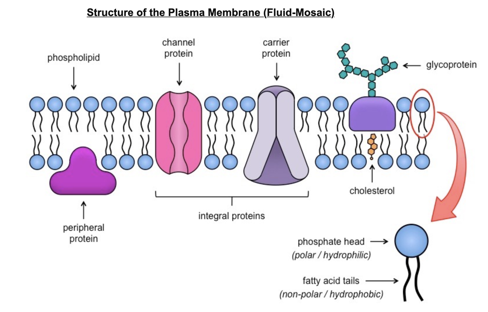
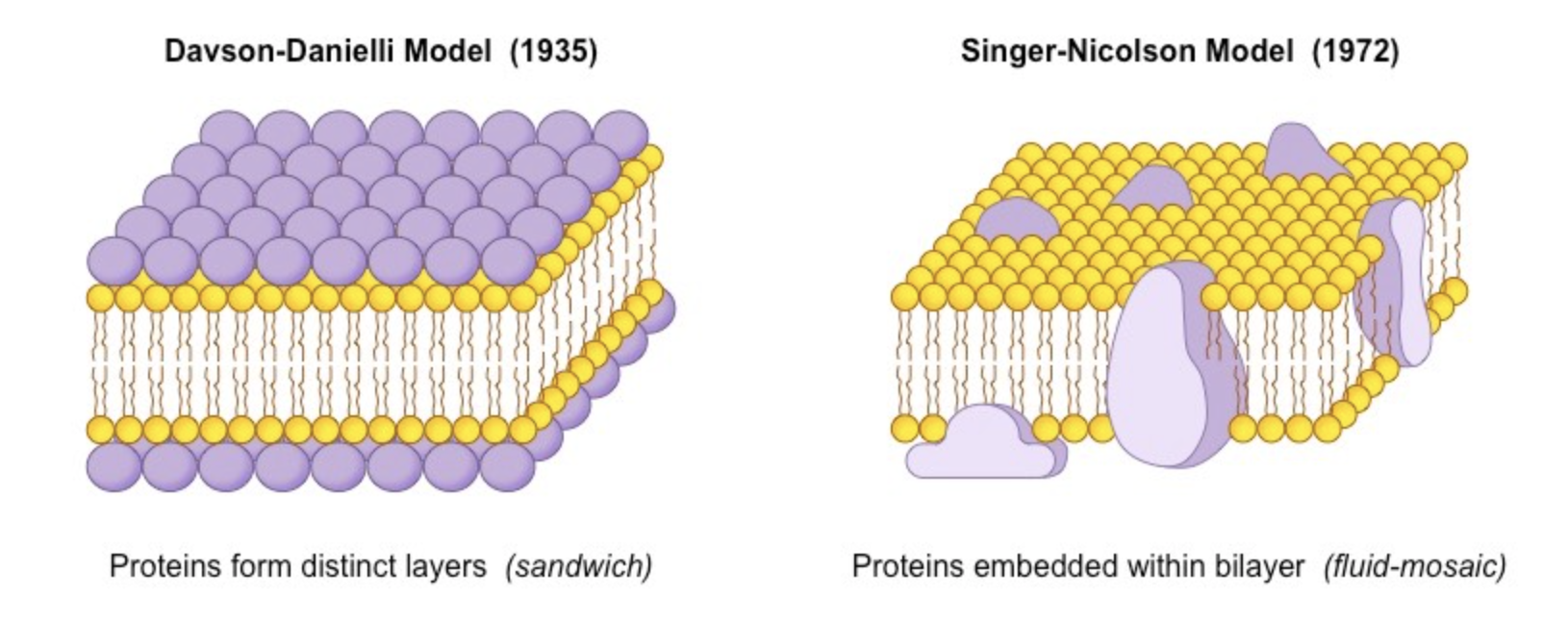
Fluid-Mosaic Model
Cell membranes are represented according to a fluid-mosaic model, due to the fact that they are:
Fluid – the phospholipid bilayer is viscous and individual phospholipids can move position
Mosaic – the phospholipid bilayer is embedded with proteins, resulting in a mosaic of components
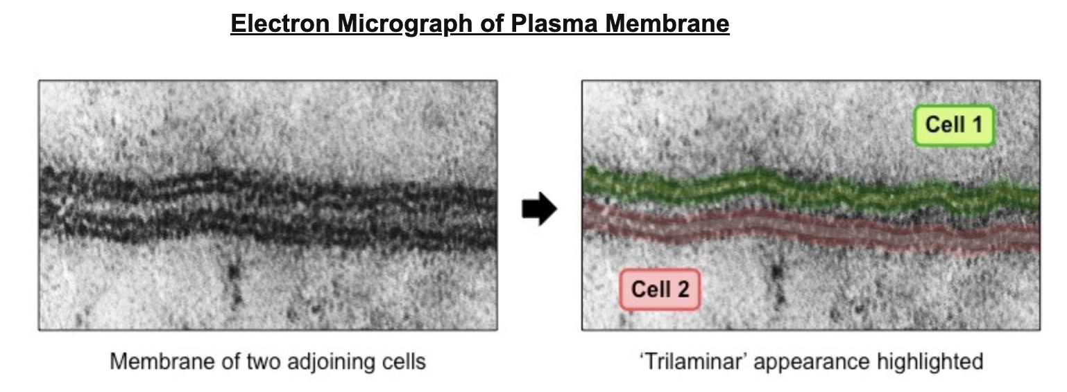
Davson-Danielli Model: Origin
Davson and Danielli made their model based of the 'trilaminar’ appearance of the cell membrane under the Electron Micrograph.
Trilaminar = 3 layers (two dark outer layers and a light inner region)
This made them deduce it was a three layer membrane
“Lipo-Protein Sandwich”
Lipid layer (lighter inner region) was sandwiched between two protein layers
The darker layers under the microscope were wrongly identified as protein layers.
Davson-Danielli Model: Problems
Assumed all membranes were of uniform thickness and had constant lipid-protein ration
Assumed all membranes would have symmetrical internal and external surfaces (not bifacial)
Did not account for the permeability of certain substances (did not recognise need for hydrophilic pores like channel proteins)
Temperatures at which membranes solifidied did not correlate with those expected under the model’s assumptions
Davson-Danielli Model: Falsification
Three Key Discoveries:
1) Membrane proteins were discovered to be insoluble in water and varied in size
Indicated hydrophobic surfaces
Varied protein sizes would not allow for a uniform and continuous layer around the outer surface of a membrane
Showed that purely protein surface were unfeasible
2) Flourecent antibody tagging of membrane proteins showed they were mobile and not fixed in place
Membranes from two different cells were tagged with red and green fluorescent markers respectively
When two cells were fused, markers became mixed throughout the membrane of the fused cell
Showed that the membrane proteins could move and did not form a static layer
3) Freeze fracturing was used to split open the membrane and reveal irregular rough surfaces within the membrane
Rough surfaces were interpreted as being transmembrane proteins
Showed that proteins were not solely localised to the outside of the membrane structure
Singer-Nicolson Model: Origin
The limitations found in the Davson-Danielli Model allowed for new understandings to be applied in the new Singer-Nicolson Model proposed in 1972
According to this model, proteins were embedded within the lipid bilayer rather than existing as separate layers
Addresses the problems and limitations found in the Davson-Danielli Model
This fluid-mosaic model remains the model preferred by scientists today (with refinements)
Key Qualities of Cellular Membranes
Semi-permeable
Only certain materials may freely cross - large and charged substances are typically blocked
Selective
Membrane proteins may regulate the passage of material that cannot freely cross
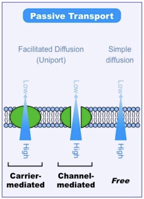
Passive Transport: Definition and Types
Movement of material along a concentration gradient (high to low)
Does not require energy expenditure (ATP hydrolysis)
Three Main Types:
Simple diffusion
movement of small or lipophilic molecules
Osmosis
movement of water molecules
Facilitated diffusion
the movement of large or charged molecules via membrane proteins
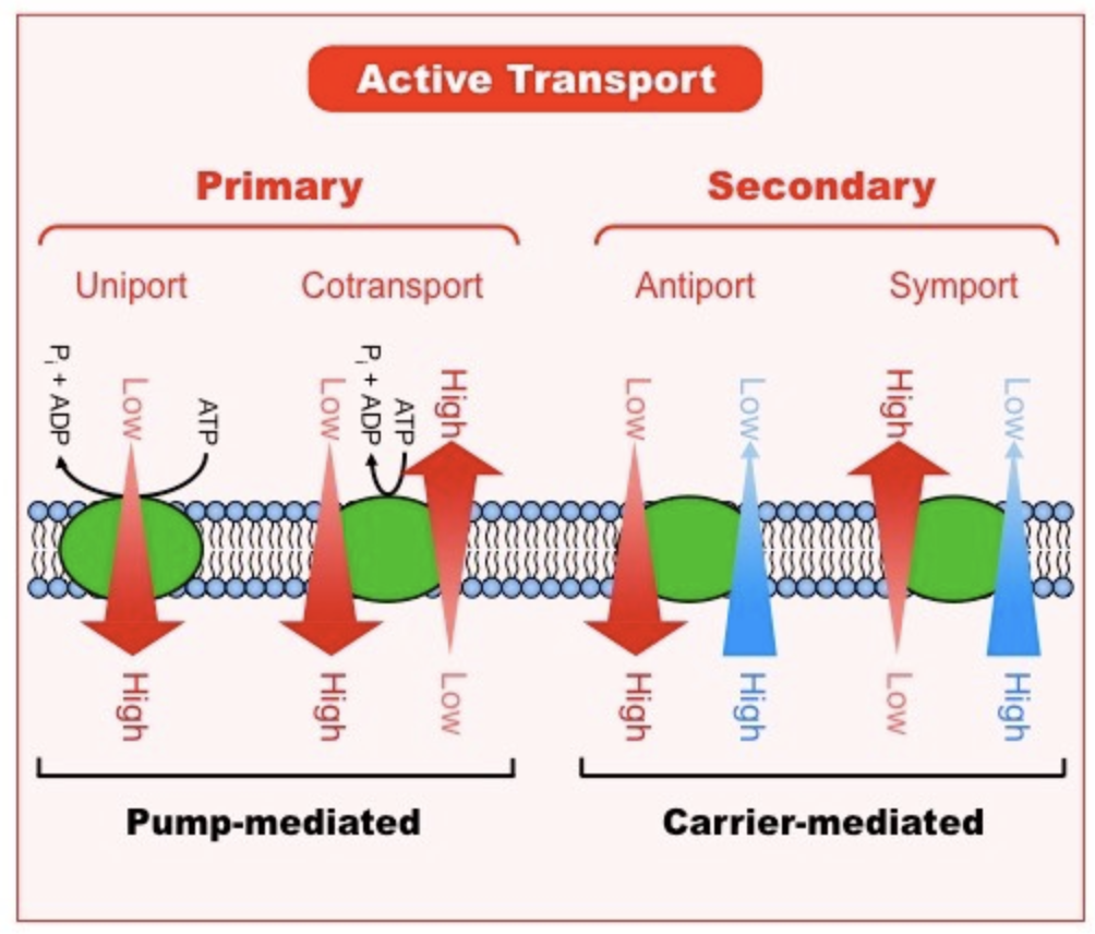
Active Transport: Definition and Types
Movement of materials against a concentration gradient (low to high)
Required expenditure of energy (ATP hydrolysis)
Two Main Types:
Primary (direct) active transport
Direct use of metabolic energy to mediate transport
Secondary (indirect) active transport
Coupling the molecule with another moving along an electrochemical gradient
Simple Diffusion: Definition and Factors
Diffusion - net movement of molecules from region of high concentration to region of low concentration
Passive directional movement and continues until equilibrium is reached
Small and non-polar (lipophilic) molecules will be able to freely diffuse across cell membranes
Factors
Temperature
Affects the kinetic energy of particles in a solution
Molecular Size
Larger particles are subjected to greater resistance within a fluid medium
Steepness of Gradient
Rate of diffusion will be greater with a higher concentration gradient
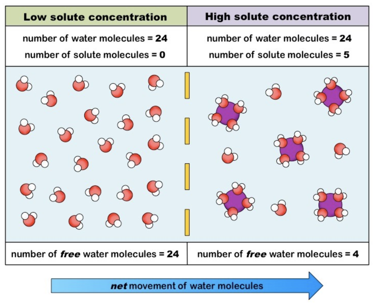
Simple Diffusion: Definition and Solute Concentration
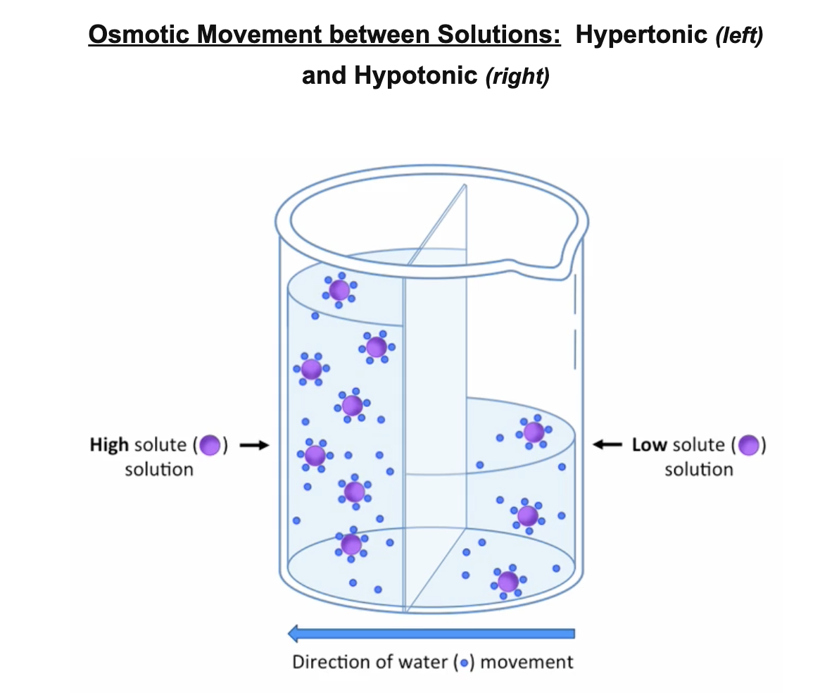
Osmosis - net movement of water molecules across a semi-permeable membrane from a region of low solute concentration to a region of high solute concentration (until equilibrium is reached).
Water is universal solvent
Will associate with and dissolve polar or charged molecules (solutes)
Because solutes cannot cross a cell membrane unaided, water moved to equalise the two solutions
Higher solute concentration = less free water molecules in solution
Osmosis is essentially the diffusion of free water molecules and hence occurs from regions of low solute concentration.
Osmolarity: Definition and Categorisation
Osmolarity - measure of solute concentration, as defined by the number of osmoles of a solute per litre of solution (osmol / L)
Categories
Solutions with relatively higher osmolarity are categorised as hypertonic
high solute concentration ⇒ gains water
Solutions with a relatively lower osmolarity are categorised as hypotonic
low solute concentration ⇒ loses water
Solutions that have the same osmolarity are categorised as isotonic
same solute concentration ⇒ no net water flow
Estimating Osmolarity (Application)
The osmolarity of a tissue may be interpolated by bathing the sample in solutions with known osmolarities
The tissue will:
lose water when placed in hypertonic solutions
gain water when placed in hypotonic solutions
Water loss or gain may be determined by weighing the sample before and after bathing in solution
Tissue osmolarity may be inferred by identifying the concentration of solution at which there is no weight change (i.e. isotonic)
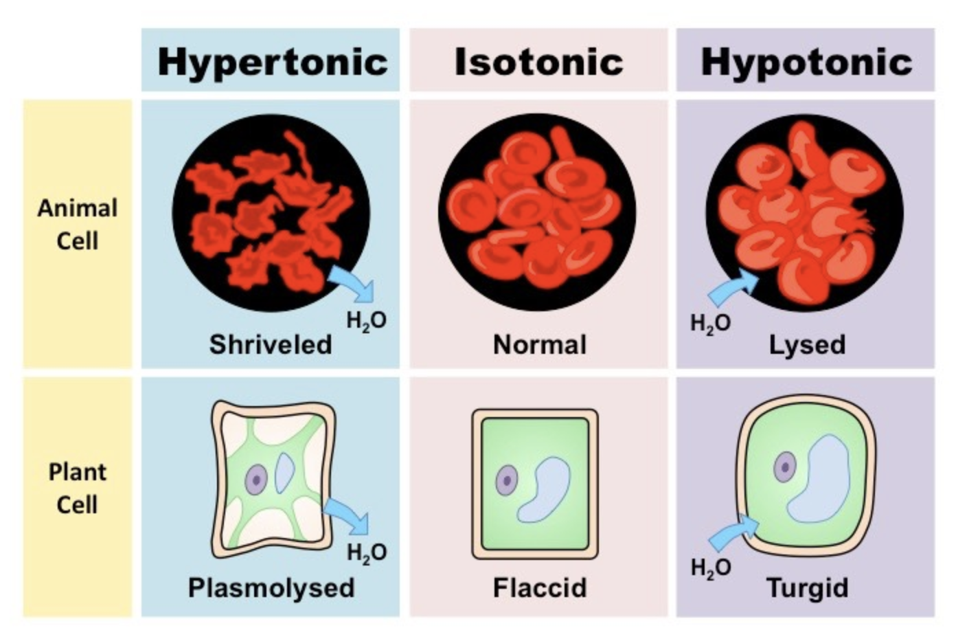
Uncontrolled Osmosis: Negative Effects
Human / Animal Cells
In hypertonic solutions:
Water will leave the cell causing it to shrivel (crenation)
In hypotonic solutions:
Water will enter the cell causing it to swell and potentially burst (lysis)
Therefore, tissues or organs to be used in medical procedures must be kept in solution to prevent cellular dessication
This solution must share the same osmolarity as the tissue / organ (i.e. isotonic) in order to prevent osmosis from occurring
Plant Tissue
In hypertonic solutions:
Cytoplasm will shrink (plasmolysis) but the cell wall will maintain a structured shape
In hypotonic solutions:
Cytoplasm will expand but be unable to rupture within the constraints of the cell wall (turgor)