KIN 223 EXAM 5 DIAGRAMS
1/19
There's no tags or description
Looks like no tags are added yet.
Name | Mastery | Learn | Test | Matching | Spaced |
|---|
No study sessions yet.
20 Terms
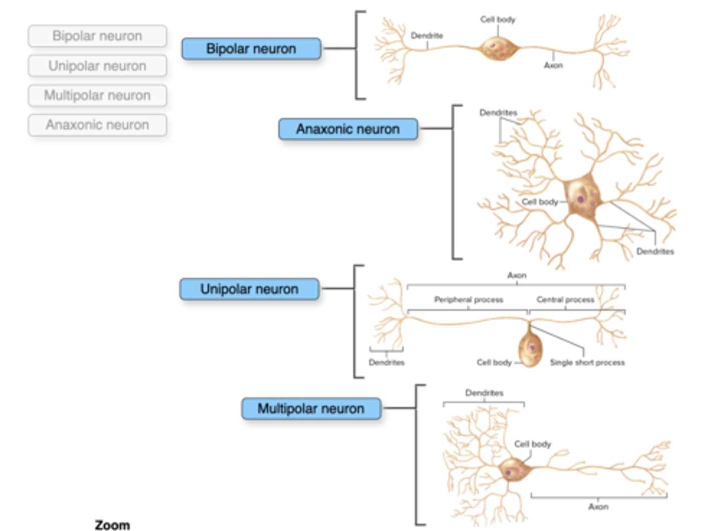
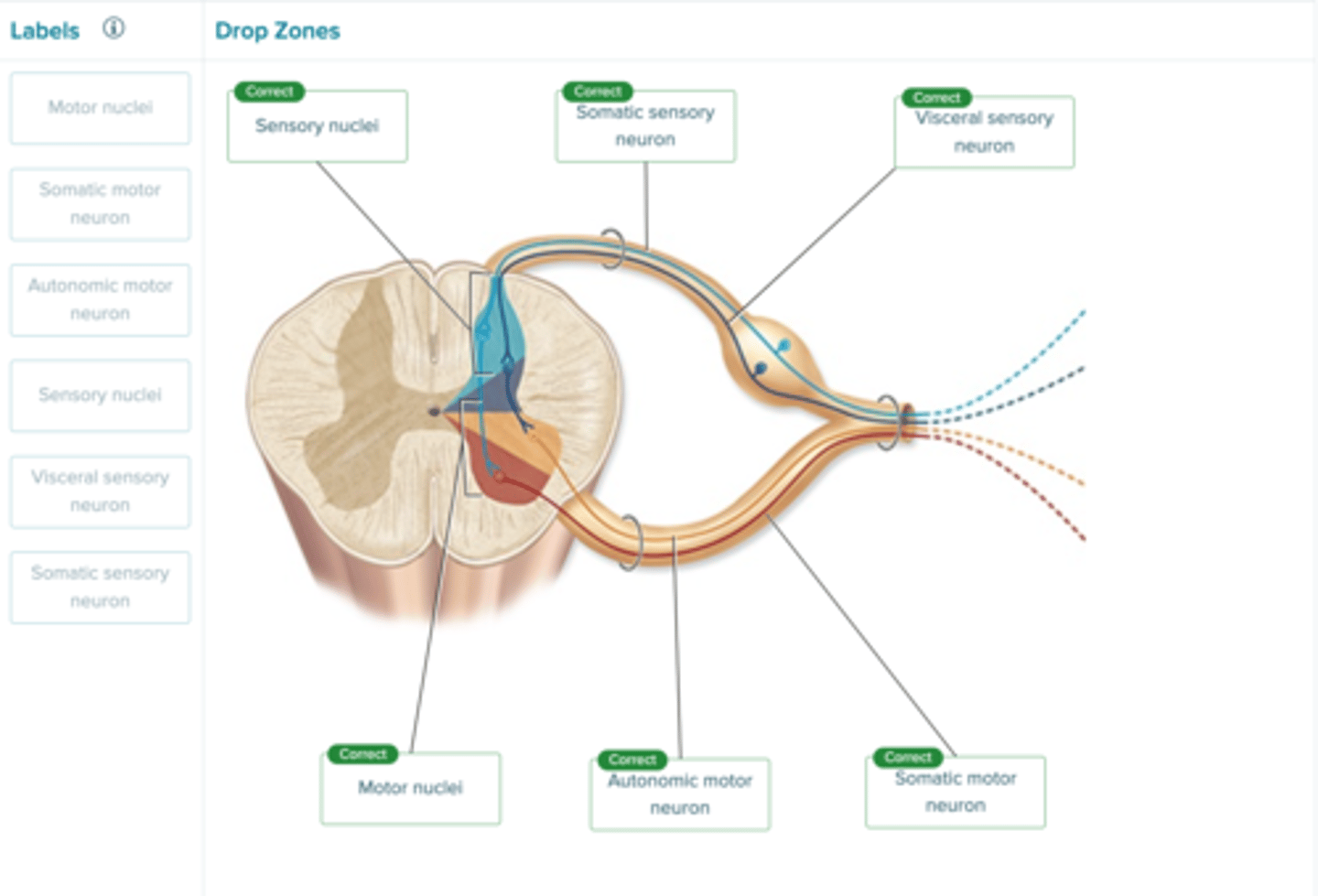
Label the figure with the items provided.
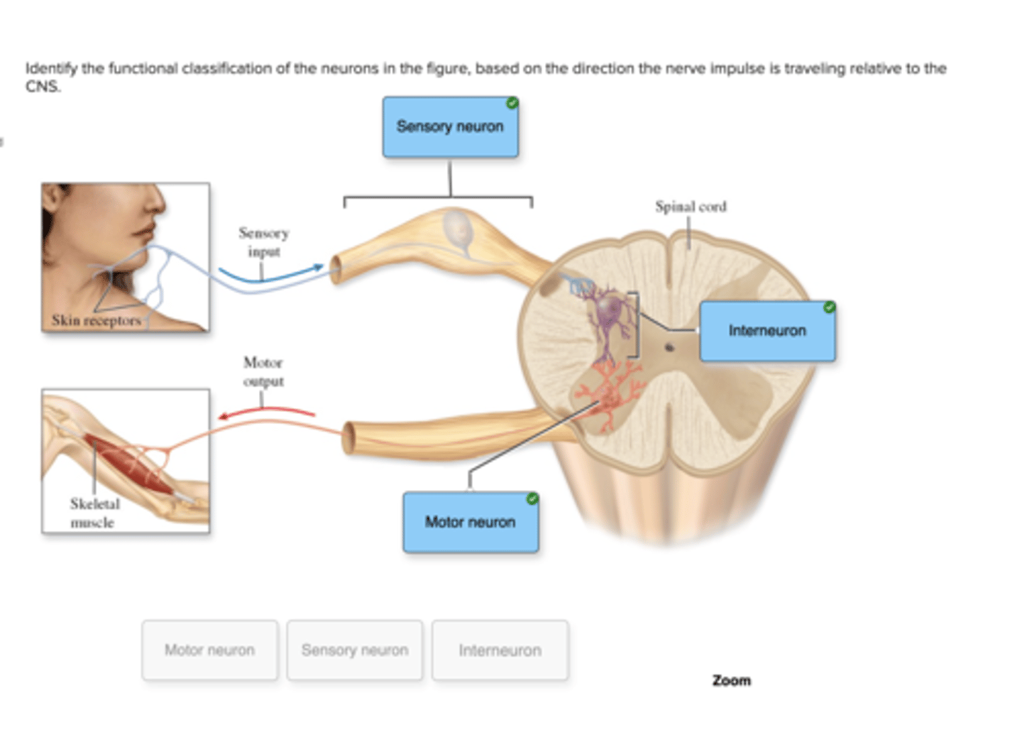
Identify the functional classification of the neurons in the figure, based on the direction the nerve impulse is traveling relative to the CNS.
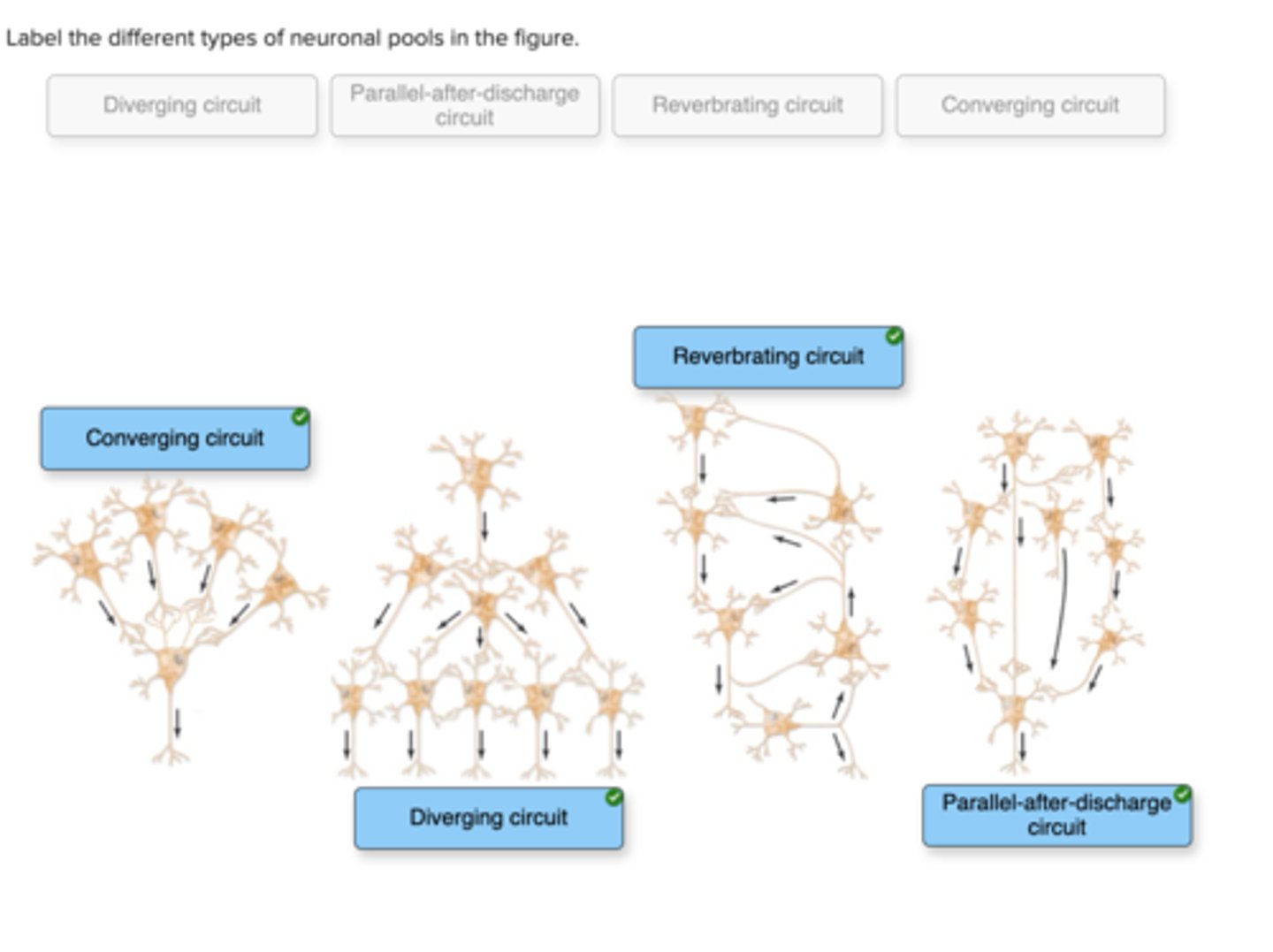
Label the different types of neuronal pools in the figure.
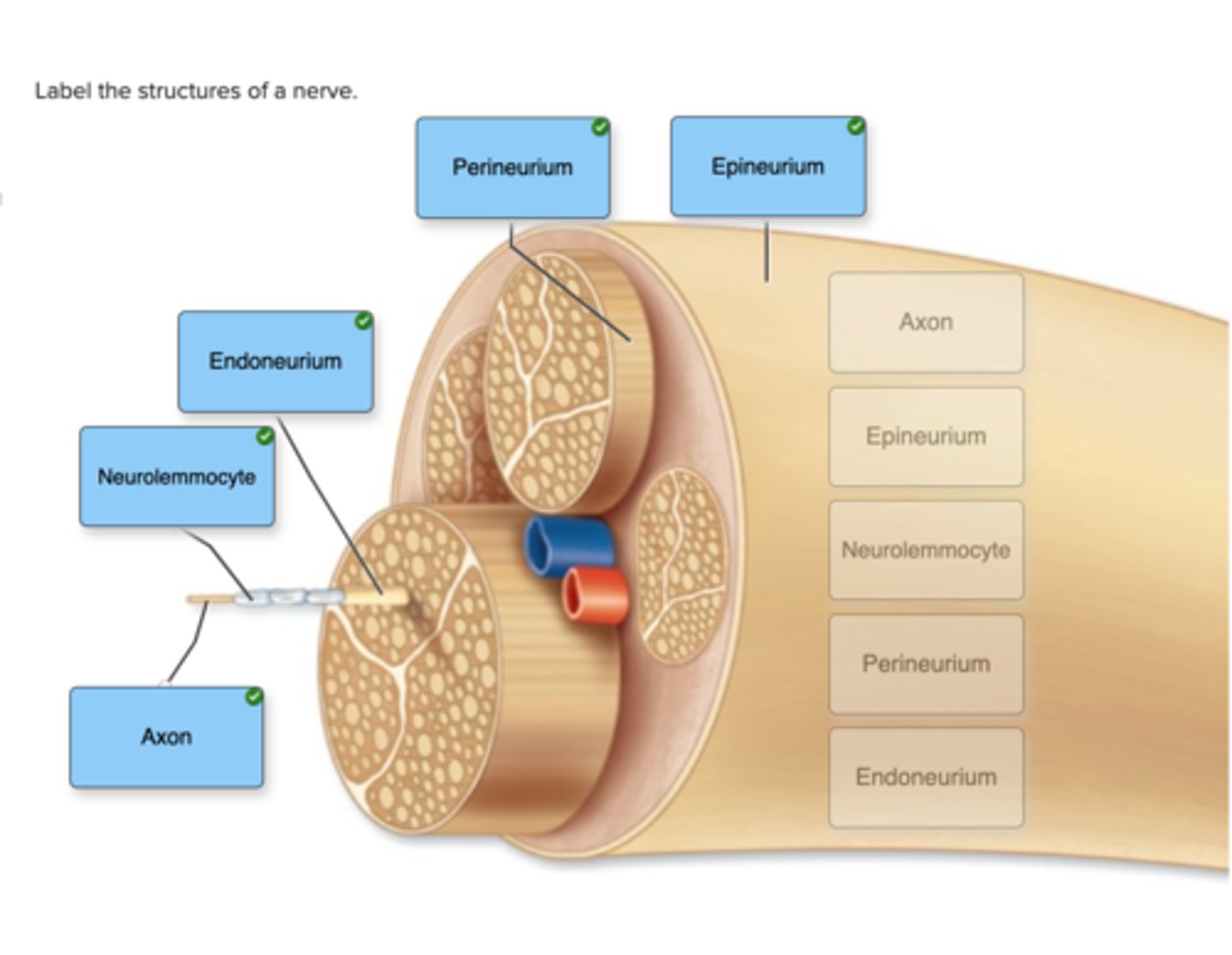
Label the structures of a nerve.
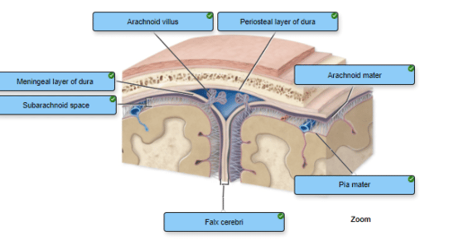
Correctly label the following meninges and associated structures.
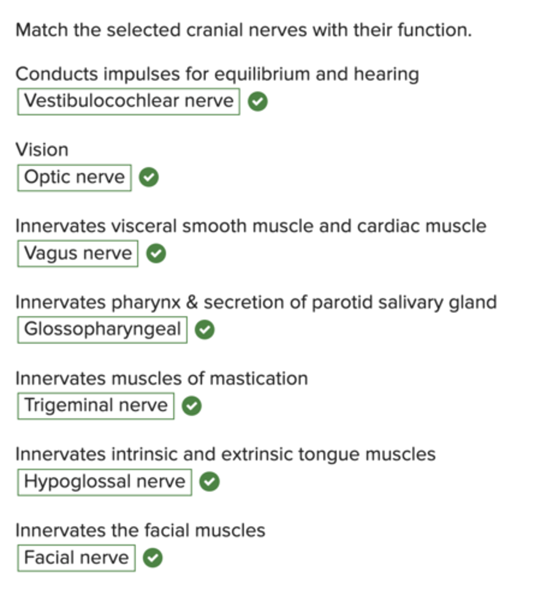
Match the selected cranial nerves with their function.
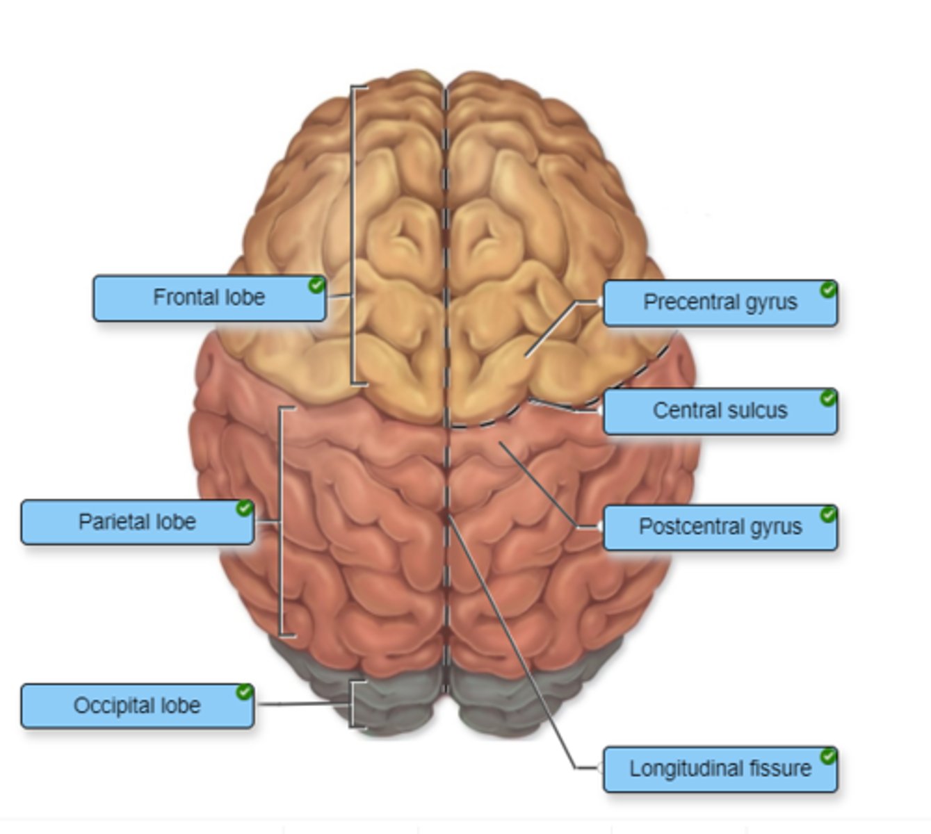
Identify the cerebral lobes on the left side of the figure. Label the additional cerebral structures on the right side of the figure.
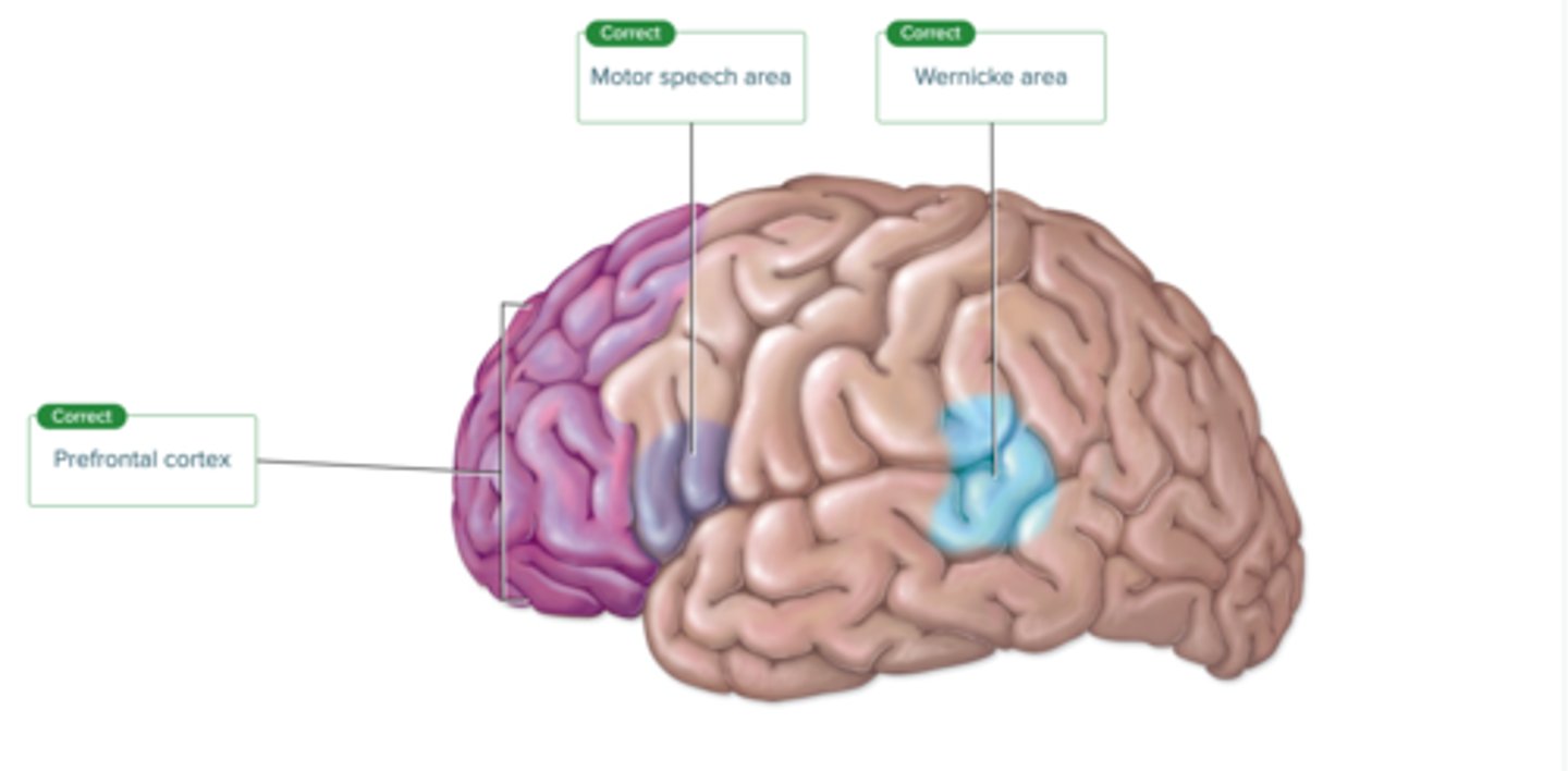
Label the regions involved in interpreting and carrying out speech information.
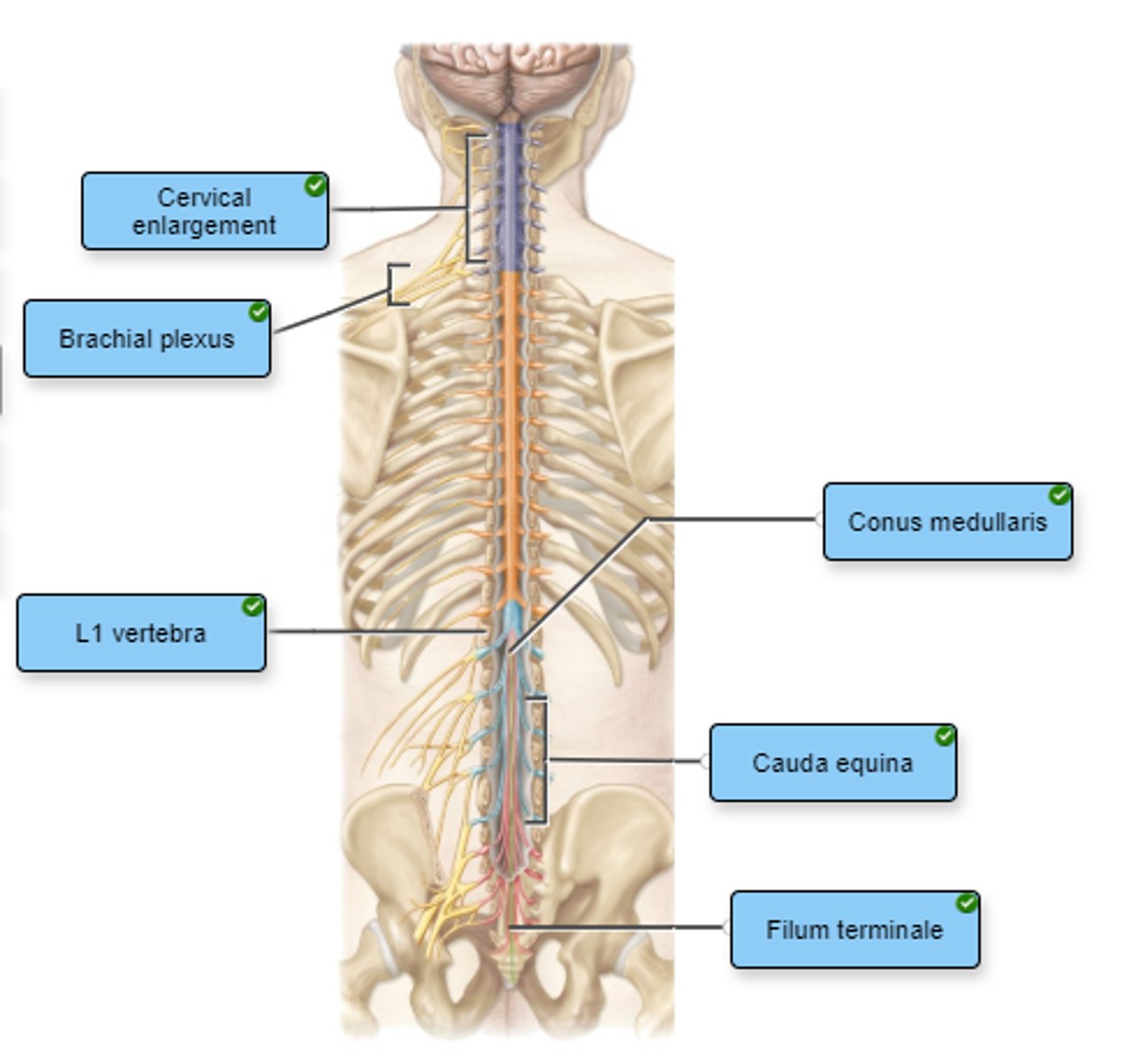
Label the structures of the spinal cord.
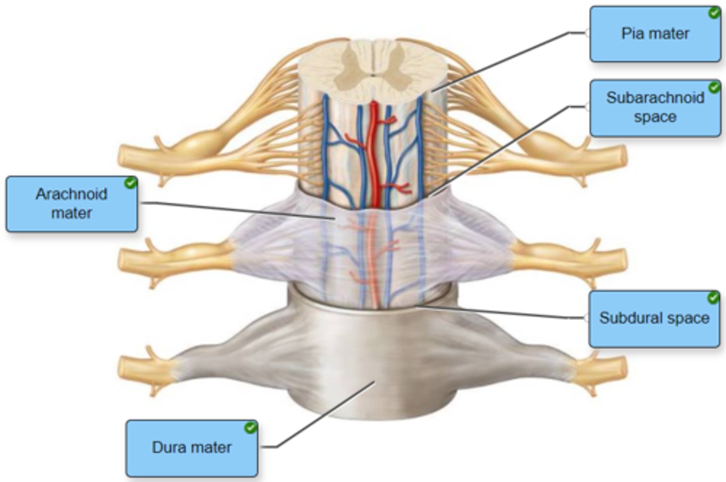
Label the spinal cord meninges and spaces.
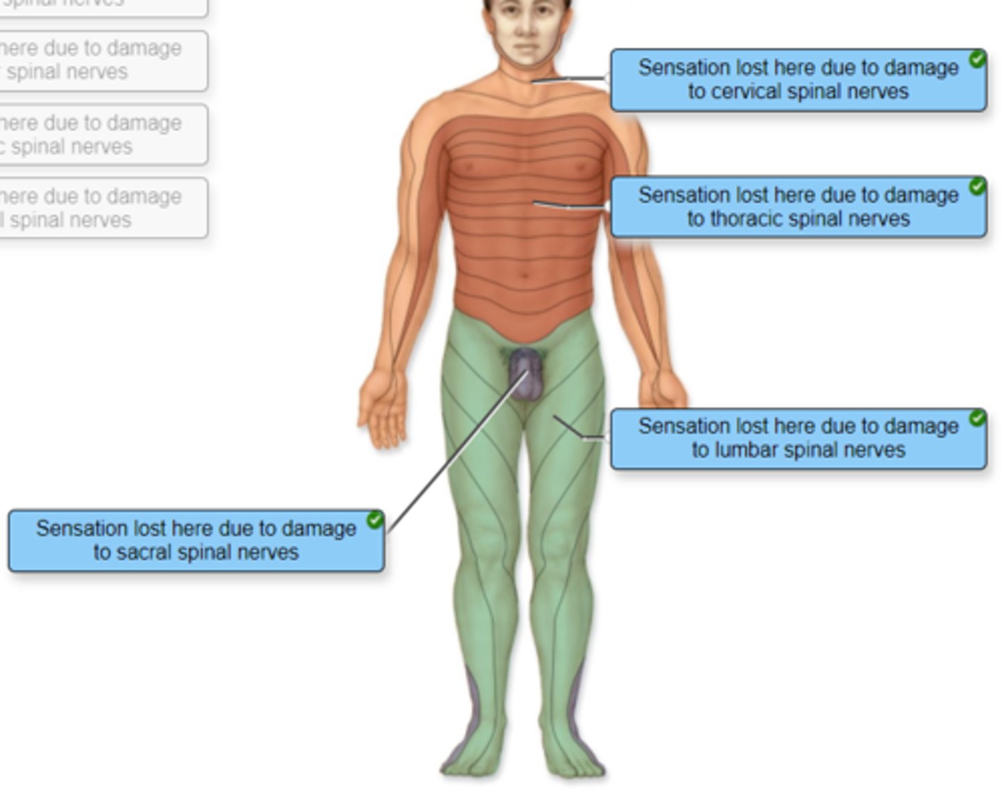
Correctly identify and label the dermatome(s) represented by the statement(s) associated with them.
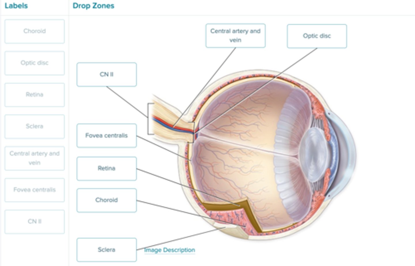
Label the figure with the items provided.
Drag and drop the labels into the appropriate location on the figure.
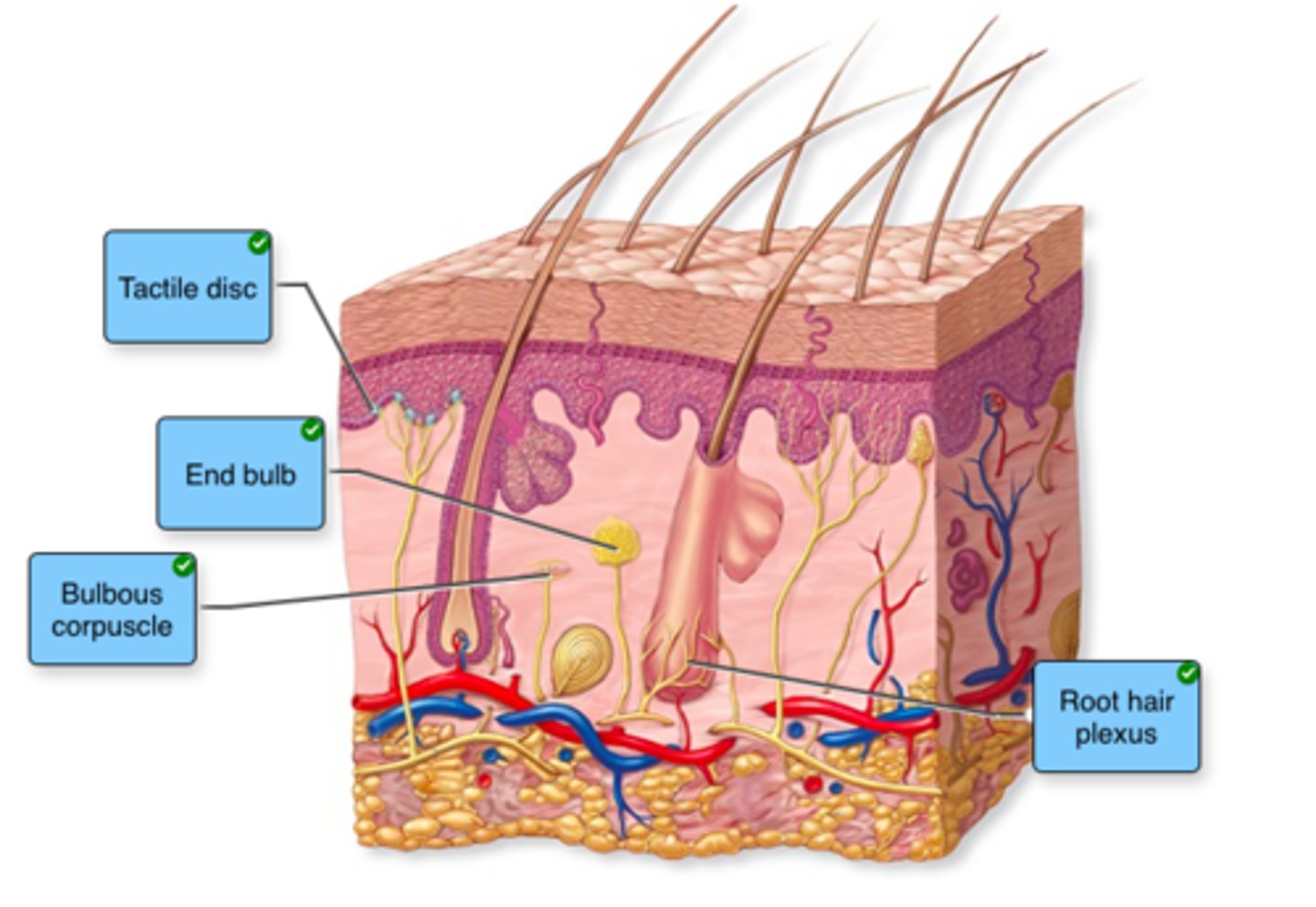
Label the type of tactile receptors in the image.
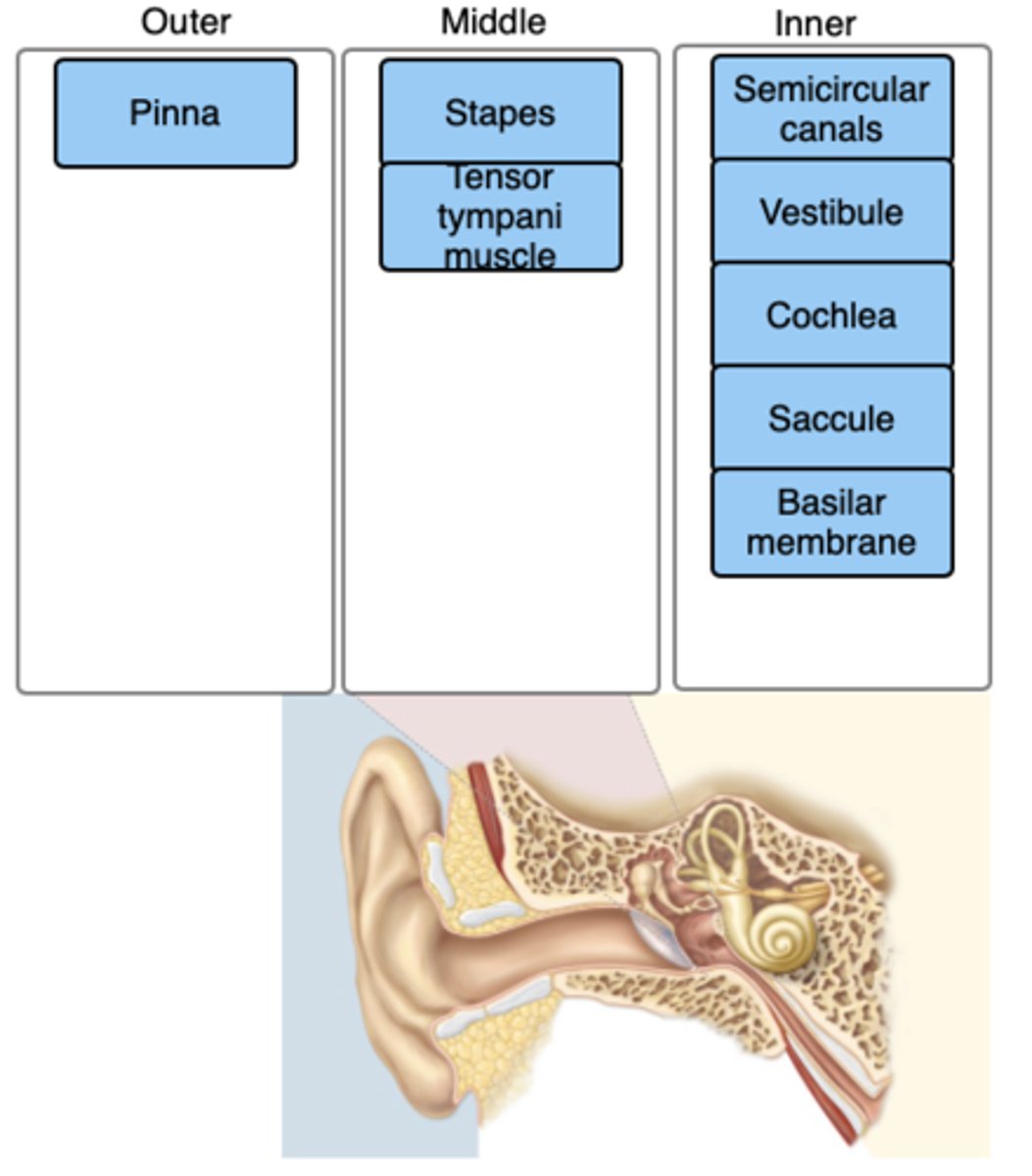
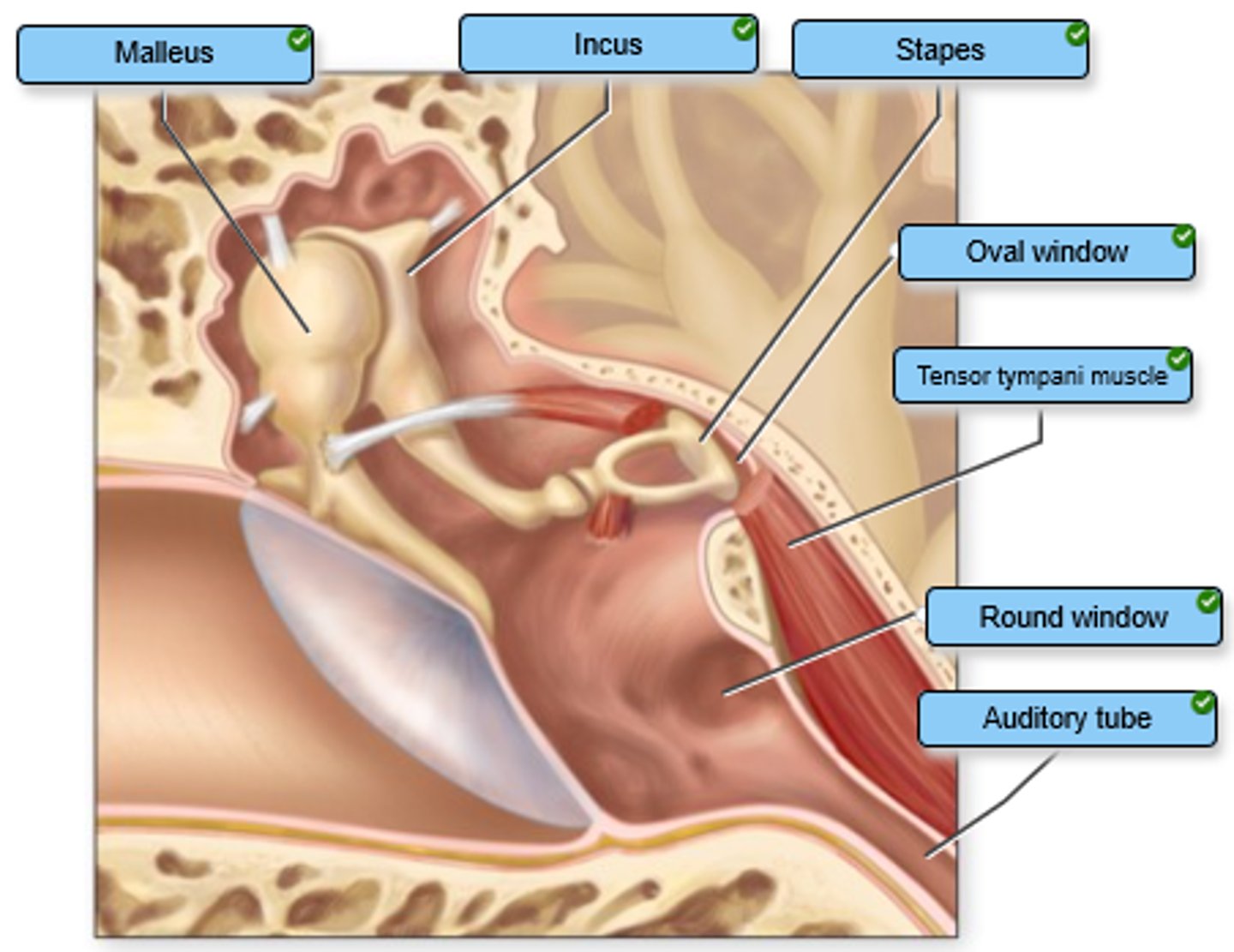
Label the structures of the middle ear.
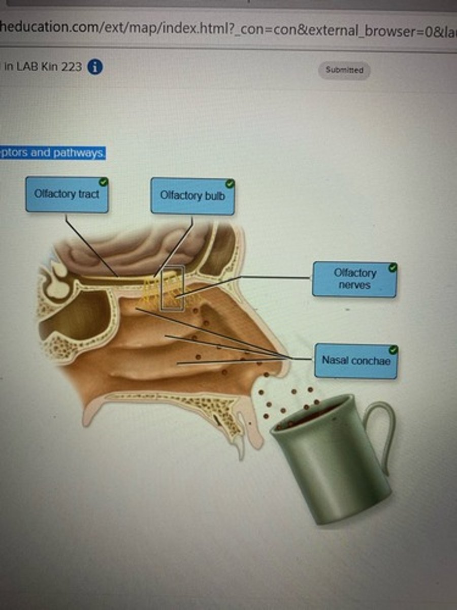
Label the olfactory receptors and pathways.
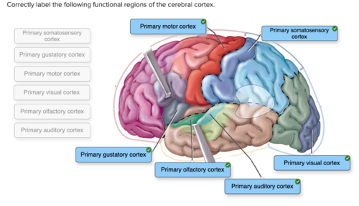
Correctly label the following functional regions of the cerebral cortex.
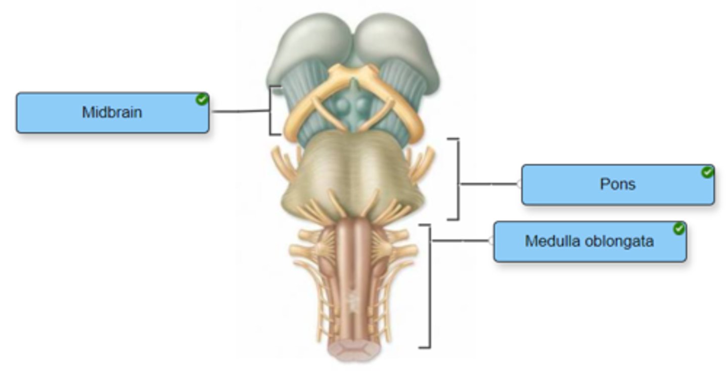
Identify the components of the brainstem.
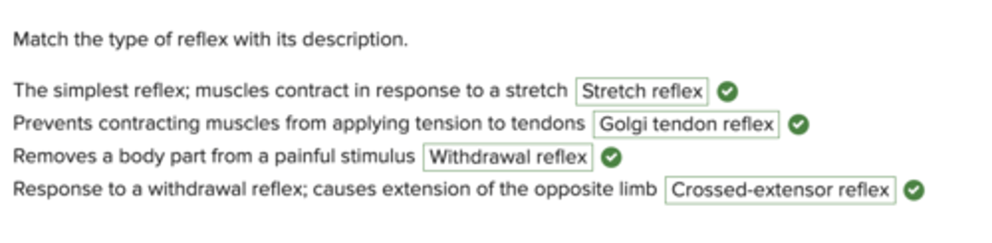
Match the type of reflex with its description.
Indicate whether the given structure is located in the outer, middle, or inner ear.