Lower Motor Neurons and Muscles and Reflex
1/32
There's no tags or description
Looks like no tags are added yet.
Name | Mastery | Learn | Test | Matching | Spaced | Call with Kai |
|---|
No analytics yet
Send a link to your students to track their progress
33 Terms
What is the Function of Lower Motor Neurons (LMNS)
They Receive commands from the Higher Motor Neurons and they directly control muscles and organs
They are efferent meaning they leave the CNS and to their target muscles
LMNS can be two types of neurons what are they?
They Can be motor neurons in the spinal cord ventral horn
Autonomic Ganglion Neurons (Pre and Post, thus forming two LMNS)
In Simple terms what is the function of the LMNS in the motor neurons in the spinal cord ventral horn
Their axons travel in the ventral roots of the spinal cord and innervate their target muscles '
This is the Somatic Motor System
What is the Function of the Autonomic Ganglion Neurons
They are Pre-Ganglionic and Post-Ganglionic, thus forming two LMNS
Their function is too innervate the smooth muscles, organs and glands
They form the Autonomic Nervous System
What are the Main Motor Neurons that are found in the Somatic Motor System (The Ventral horn of the Spinal Cord)
These are the Alpha Motor Neurons

What is a Motor Unit
This comprises of a Motor Neuron and all the muscles fibers or Muscle Cells it will innervate
One Motor Neuron + the Muscle cells it innervates
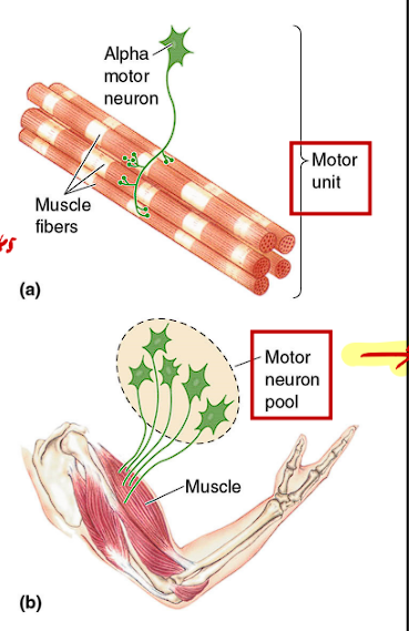
What is a Motor Neuron pool
This is all the Motor Neurons that are needed to innervate a single muscle (Bicep)
All the Motor Neurons → One Muscle (Bicep)
The Ventral Root/Horn is the motor part of the spinal cord
yes this is true and important
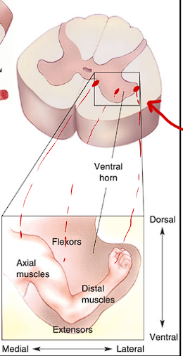
The Ventral Horn of the Spinal Cord contains what type of representation
It contains a somatotopic Representation
Distal Muscles such as the Digits are represented in the peripheral areas of the Ventral Horn
The Axial and central muscles such as the core are represented in the more central area of the Ventral Horn
What are the two type of muscles
Smooth Muscles
Striated Muscles
What are some examples of Smooth Muscle
Muscles of the digestive tract, arteries and related structures
What are some examples of striated muscle
Cardiac muscle, skeletal muscles.
Striated due to myosin and actin filaments
What are the three kind of skeletal muscle fibers
Red Fibers (Type I)
White Fibers (Type IIB)
Intermediate Fibers (Type IIA)
What are some Traits of the Red Fibers (Skeletal Muscle)
They are red due to their abundance in mitochondria
They are thin
They contract weakly and slowly
Thus they are able to sustain contractions for a longer period of time
They are composed of slow motor units
Example - Long Distance Runners
What are some Traits of the White fibers
They are larger
They use aerobic metabolism
They contract in brief and powerful twitches:
They fatigue rapidly
They are composed of fast motor units
example - A boxer, sprinter
What are the Intermediate fibers for (Skeletal Muscle)
They share properties between red and white fibers
Axial Muscles pertain to
The Core, The trunk and the abs
Distal Muscles pertain to
Hands, feet, digits
Proximal muscles pertain to
Shoulder, Elbow, pelvis (areas that are the first point of joint connection)
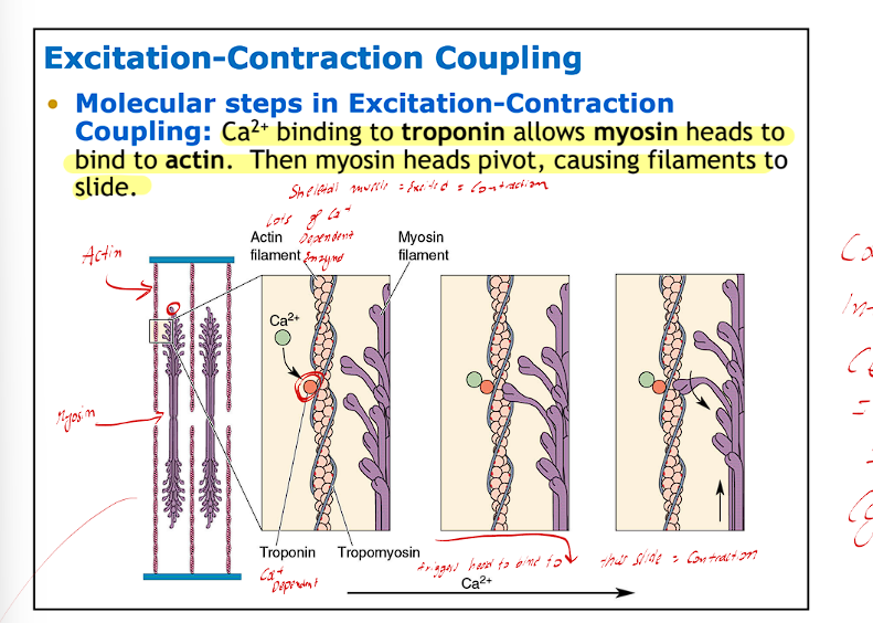
In Simple Terms what is Excitation-Contraction Coupling (You will come back to this)
Alpha motor neurons will release ACH
The Muscle receptors will receive this and will open ACH Receptors
This will cause the opening of Ion Channels
Thus an influx of Na+ and Ca+
This will depolarize the muscle allowing for the opening of V-G Ca2+ Channels'
this will further cause activation of the SR (Holds more intracellular Ca2+)
This is what causes the sliding mechanism for the Actin and Myosin filaments (Contraction)
What is a reflex
It is an involuntary, specific, stereotyped motor response to a sensory input
Most reflexes are very simple and will involve what?
1 Sensory neuron from the DRG (afferent) 1 Efferent Motor Neurons, and 1 or more interneurons (for reciprocal inhibition)
The Simplest reflex is what? what does this mean
The Simplest reflex are monosynaptic, meaning they involve two neurons (one sensory and one motor)
What are Supra-Spinal Mechanisms, what is their job
This is how reflexes can be suppressed
e.g. - food that you cooked is super hot and naturally, or by reflex you will drop is, however since you made it for a loved one it will not be instantly dropped but with a certain time
Myotatic Reflex / Stretch Reflex / Tendon Reflex
Traits'
It will cause the stretch of muscles and tendons
Muscle spindles that detect muscle length will notice stretch and will send info afferently to the ventral horn
This will activate the alpha motor neurons to efferently cause the same group of muscles to contract
The muscle that stretches will be contracted (via Extrafusal muscle fibers
This is a simple reflex (Monosynaptic)
Examples
Patellar Tendon Reflex
Achilles Tendon Reflex
The Extrafusal muscle fibers used are innervated by the Alpha Motor Neurons
The Muscle spindles used are innervated by the Gamma (y) motor neurons
Reciprocal Inhibition during the myotatic stretch reflex
The same sensory info that is sent to the ventral horn will go to a different set of Alpha Motor neurons via inhibitory interneurons that will produce inhibitory signals to these new alpha motor neurons to control the antagonistic muscles to relax
If the synergist muscle contracts, the antagonist muscle must contract
Pathway for Stretch Reflex
Stretch of Muscle Spindle
Activate Type IA and type II sensory fibers
Info carried to spinal cord
excitatory synapse to Alpha Motor Neurons
Carried to extrafusal muscle fibers, causing synergist to contract
Reverse/Inverse Myotatic Reflex (Autogenic Inhibition)
This shows how muscle tension can lead to inhibition of motor neurons
Traits
This is done through the Golgi Tendon Organs which are specialized to signal information on muscle tension
They are inhibitory to the muscles they innervate
They send info via IB afferent sensory fibers and result in feedback control to prevent excessive stretch
Regulate Muscle tension within an optimal range
Damage to this causes Clasp-Knife response -kind of like a army knife, hard to push knife down initially, but suddenly goes right back into place
Flexor Reflex/Withdrawal Reflex
It is often based on painful stimuli that can elicit coordinated withdrawal reflexes
Traits:
The flexor Reflex is initiated by cutaneous receptors and involves a whole limb, and thus transduces through multiple spinal cords (thus polysynaptic reflex)
In the Stretch Reflex the Alpha motor neurons will interact with extensor muscles, in this case, they will interact with flexor muscles
Example - Left side noxious stimuli
causes the left side to be activated (the left leg will contract)
The extensor for this leg is inactive
It can be inhibited by the supra-spinal mechanism
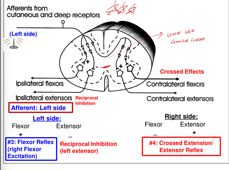
Reciprocal inhibition and crossed effects in reflex
Reciprocal inhibition occurs in all types of reflexes
Reflex activity in a given muscle produces similar activity in its ipsilateral synergists and the opposite activity in its ipsilateral antagonists
This is important in the Crossed Extensor Reflex
This is important for walking and maintaining balance
A flexor reflex on one side will cause the opposite pattern activity in the contralateral limb
ex - Stepping on a sharp object with the left foot left foot withdrawal
(Flexor reflex), while the right leg extends to support the body (Crossed
extension reflex).
Crossed-extensor reflex: Activation of extensor muscles and inhibition of flexors on the opposite side
What is the sarcoplasimic reticulum
The sarcoplasmic reticulum is a specialized endoplasmic reticulum in muscle cells that stores calcium ions (Ca2+).
During excitation-contraction coupling, the sarcoplasmic reticulum releases stored Ca2+ into the muscle cell's cytoplasm following depolarization. This influx of calcium initiates muscle contraction by enabling the sliding mechanism between actin and myosin filaments.
Excitation-Contraction Coupling further
The motor neurons release Acetylcholine (ACh). ACh produces large EPSP in the post- synaptic muscle fiber (aka. End- Plate Potential; EPP) The summation of EPPs, or when an EPP overcomes the threshold, then EPP evokes muscle action potential. Muscle Action potential: a. cause Ca2+ influx through plasma voltage- gated calcium channels; then b. triggers Ca2+ release from SR (sarcoplasmic reticulum). Muscle fiber (cell) contracts. Ca2+ reuptake then the fiber relaxes.
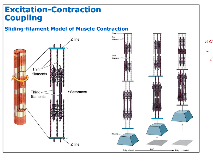
What is the molecular basis for Muscle Contraction (Z line stuff)
Z Lines - a division of myofibril into segments by disks
Sarcomere: Two Z lines and the myofibril
Thin Filaments are a series of bristles
Thick Filaments are found between and among thin filaments
The Binding of Ca2+ to Troponin is what causes the myosin to bind to actin (Contraction-sliding)